Endoscopic ultrasound-guided drainage of a right liver fistula caused by percutaneous thermoablation
Angelica Toppeta, Jean-Philippe Ratone, Solene Hoibian, Yanis Dahel, Antoine Assaf, Marc Giovannini, Fabrice Caillol

Abstract
Genes, proteins, chemicals, diseases, species, mutations and cell lines named across the full text — each resolved to its canonical identifier and authoritative record.
Click any figure to enlarge with its caption.
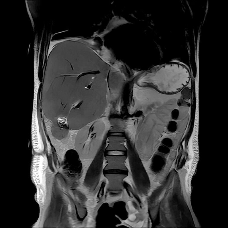 Fig. 1
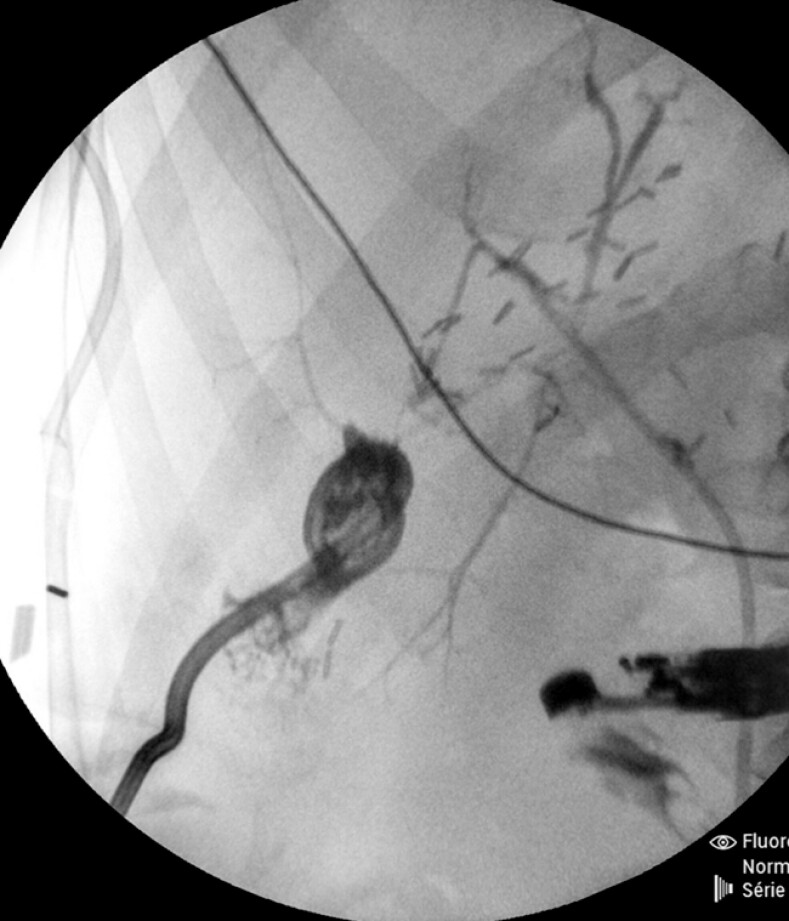 Fig. 2
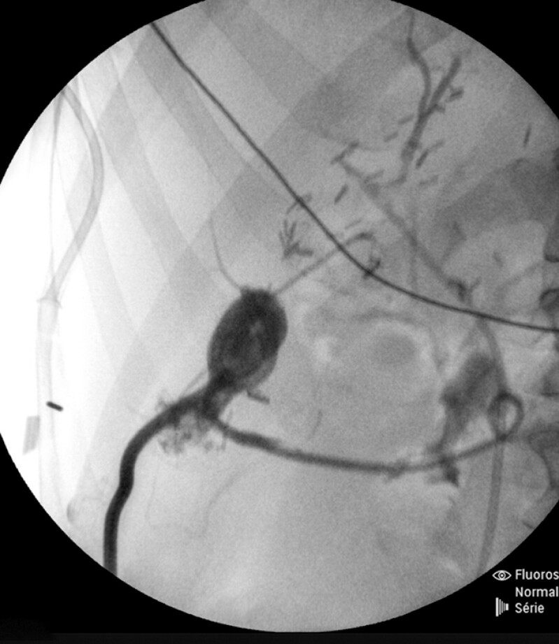 Fig. 3
Peer Reviews
No public reviews on file for this paper yet. If you reviewed it on a platform where reviews are public (OpenReview, ICLR, NeurIPS, ICML), you can paste yours below so the community can read it here.
Videos
No videos yet. Explain this paper in a talk, walkthrough, or lecture? Add one.
Taxonomy
TopicsGallbladder and Bile Duct Disorders · Biliary and Gastrointestinal Fistulas · Esophageal and GI Pathology
We present a case of a 35-year-old man who developed a biloma in the right liver following thermoablation for liver metastasis. His medical history included distal splenopancreatectomy for grade 2 neuroendocrine tumors, followed by left hepatectomy for liver metastasis 5 years before. The patient recently underwent chemotherapy and a few sessions of percutaneous thermoablation for liver metastases. The biloma was treated via percutaneous drainage ( Fig. 1 ). However, after noting that the biloma was only partially resolved, a biliary fistula was identified. Endoscopic retrograde cholangiopancreatography was attempted, but the bile ducts were atrophied; thus, cannulation of the target bile duct failed. Neither sphincterotomy nor plastic stent placement in the right liver resolved the fistula. After multidisciplinary discussions, we opted to internalize the biliary fistula via endoscopic ultrasound (EUS)-guided drainage. This approach was selected based on research indicating that the right liver is accessible via EUS 1 2 3 . A curved linear therapeutic EUS-scope was introduced into the duodenum under fluoroscopy. Contrast and saline solution were injected into the fistula through a percutaneous drain to delineate the trajectory under EUS and fluoroscopy ( Fig. 2 ). The fistula was punctured from the duodenum using a 19-G needle, and a 0.025-in guidewire was inserted. The tract was first dilated using a 6-Fr cystotome and then a 4-mm biliary balloon catheter. A double pigtail stent was inserted into the fistula and duodenum. Minor bleeding occurred but ceased spontaneously. Contrast was injected via the percutaneous drain, and successful drainage of the contrast agent into the duodenum confirmed correct placement of the stent ( Fig. 3 ). One week later, a second double pigtail stent was placed parallel to the first ( Video 1 ). The percutaneous drain was removed 48 hours later. This video shows a valid EUS-guided alternative for internalizing a biliary fistula from the duodenum when standard methods fail.
IRM image showing a biliary fistula in the right hepatic lobe and biloma.
Fluoroscopy image of the biliary fistula after contrast agent injection via the percutaneous drain.
Fluoroscopy image showing contrast medium draining into the duodenum.
EUS-guided drainage of a right liver fistula from the duodenum with two double pigtail stents.Video 1
Endoscopy_UCTN_Code_TTT_1AS_2AH
The reference list from the paper itself. Each links out to its DOI / PubMed record.
- 1Nakai Y Matsubara S Mukai T Drainage for fluid collections post pancreatic surgery and acute pancreatitis: similar but different?Clin Endosc 20245773574610.5946/ce.2023.25438756067 PMC 11637669 · doi ↗ · pubmed ↗
- 2Caillol F Rouy M Pesenti C Drainage of the right liver using EUS guidance Endosc Ultrasound 2019801 S 50S 5610.4103/eus.eus_52_1931897380 PMC 6896427 · doi ↗ · pubmed ↗
- 3Shah J Jena A Singh AK Endoscopic Ultrasound (EUS)-guided Drainage of Caudate Lobe Abscess: A Single Center Experience Surg Laparosc Endosc Percutan Tech 20233368268710.1097/SLE.000000000000122937725821 · doi ↗ · pubmed ↗