A Typical Dermoscopic Pattern of PLEVA
Mahesh Mathur, Sumit Paudel, Supriya Paudel, Sambidha Karki, Sandhya Regmi, Nabita Bhattarai

TL;DR
The paper introduces a new dermoscopic pattern for diagnosing PLEVA, a skin condition, offering a non-invasive alternative to biopsies.
Contribution
The paper presents a novel dermoscopic pattern specific to PLEVA for improved diagnosis.
Findings
A new dermoscopic pattern for PLEVA was identified.
This pattern can help diagnose PLEVA non-invasively.
Skin biopsy is less effective for PLEVA diagnosis due to non-specific features.
Abstract
PLEVA is a benign inflammatory disorder. Dermoscopy is a non‐invasive diagnostic modality. Thus, the new dermoscopic pattern mentioned in our literature can aid in the diagnosis of the disease, unlike the skin biopsy, which is an invasive modality and shows non‐specific features in PLEVA.
Genes, proteins, chemicals, diseases, species, mutations and cell lines named across the full text — each resolved to its canonical identifier and authoritative record.
Click any figure to enlarge with its caption.
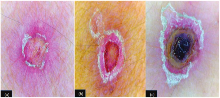 Figure 1
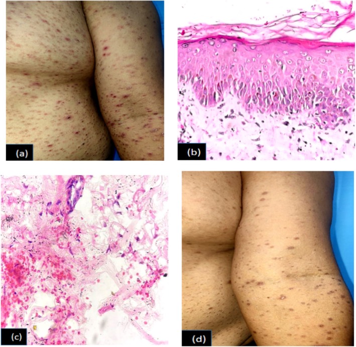 Figure 2
Peer Reviews
No public reviews on file for this paper yet. If you reviewed it on a platform where reviews are public (OpenReview, ICLR, NeurIPS, ICML), you can paste yours below so the community can read it here.
Videos
No videos yet. Explain this paper in a talk, walkthrough, or lecture? Add one.
Taxonomy
TopicsAutoimmune Bullous Skin Diseases · Dermatological and Skeletal Disorders · Vascular Tumors and Angiosarcomas