Reversible Cortical Visual Impairment in an Adolescent Due to a Posterior Fossa Arachnoid Cyst: A Case Report
Jelena Škunca Herman, Dario Josip Živković, Ivana Orešković, Lana Knežević, Maja Malenica Ravlić, Blanka Doko Mandić, Goran Marić, Ante Vukojević, Hrvoje Sliepčević, Mia Zorić Geber, Vladimir Kalousek, Zoran Vatavuk

TL;DR
A 16-year-old girl experienced reversible vision loss due to a brain cyst, which improved after treatment.
Contribution
This is the first reported case of reversible cortical visual impairment caused by a posterior fossa arachnoid cyst in an adolescent.
Findings
The patient's vision rapidly recovered after cystoperitoneal drainage.
Functional tests confirmed bilateral cortical visual impairment without damage to the anterior visual pathways.
The case highlights posterior fossa lesions as a potential cause of unexplained bilateral visual loss.
Abstract
Background: Arachnoid cysts are typically benign and asymptomatic, but large cysts can exert a mass effect on adjacent neural structures. Based on the available literature, no cases of cortical visual impairment (CVI) in an adolescent caused by posterior fossa arachnoid cysts have been reported. Case presentation: We report the case of a previously healthy 16-year-old girl who presented with sudden and rapidly progressive bilateral visual loss due to a large retrocerebellar arachnoid cyst. She reported blurred vision, tunnel vision-like, and decreased visual acuity. Although neuro-ophthalmologic and imaging workup revealed no damage to the anterior visual pathways, she exhibited progressive visual decline. Functional tests confirmed bilateral cortical visual impairment: pattern-reversal visual evoked potentials (VEPs) showed preserved and symmetric P100 latencies and amplitudes, while…
Genes, proteins, chemicals, diseases, species, mutations and cell lines named across the full text — each resolved to its canonical identifier and authoritative record.
Click any figure to enlarge with its caption.
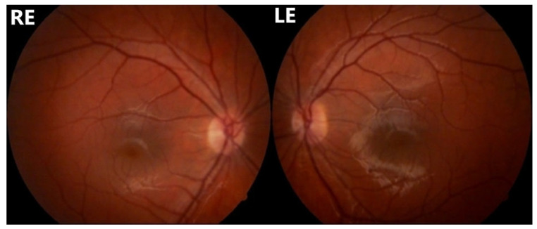 Figure 1
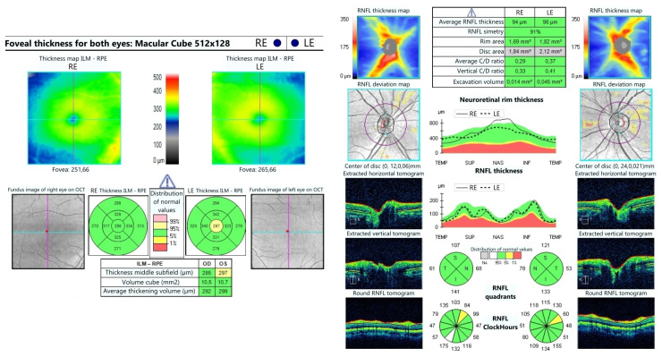 Figure 2
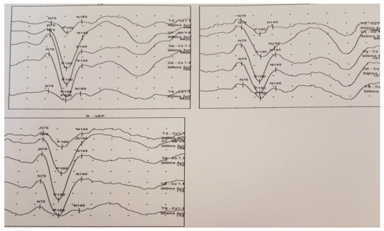 Figure 3
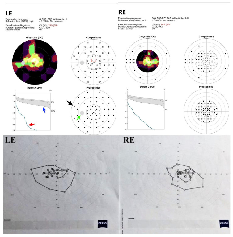 Figure 4
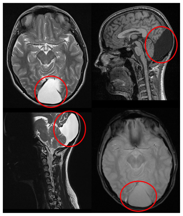 Figure 5
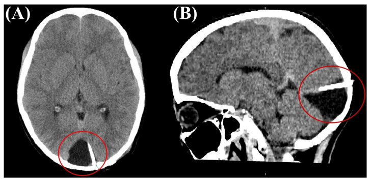 Figure 6
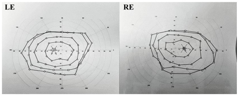 Figure 7
Peer Reviews
No public reviews on file for this paper yet. If you reviewed it on a platform where reviews are public (OpenReview, ICLR, NeurIPS, ICML), you can paste yours below so the community can read it here.
Videos
No videos yet. Explain this paper in a talk, walkthrough, or lecture? Add one.
Taxonomy
TopicsCerebrospinal fluid and hydrocephalus · Cerebral Venous Sinus Thrombosis · Vascular Malformations Diagnosis and Treatment