Assessment of Retinal Microcirculation in Primary Open-Angle Glaucoma Using Adaptive Optics and OCT Angiography: Correlation with Structural and Functional Damage
Anna Zaleska-Żmijewska, Alina Szewczuk, Zbigniew M. Wawrzyniak, Maria Żmijewska, Jacek P. Szaflik

TL;DR
This study shows that changes in retinal blood vessels can be detected early in glaucoma and correlate with disease severity, offering better tools for early diagnosis and monitoring.
Contribution
The study introduces combined use of adaptive optics and OCT angiography to detect early vascular changes in glaucoma, linking them to structural and functional damage.
Findings
Glaucoma eyes showed thicker arteriole walls and narrower lumens compared to healthy eyes.
OCTA revealed reduced vessel densities and enlarged FAZ areas in glaucoma patients.
Vessel densities correlated positively with structural OCT parameters and negatively with visual field deficits.
Abstract
Background: This study aimed to evaluate retinal arteriole parameters using adaptive optics (AO) rtx1™ (Imagine Eyes, Orsay, France) and peripapillary and macular vessel densities with optical coherence tomography angiography (OCTA) in eyes with different stages of primary open-angle glaucoma (POAG) compared to healthy eyes. It also investigated the associations between vascular parameters and glaucoma severity, as defined by structural (OCT) and functional (visual field) changes. Methods: Fifty-seven eyes from 31 POAG patients and fifty from 25 healthy volunteers were examined. Retinal arteriole morphology was assessed using the AO rtx1™-fundus camera, which measured lumen diameter, wall thickness, total diameter, wall-to-lumen ratio (WLR), and wall cross-sectional area. OCTA was used to measure vessel densities in superficial (SCP) and deep (DCP) capillary plexuses of the macula and…
Genes, proteins, chemicals, diseases, species, mutations and cell lines named across the full text — each resolved to its canonical identifier and authoritative record.
Click any figure to enlarge with its caption.
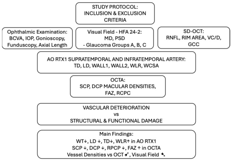 Figure 1
Peer Reviews
No public reviews on file for this paper yet. If you reviewed it on a platform where reviews are public (OpenReview, ICLR, NeurIPS, ICML), you can paste yours below so the community can read it here.
Videos
No videos yet. Explain this paper in a talk, walkthrough, or lecture? Add one.
Taxonomy
TopicsGlaucoma and retinal disorders · Retinal Imaging and Analysis · Retinal Diseases and Treatments