Regeneration of Biomechanically Functional Tendon Tissue Following Injection of Uncultured, Autologous, Adipose-Derived Regenerative Cells into Partial Achilles Tendon Defects in Rabbits
Christoph Schmitz, Christopher Alt, Tobias Wuerfel, Stefan Milz, Jacqueline Dinzey, Ashley Hill, Katie J. Sikes, Lindsey H. Burton, Jeremiah Easley, Holly L. Stewart, Christian M. Puttlitz, Benjamin C. Gadomski, Kevin M. Labus, David A. Pearce, Nicola Maffulli, Eckhard U. Alt

TL;DR
Injecting unmodified fat-derived cells into rabbit tendon injuries promotes tissue regeneration and improves biomechanical properties compared to untreated injuries.
Contribution
This study demonstrates that uncultured, autologous adipose-derived regenerative cells can regenerate functional tendon tissue in a rabbit model.
Findings
UA-ADRC-treated tendons showed new connective tissue formation, indicating regeneration rather than scarring.
Biomechanical testing revealed higher viscoelasticity in UA-ADRC-treated tendons compared to controls.
The results suggest UA-ADRC therapy could be a viable treatment for partial tendon tears.
Abstract
Current treatment strategies for partial tendon tears often lack the capacity to promote true tissue regeneration and improve long-term clinical outcomes. This study tested the hypothesis that treatment of a partial defect in the rabbit common calcaneus tendon (CCT) with uncultured, unmodified, autologous, adipose-derived regenerative cells (UA-ADRCs) enables regenerative healing without scar formation. A full-thickness, 3 mm defect was produced in the midsubstance of the right gastrocnemius tendon, a component of the CCT, in adult female New Zealand white rabbits. Animals received either an injection of 28.3 × 10⁶ UA-ADRCs in 0.5 mL Ringer’s lactated solution (RLS) or saline, or RLS or saline alone as sham treatment. Tendons were analyzed 4 or 12 weeks post-treatment using histology, immunohistochemistry and non-destructive biomechanical testing. UA-ADRC-treated tendons showed newly…
Genes, proteins, chemicals, diseases, species, mutations and cell lines named across the full text — each resolved to its canonical identifier and authoritative record.
Click any figure to enlarge with its caption.
 Figure 1
 Figure 2
 Figure 3
 Figure 4
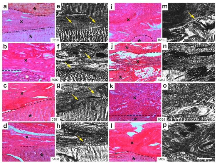 Figure 5
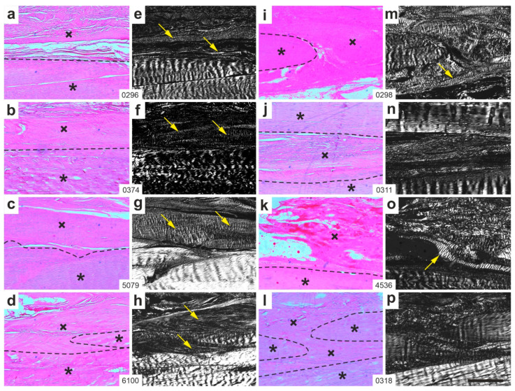 Figure 6
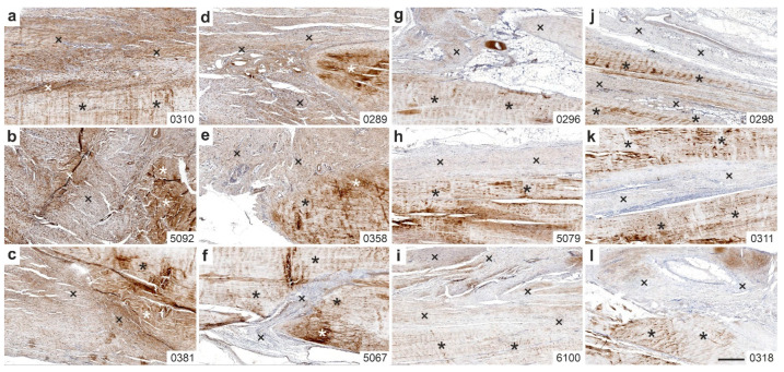 Figure 7
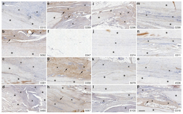 Figure 8
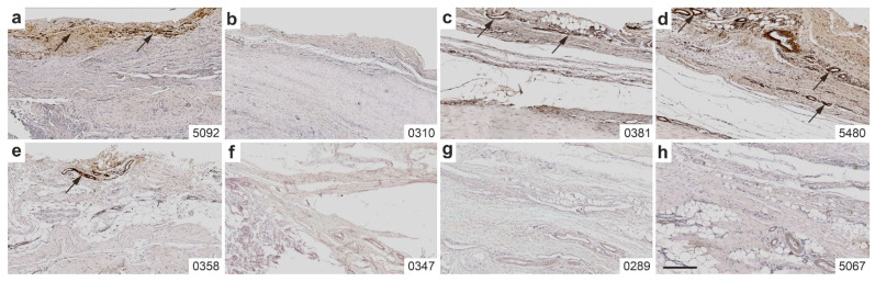 Figure 9
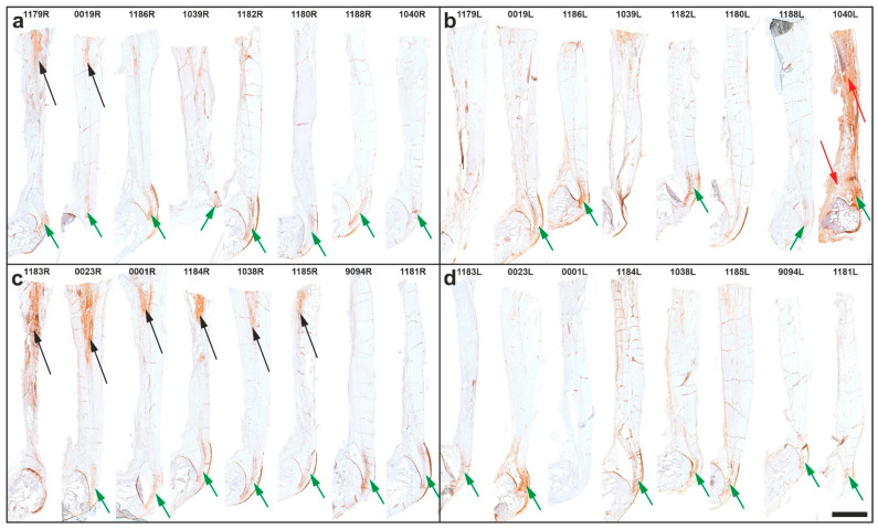 Figure 10
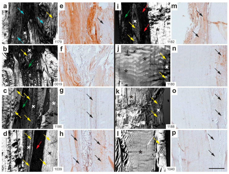 Figure 11
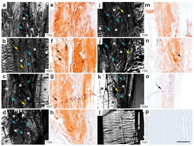 Figure 12
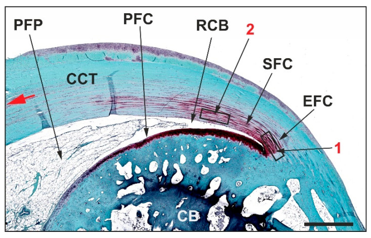 Figure 13
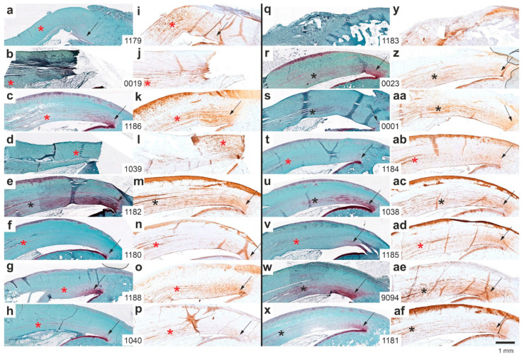 Figure 14
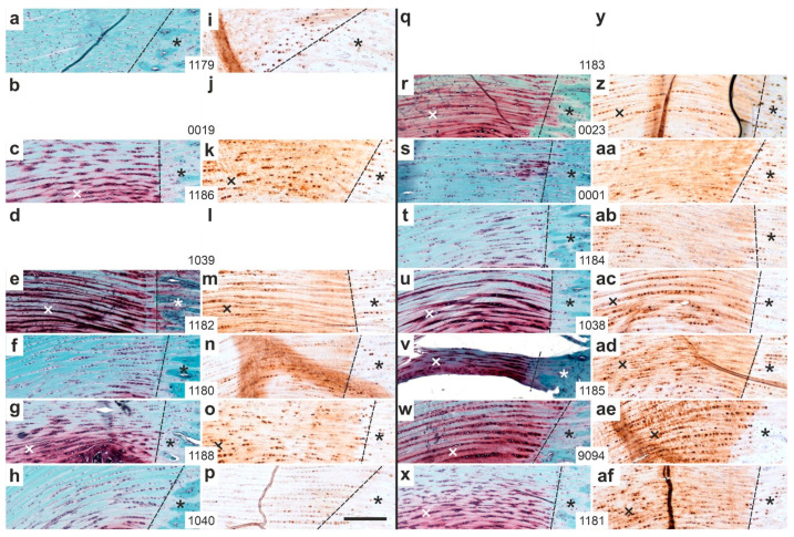 Figure 15
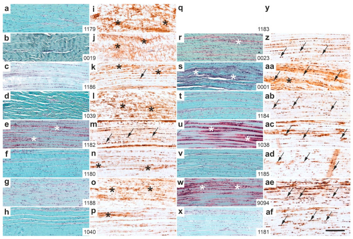 Figure 16
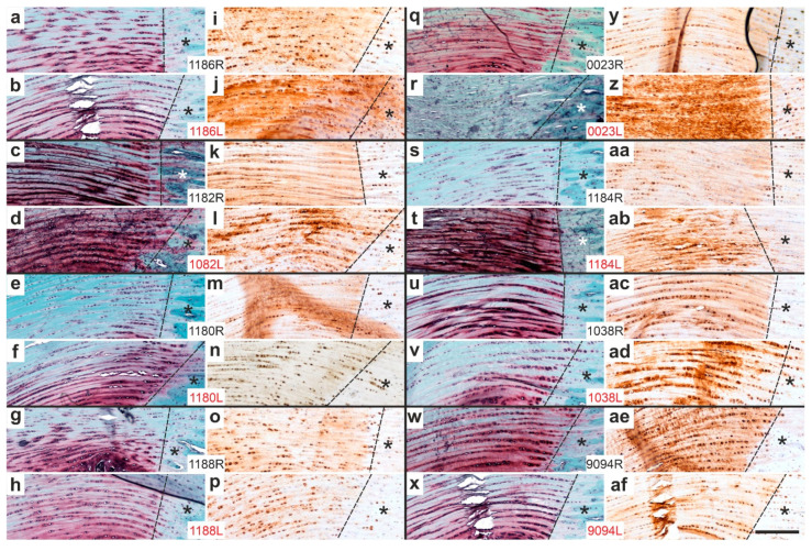 Figure 17
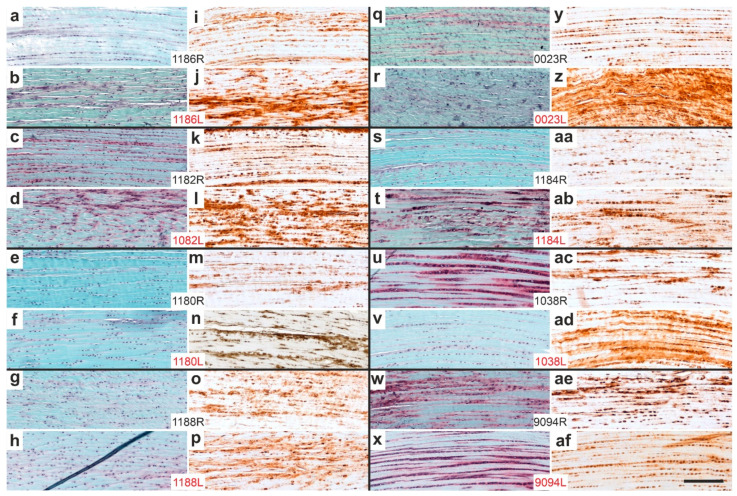 Figure 18
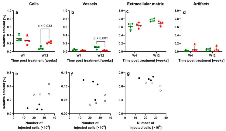 Figure 19
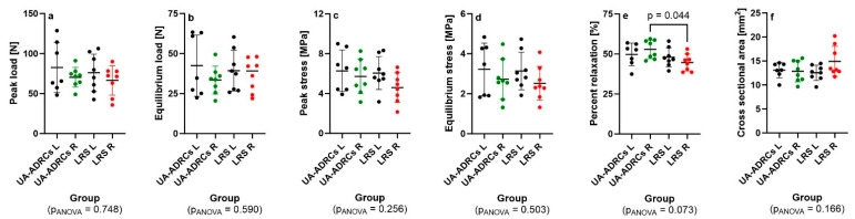 Figure 20
 Figure 21
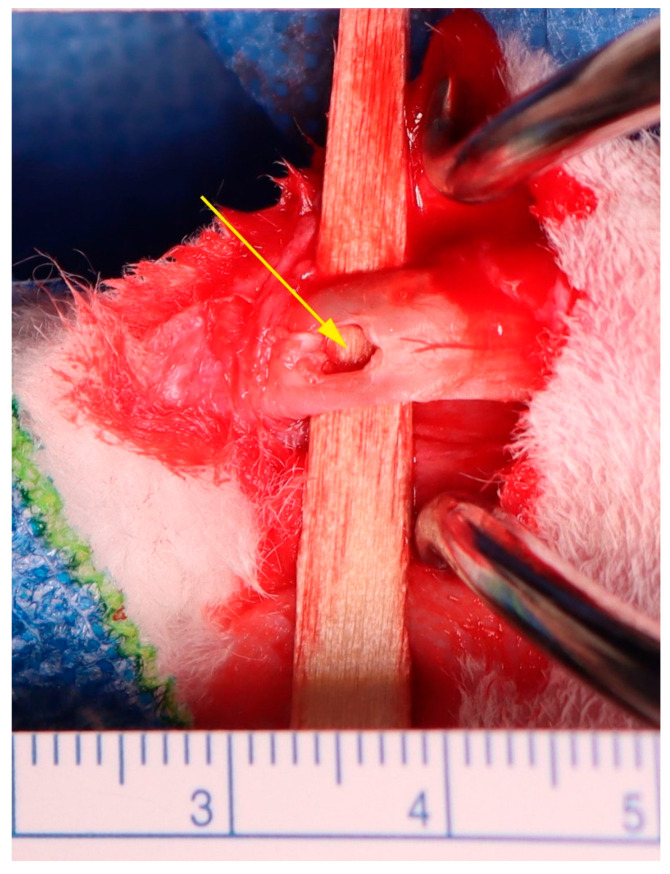 Figure 22
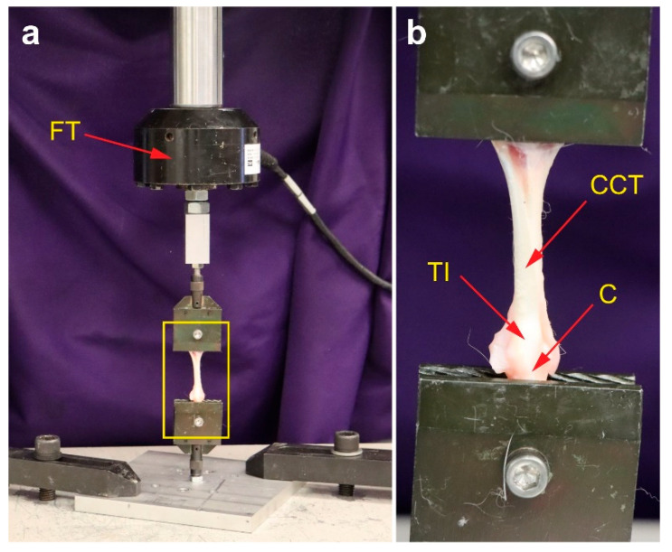 Figure 23
Peer Reviews
No public reviews on file for this paper yet. If you reviewed it on a platform where reviews are public (OpenReview, ICLR, NeurIPS, ICML), you can paste yours below so the community can read it here.
Videos
No videos yet. Explain this paper in a talk, walkthrough, or lecture? Add one.
Taxonomy
TopicsTendon Structure and Treatment · Wound Healing and Treatments · Sports injuries and prevention