Platelet-Rich Plasma (PRP) Mitigates Silver Nanoparticle (AgNP)-Induced Pulmonary Fibrosis via iNOS/CD68/CASP3/TWIST1 Regulation: An Experimental Study and Bioinformatics Analysis
Shaimaa R. Abdelmohsen, Ranya M. Abdelgalil, Asmaa M. Elmaghraby, Amira M. Negm, Reham Hammad, Eleni K. Efthimiadou, Sara Seriah, Hekmat M. El Magdoub, Hemat Elariny, Islam Farrag, Nahla El Shenawy, Doaa Abdelrahaman, Hussain Almalki, Ahmed A. Askar, Marwa M. El-Mosely

TL;DR
This study shows that platelet-rich plasma (PRP) can reduce lung damage caused by silver nanoparticles in rats, with effects similar to a common anti-inflammatory drug.
Contribution
The study is the first to demonstrate PRP's antiapoptotic and antifibrotic effects in AgNP-induced pulmonary fibrosis in an experimental rat model.
Findings
PRP significantly reduced body weight loss and hydroxyproline levels in AgNP-treated rats.
PRP decreased CASP3 and TWIST1 gene expression, mitigating AgNP-induced lung fibrosis.
PRP improved histopathological and immunohistochemical changes comparable to dexamethasone.
Abstract
Platelet-rich plasma (PRP) has become an increasingly valuable biologic approach for personalized regenerative medicine because of its potent anti-inflammatory/healing effects. It is thought to be an excellent source of growth factors that can promote tissue healing and lessen fibrosis. Although this treatment has demonstrated effectiveness in numerous disease areas, its impact on pulmonary fibrosis (PF) caused by silver nanoparticles (AgNPs) via its antiapoptotic effects remains to be explored. AgNPs were synthesized biologically by Bacillus megaterium ATCC 55000. AgNP characterization was carried out via UV–Vis spectroscopy, X-ray diffraction (XRD), dynamic light scattering (DLS), transmission electron microscopy (TEM), and scanning electron microscopy (SEM) imaging to reveal monodispersed spheres with a mean diameter of 45.17 nm. A total of 48 male Wistar rats divided into six…
Genes, proteins, chemicals, diseases, species, mutations and cell lines named across the full text — each resolved to its canonical identifier and authoritative record.
Click any figure to enlarge with its caption.
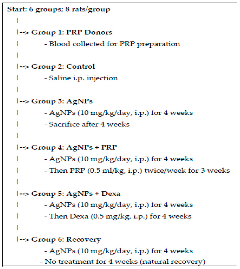 Figure 8
Peer Reviews
No public reviews on file for this paper yet. If you reviewed it on a platform where reviews are public (OpenReview, ICLR, NeurIPS, ICML), you can paste yours below so the community can read it here.
Videos
No videos yet. Explain this paper in a talk, walkthrough, or lecture? Add one.
Taxonomy
TopicsExtracellular vesicles in disease · Inhalation and Respiratory Drug Delivery
1. Introduction
1.1. Background
Pulmonary fibrosis (PF) is an incurable and progressive condition leading to lung adenocarcinoma or squamous cell carcinoma [1]. PF affects the flexibility of the alveoli and is characterized by infiltration of inflammatory cytokines and apoptosis, which might result in tissue hypoxia [2] and increased damage. The first-line treatment for PF is oral administration of glucocorticoids (dexamethasone (Dexa)), which are anti-inflammatory and antifibrotic [2]. Glucocorticoids cause unfavorable adverse reactions and impede the disease’s clinical progression; therefore, safe alternative treatments are a top priority. Moreover, if these alternative treatment options are natural, they would be better.
The “personalized regenerative medicine (PRM)” umbrella, rather than the one-size-fits-all treatment [3,4], involves the use of platelet-rich plasma (PRP). PRP has potential therapeutic effects through altering cellular activities [5], increasing cell proliferation, and enhancing cellular reconstruction in chondrocytes [6], fibroblasts [7], endometrial cells [8], and corneal cells [9]. PRP is currently a favorable, popular treatment option for many medical conditions because of its biosafety and autologous nature as well as its ease of collection and application [10].
Recently, nanotechnology has been used in a wide range of life sectors, such as medicine, diagnosis, pharmaceuticals, and beauty products [11,12]. Silver (Ag) nanoparticles (NPs) are widely used in electronics, food packaging, fabrics, water purification systems, and biomedical applications [13,14]. However, AgNPs interact with biological targets because of their small size (1–100 nm), making them easier than drugs of comparable sizes. This enables them to cross the normal plasma membrane barrier [15], contributing to the use of AgNPs for wound care, catheters, and dental supplies [16], as well as other uses, but, unfortunately, would contribute to their potential toxic effects [17,18].
1.2. Problem Statement/Hypothesis
One of the crucial organs exposed to NPs is the lung [19]. Insoluble NPs contribute to tissue damage, inflammation, and lung tumors, according to different reports [20,21]. To avoid the adverse effects of glucocorticoid use for PF treatment or AgNP-induced lung fibrosis injury, it is crucial to identify an alternative effective, safe, and more environmentally friendly, personalized treatment [22].
1.3. Aim
We examined whether PRP had better or comparable antifibrotic/antiapoptotic and regenerative abilities in the AgNP-induced PF experimental model than Dexa did. Furthermore, whether PRP mitigates AgNP-induced PF via the regulation of the fibrosis/apoptosis axis was experimentally investigated via bioinformatics analysis. The findings of the present study could lead to an innovative, personalized, relatively safe treatment option for PF.
2. Results
2.1. AgNP Characterization Results
As explained in Figure 1a, the UV–visible spectrum of the synthesized AgNPs presented a maximum absorbance (1.312 abs) at 401.5 nm. The average particle size, as determined by dynamic light scattering (DLS), is 53.7 nm, as illustrated in Figure 1b.
The transmission electron microscopy (TEM) results demonstrated that the size of the AgNPs ranged from 26.47 nm to 63.25 nm, with an average main diameter of 45.17 nm, as shown in Figure 1(c1,c2).
By scanning electron microscopy (SEM) imaging, the AgNPs were observed to have a spherical shape, all within the nanoscale range (Figure 1d).
The diffraction pattern produced by X-ray diffraction (XRD) provided information about the crystalline nature of the synthesized AgNPs (Figure 1e). The diffraction peaked at 2θ values of 38.1°, 44.3°, 64.4°, and 77.4°, corresponding to the 111, 200, 220, and 311 planes, respectively, of the face-centered cubic (fcc) structure of silver (JCPDS No. 04-0783). It is noteworthy to mention that no peaks related to impurities were observed.
2.2. Bioinformatics/In Silico Analysis (Figure 2)
2.2.1. DEGs from Online Datasets in Lung Cancer
To retrieve relevant gene expression data related to PF, we accessed the UCSC Xena Browser (https://xenabrowser.net, accessed on 8 May 2025) to select the dataset (TCGA lung adenocarcinoma or lung squamous cell carcinoma). The first variable was phenotypic sample types, such as primary tissue vs. normal solid tumors (https://xenabrowser.net/heatmap/, accessed on 8 May 2025) to compare the expression of different genes in available online datasets via the Xena Differential Gene Expression Analysis Pipeline (https://github.com/ucscXena, accessed on 8 May 2025) to perform Differential Gene Expression (DGE) analysis and further downstream analyses. The second variable was antiapoptotic gene expression (Figure 2a). Converting gene expression values into a principal component 3D plot for PCA is shown in Figure 2b. Moreover, the genes with their Log2FC, average expression, and p value are attached as a Supplementary Excel File.
2.2.2. Data Sourcing
TWIST1 is a protein-coding gene located on chromosome 7; 7p21.1 (−1 strand) and via the Clinical Interpretations of Variants in Cancer (CiViC) v2.2.33 (https://civicdb.org/features/5964/summary, 13 December 2023). TWIST1 inhibits the hypoxia-inducible factor-2-alpha (HIF-2-alpha) transcription factor network, cytokine signaling in the immune system, disease, immune system, generic transcription pathway, signaling by interleukins, disease signal transduction by growth factor receptors and second messengers, interleukin-4 and interleukin-13 signaling, RNA polymerase II transcription, gene expression (transcription), regulation of RUNX2 expression and activity, and more biological processes/pathways from biocarta, KEGG, pid, gene ontology (GO) (https://platform.opentargets.org/target/ENSG00000122691, accessed on 8 May 2025). and reactome sources.
2.2.3. Data Processing Results
Gene–Gene and Protein–Protein Interactions (PPIs)
TWIST1 is expressed in and related to myeloid dendritic cells and memory T-regs, where it has antiapoptotic effects. TWIST1 interacts with the tumor suppressor protein transcription factor p53 (which is inhibited by acetylsalicylic acid) and regulates p53 target gene expression (Figure 2c) via BioGRID^4.4,^ as confirmed via UCSC (http://genome.ucsc.edu/cgi-bin/hgGeneGraph?gene=TWIST1&1=OK&supportLevel=pwy&hideIndirect=on&geneCount=20&hgsid=2545836050_aoRiUiKKxAWYBfX8MdRsxB6DAq1A&geneAnnot=drugbank&geneCount=15&1=OK, accessed on 8 May 2025) (Figure 2d) for gene interactions and pathways from curated databases and text mining. This finding was confirmed via the Comparative Toxicogenomics Database (ctd) for gene–gene interactions shown in Figure 2e, where the ctd database integrates gene–gene and protein–protein interactions from BioGRID (https://ctdbase.org/detail.go?type=gene&acc=7291, accessed on 8 May 2025).
Relative expression heatmaps of the TWIST1, CD68, NOS2, and CASP3 genes within the lung tissue/cells from the Human Universal Single-cell Hub (http://husch.comp-genomics.org/#/info_tissue/Lung, accessed on 8 May 2025) (Figure 2f).
Finally, as shown in Figure 2g, via STRING v12.0, the link between NOS2, CD68, and TWIST1 through CASP3 was confirmed.
2.3. Changes in Body Weight (BWt) in the Different Groups
Figure 3a shows that a lower BWt gain was observed in the AgNP group than in the control group (p < 0.001). A greater BWt gain was detected in the AgNP + PRP group than in the AgNP group (p < 0.001).
Notably, no mortality, gross effects, or significant differences in nourishment were observed during the study period in any of the rats that were administered AgNPs compared with those in the control group. However, treated rats presented a marked decrease in activity.
2.4. Levels of the Lung Tissue Fibrosis Marker Hydroxyproline (HP) in the Different Groups
As depicted in Figure 3b, the HP level was greater (p < 0.001) in the AgNP group than in the control group by approximately 4.3-fold. A lower HP level of approximately 49% was observed in the AgNP + Dexa group than in the AgNP group. A substantial (p < 0.001) decrease in the HP level of approximately 40% was also observed in the AgNP + PRP group compared with the AgNP group.
2.5. PRP Alleviates the Expression of the Apoptotic Marker CASP3 and the Fibrosis Indicator TWIST1
As shown in Figure 3c, the level of CASP3 was 5.6-fold greater in the AgNP group than in the control group. Moreover, significant downregulation (p < 0.001) was observed in the AgNP + PRP and AgNP + Dexa groups, with reductions of approximately 52% and 61%, respectively, compared with those in the AgNP group. A similar trend was noted for TWIST1 (Figure 3d), which was significantly elevated by approximately 6.5-fold in the AgNP group compared with the control group. In contrast, the TWIST1 level was significantly lower in the AgNP + PRP and AgNP + Dexa groups than in the AgNP group by approximately 60.5% and 53.6%, respectively.
These findings suggest that PRP has antiapoptotic and antifibrotic potential against AgNP-induced PF.
2.6. Histological Studies Results
2.6.1. Hematoxylin and Eosin (H&E) Staining
The results of the H&E staining used to examine the lung tissue in the different groups are shown in Figure 4a–l. Lung sections from the AgNP and recovery groups revealed a loss of the normal architecture of the lung tissue in the form of numerous and enormous hemorrhagic areas, destroyed and narrowed alveoli with noticeable thickening of the interalveolar septa, and extravasated red blood cells (RBCs) within the lung interstitium. Large alveolar macrophages with vacuolated acidophilic cytoplasm, congested dilated pulmonary blood vessels, extensive lymphocytic infiltration, and epithelial desquamation were observed in a few bronchioles. Compared with the AgNP group, the AgNP + PRP group presented significant structural improvement, and the typical lung architecture of the control group was restored.
Dexa therapy in the AgNP + Dexa group appeared to decrease the thickness of the interalveolar septa. Compared with those in the AgNP group, there was also less cellular infiltration and a wider alveolar lumen. However, several congested blood vessels and interalveolar septal thickening still occur in some regions.
2.6.2. Sirius Red Stain
Small strands of collagen fibers could be observed in the thin interalveolar septum surrounding the blood vessels, around the pulmonary bronchioles, and around the alveoli in Sirius Red-stained slices from the control group. The collagen fiber distribution and density clearly increased in some of the AgNP-treated and recovery groups. The thickened interalveolar septa, the area around the bronchioles, and the area around the clotted blood all showed signs of interstitial fibrosis, as shown in Figure 5. In contrast, lung tissue sections from the AgNP + PRP and AgNP + Dexa groups presented reduced collagen fiber deposition around blood vessels, pulmonary bronchioles, alveoli, and alveolar sacs, as well as thin interalveolar septa. This reduction was more pronounced in the AgNP + PRP group, which closely resembled that of the control group. These findings align with the results of the biochemical analysis, indicating the downregulation of the TWIST1 gene and the lower HP content in lung tissues.
2.7. Immunohistochemistry Results
2.7.1. iNOS
Figure 6 shows the presence of a few cytoplasmic immunoreactions of iNOS in the bronchioles and lung interstitial cells in the control group. Compared with the control group, both the AgNP-treated group and the recovery group presented strong positive dark brown iNOS immunoreactions that revealed apoptotic alterations in the lung tissue and were evidently apparent in the cytoplasm of the alveolar epithelial cells, interalveolar septa, interstitial cell populations, and bronchiolar epithelial lining.
Like the control group, the AgNP + PRP group presented a few and sparse cytoplasmic immunoreactions of iNOS among the cells of the lung interstitium and bronchioles. There was a noticeable decrease in iNOS expression in the AgNP + Dexa group. A weak positive brown cytoplasmic immunoreaction for iNOS was found in the pulmonary interstitial cells, cells of the alveolar epithelium, and the bronchiolar epithelial lining in the AgNP + PRP and AgNP + Dexa groups.
2.7.2. Alveolar Macrophage IHC Response to Cluster of Differentiation 68 (CD68)
Only a small percentage of the interalveolar macrophages within the control group exhibited a slightly positive cytoplasmic immunoreaction to CD68, as depicted in Figure 7. However, compared with those in the control group, the interalveolar septa and bronchial wall of the AgNP-treated and recovery groups presented a significant number of cells with positive cytoplasmic immunoreaction. The AgNP + PRP group presented a considerable decrease in positively stained CD68 immunoreactivity, while it was still similar to that of the control group. Compared with the AgNP + Dexa group, the AgNP + Dexa group presented a discernible decrease in the number of strongly stained CD68 immunoreactions. Immunoreactions manifested as mildly positive cytoplasmic immunoreactions in cells of the interalveolar septa.
2.7.3. Morphometric Analysis
Figure 3e shows that the AgNP group and the recovery group had significantly thicker interalveolar septa, on average, by approximately 23- and 18-fold, respectively, than did the control group under H&E staining (p ˂ 0.001). In terms of morphometric aspects, the AgNP + PRP group and the AgNP + Dexa group showed substantial improvements compared with the AgNP group.
3. Discussion
Currently, there are a variety of effective treatments for PF, including pirfenidone, N-acetylcysteine, and prednisone, which suppress collagenase activity and alveolar type II pneumocyte proliferation [23], increasing the risk of developing pneumonia or pulmonary tuberculosis [24] and lung cancer [25]. Therefore, finding safe alternative treatments for PF or lung injury from interactions with environmental NP exposure [25] is necessary.
BWt is frequently employed as a sensitive marker of chemical toxicity [26]. Our results were consistent with those of El-Naggar et al. (2021) [27], Tiwari et al. (2011) [28], Yin et al. (2015) [29], and Xia et al. (2014) [30], who demonstrated that administering AgNPs at concentrations of 20 and 40 mg/kg decreased the BWt. However, no apparent changes in BWt were reported by Ma et al. (2020) [31] when rats were exposed to AgNPs (200 μL, 1 mg/mL), through a single intratracheal instillation. On the other hand, the BWt of the rats increased upon exposure to AgNPs at doses of 100, 1000, and 5000 mg/kg per day for seven, fourteen, and twenty-one days, respectively, in one study [32].
Compared with the AgNP and AgNP + Dexa groups, the PRP (0.5 mL kg^−1^ BWt i.p.) group presented significantly greater BWt values. These results support the findings of Xu et al. (2018) [33] and Tong et al. (2018) [34], who demonstrated that PRP reduced weight loss in diabetic rats. The effectiveness of this therapy lies in its content of growth factors and proteins that mimic and assist physiological tissue reconstruction [35] through cell cycle progression by promoting cyclin expression [36] and its proliferative effect on preadipocytes [37].
HP was used as a PF marker to assess the effect of PRP on AgNP-induced PF [27,38]. Compared with those in the control group, the HP levels in the AgNP-treated group significantly increased. An improvement in lung tissue architecture and a dramatic reduction in the distribution of collagen fibers were revealed by histological assessment of lung tissue sections from PRP-treated rats. Dexa treatment reduced HP levels more than PRP did; however, both treatments showed comparable efficacies, which is in line with the findings of Salem et al. (2018) [39,40]. PRP supports the body’s innate healing mechanisms by delivering platelets and various CDs to the injured location and attracting stem cells as an initial response to the injury [41]. These proteins are known to maintain membrane integrity, promote endothelial development, decrease membrane permeability, activate intracellular signaling pathways, and trigger regenerative process transcription [42,43,44]. Notably, owing to their small size, AgNPs are able to infiltrate the intracellular environment, where they can prevent cellular development and trigger apoptosis and necrosis [45,46,47,48].
The degree of lung damage caused by subchronic exposure to AgNPs was also assessed at the molecular level by analyzing the activation of the CASP3 and TWIST1 genes. The current study demonstrated that following AgNP administration, the CASP3 and TWIST1 genes were significantly upregulated compared with those in the control group. PRP injection caused significant downregulation of the CASP3 and TWIST1 genes, similar to Dexa therapy. Our findings were consistent with those of Sekerci et al. (2017), who reported that PRP lowered CASP3 levels in rats [49].
The histological analysis of lung tissues in this work confirmed that PRP had a reasonable antiapoptotic effect. CASP3 overexpression is interpreted as an increase in apoptosis [50]. Exposure to NPs reduces mitochondrial activity, which leads to an increase in apoptosis [51]. The release of cytochrome c from the mitochondria activates CASP3, which causes the creation of an apoptosome [52]. Caspases such as CASP3 may be activated by oxidative stress, which enhances cell death [53]. AgNPs cause apoptosis via the caspase-dependent mitochondrial pathway, altering cell dynamics, damaging the cell membrane, inactivating ATPase, producing excessive reactive oxygen species, and activating the caspase cascade [54,55].
Hunyenyiwa et al. (2021) reported that TWIST1 and other E-box transcription factor motifs were significantly upregulated in the PF of bleomycin-injured mice [56]. This upregulation was also associated with increased collagen synthesis, which aligns with our results. TWIST1 is related to TP53, hypoxia inducible factor, and other transcription factors that are related to apoptosis, autophagy, and death, as revealed by our bioinformatics results.
Moreover, the current work revealed that AgNP-treated lung sections presented increased iNOS expression due to the production of proinflammatory factors that catalyze arginine breakdown and nitric oxide production, leading to severe oxidant (ONOO) damage and DNA and cytomembrane oxidation [57,58].
Compared with that of the control group, the average quantity of CD68+ immunoreactive cells in the lung sections of the AgNP group was notably greater. This could be explained as a compensatory mechanism to protect normal lung function from the risk of the inflammatory cascade [59]. Compared with that in the AgNP-treated group, a significant improvement in the histological architecture of the lung tissues was observed in the AgNP + PRP group. This occurred because the growth factors in PRP and platelet stimulation promote stem cell proliferation, leading to lung tissue regeneration [60], an enhanced natural healing process [61], and increased cyclic adenosine monophosphate (cAMP) levels, which are known for their antifibrotic effects [62].
The molecular, histological, and IHC results were confirmed by the STING database results connecting CD68 and NOS2 to TWIST1 through CASP3.
4. Materials and Methods
4.1. Drugs, Chemicals, Reagents, Antibodies, and Kits
Dexamethasone injectable vials containing 8 mg/2 mL (Pharm. Inco. Amryia, Cairo, Egypt). Bacillus megaterium ATCC 55000 culture, PVDF filter, silver nitrate solution, deionized water, sodium citrate, phosphate-buffered saline (PBS), calcium chloride (CaCl_2_), and sodium pentobarbital were used. The other substances or reagents/solvents were of the highest quality. Rat HP ELISA kits (Hangzhou East Biopharma Co., Ltd., Hangzhou, China), an RNA Qiagen tissue extraction kit (Qiagen, Hilden, Germany), a high-capacity cDNA reverse transcription kit (Fermentas, Thermo Fisher Scientific, Waltham, MA, USA), SYBR Green Master Mix (Applied Biosystems, Foster City, CA, USA), an iNOS-recognizing primary antibody (rat monoclonal antibody, 1:500 dilution; Transduction Laboratories, San Diego, CA, USA), and a CD68-recognizing primary antibody (mouse monoclonal antibody, 1:200 dilution; NCL-L-CD68; Leica Biosystems, Benton La, Newcastle Ltd., Balliol Business Park, Benton Ln, Newcastle upon Tyne, UK) were used.
4.2. AgNP Biosynthesis
Bacillus megaterium ATCC 55000 cultures were subcultured on nutrient broth media in conical flasks and incubated with shaking aerobically at 37 °C for 48 h. After the incubation period, the bacterial cells were removed from the suspension by filtration through a 0.44 _m PVDF filter; then, they were centrifuged at 10,000 rpm to remove occasional bacterial cells and macromolecules. An aqueous solution of 1 mM silver nitrate solution (50 mL) was mixed with bacterial supernatants (50 mL), and the pH was adjusted to 8.5. The mixture was incubated in a rotary shaker at 37 °C and 200 rpm in the dark for 24 h. Control experiments were performed with uninoculated media and silver nitrate solution to determine the role of bacteria in NP synthesis. Silver ion reduction was examined by sampling approximately 2 mL of the solution at time intervals and monitoring the UV–Vis spectra via a UV–Vis spectrophotometer (JASCO V-560) (Tokyo, Japan). During each reaction, a change in vessel color was observed, resulting in the formation of a white suspension. The AgNPs were further centrifuged at 12,000 rpm for 30 min, and the collected precipitate pellet was dried and weighed.
4.3. The AgNP Suspension Was Purified via Centrifugation [63,64]
Centrifugation: The synthesized AgNPs were pelleted by subjecting the suspension to high-speed centrifugation at 12,000 rpm for 30 min. This step concentrates the nanoparticles by separating them from the reaction medium.
Supernatant removal: The supernatant, which contains unreacted precursors, soluble impurities, and any residual biological materials (e.g., proteins or enzymes), was carefully discarded to minimize contamination.
Pellet resuspension: The resulting nanoparticle pellet was resuspended in distilled water.
Washing steps: To ensure thorough purification, the centrifugation and resuspension steps were repeated 2–3 times. This iterative washing helps remove residual contaminants and stabilizes the AgNPs in the desired medium. Finally, the collected precipitate pellet was dried and weighed.
4.4. AgNP Characterization
Characterization of the AgNPs was performed by using a JASCO V-560 UV–Vis spectrophotometer (Tokyo, Japan) at wavelengths ranging from 200 to 900 nm and at a resolution of 1 nm with cell-free supernatant without the addition of zinc nitrate as a baseline blank (negative) for autozero support. For the particle size investigation, the samples were diluted tenfold (10×) prior to analysis with deionized water before estimation. The morphology and size of the manufactured AgNPs were determined via DLS, TEM, and scanning electron microscopy SEM (JEOL JEM-100 CX, Peabody, MA, USA).
TEM imaging was scouted by a drop covering the AgNPs on the carbon-coated TEM layers. The samples were subsequently centrifuged before XRD examination, after which they were transferred to the recovered precipitate, after which they were subsequently centrifuged and drained below vacuity. XRD was performed with a Shimadzu instrument (XRD-6000 line; Tokyo, Japan). The estimations included stress investigations, remaining austenite quantification, crystallite capacity, crystallinity considerations, and material examination through overlaid XRD models [65].
4.5. Bioinformatics/In Silico Analysis
4.5.1. DGE of the Different Genes from Online Datasets in Lung Cancer
To retrieve relevant gene expression data related to PF, we accessed the UCSC Xena Browser (https://xenabrowser.net, accessed on 8 May 2025).
4.5.2. Data Sourcing
According to Online Mendelian Inheritance in Man^®^ (https://omim.org/entry/601622, accessed on 8 May 2025), according to the ontology search (OLS) ebi.ac.uk (https://www.ebi.ac.uk/ols4/ontologies/ncit/classes/http%253A%252F%252Fpurl.obolibrary.org%252Fobo%252FNCIT_C39917, accessed on 8 May 2025) and an orphaned (https://www.orpha.net/en/disease/gene/TWIST1?name=twist1&mode=gene, accessed on 8 May 2025) as well as cancer network genes (CNGs) (http://network-cancer-genes.org/query.php, accessed on 8 May 2025) DECIPHER GRCh38 genome browser (https://www.deciphergenomics.org/browser#q/TWIST1/location/7:18944313-19194313, accessed on 8 May 2025). The Clinical Interpretations of Variants in Cancer (CiViC) v2.2.33 (https://civicdb.org/features/5964/summary TWIST1 processes/pathways, accessed on 13 December 2023) from biocarta, KEGG, pid, gene ontology (GO) (https://platform.opentargets.org/target/ENSG00000122691, accessed on 8 May 2025), and reactome sources.
4.5.3. Data Processing
Protein–protein interactions (PPIs). Through the STRING 12.0 database, the links among CD68, NOS2, TWIST1, and CASP3 were explored (https://string-db.org/ accessed on 8 May 2025).
4.6. PRP Preparation
PRP was prepared via the double centrifugation tube method [66]. Three milliliters of blood were drawn from the retro-orbital plexus of the donor rat group. After the rats were given a sodium pentobarbital injection (30 mg kg^−1^), the whole blood was collected in a 0.3-millilitre 3.2% sodium citrate tube and centrifuged at 1500× g for 15 min at room temperature. The supernatant plasma and buffy coat were transferred to a sterile tube and subjected to a second centrifugation at 2800× g for 7 min to concentrate the platelets. The platelet-poor plasma (PPP) was partially removed, leaving 1 mL of PRR above the pellet. The platelet concentration was confirmed to be >1,000,000 platelets/μL via hematologic analysis.
The initial centrifugation ran for 10 min at 1600 rpm. Without damaging the buffy coat, the section directly above it was retrieved. Two portions were obtained from the second centrifugation, which was performed for ten minutes at 2000 rpm. The top portion included PPP, and the bottom portion contained the platelet button. The platelet pellet was resuspended in PBS (1:1) after the PPP was removed. A hematology analyzer (MICROS abc LC-152; Horiba Ltd. (Kyoto, Japan), with a minimum of one million cells per microliter, was used [67]. PRP was instantly activated by calcium chloride (CaCl_2_) (0.8 mL PRP + 0.2 mL 10% CaCl_2_) [39].
4.7. Experimental Design
4.7.1. Sample Size and Study Power
For scientific validity as well as animal welfare, a pilot study was conducted. The differences in the BWt mean and standard deviation (SD) values at a power of 80% were used to minimize the number of animals used, and an alpha level of 0.05 (http://www.lasec.cuhk.edu.hk/sample-size-calculation.html, accessed on 8 May 2025) was used for 8 rats/group.
Animals: Male albino Wistar rats (weighing 160 ± 30 g) were provided by Helwan Breeding Farm, Helwan, Egypt. The animals were kept in an animal care facility under controlled temperature (25 ± 2 °C) and humidity (54 ± 5%) and on a 12 h/12 h light/dark cycle, with free access to tap water and standard pelleted animal food provided ad libitum. Where, a typical laboratory chow diet was provided in clean, well-ventilated wire-mesh cages (25 × 30 × 25 cm) with 2 rats in each cage. The animal facility is located at the Faculty of Medicine (Girls), Al-Azhar University, Cairo, Egypt.
4.7.2. Experimental Protocol (Figure 8)
Predefined, well-characterized AgNPs were administered to rats at a dose of 10 mg kg^−1^ BWt via an intraperitoneal (i.p.) injection once daily for four successive weeks. The AgNP dose was selected because of its ability to produce considerable subchronic toxicity in albino rats, according to the study by Yousef et al. in 2012 [68].
Half a milliliter per kg BWt of the produced PRP was injected i.p. twice a week for three weeks, 24 h after the last AgNP dose [69].
The i.p. route was selected for administering AgNPs in this study because of its significantly greater precision and consistency than other routes of potentially hazardous exposure, ensuring subchronic toxicity [70].
The rats were randomly divided into six groups with 8 rats/group, as shown in Figure 8.
Group 1 (PRP donors) served as blood donors for the preparation of the PRP,
Group 2 (control) rats were used as controls (saline) (negative control).
Group 3 (AgNP) rats were subjected to i.p. injection of AgNPs (10 mg kg^−1^ BWt/day) for four weeks and then sacrificed [68],
Group 4 (AgNP + PRP) rats received an i.p. injection of AgNPs (10 mg kg^−1^ BWt/day) for four weeks followed by an i.p. injection of PRP at a dose of 0.5 mL kg^−1^ BWt twice weekly for three weeks, 24 h after the last AgNP dose.
Group 5 (AgNPs + Dexa) rats received AgNPs similar to those in group 3, followed by Dexa i.p. at a dose of 0.5 mg kg^−1^ BWt for four weeks, according to Chen et al., 2006 [22].
Group 6 (recovery) rats were given an i.p. injection of AgNPs (10 mg kg^−1^ BWt/day) for four weeks and left untreated for four weeks until the end of this study.
Experimental design and protocol flowchart.
4.8. Body Weight
To determine BWt changes, the animals were weighed before and at the end of the experimental protocol.
4.9. Biochemical Assays
At the end of the experiment, the rats were euthanized via sodium pentobarbital injection (30 mg/kg), after which they were sacrificed by dislocation. The lungs were then processed for biochemical investigations of HP per lung and quantitative real-time polymerase chain reaction (qRT–PCR) to detect CASP3 (Qiagen, Cat No. QT00088196) and TWIST1 (Qiagen, Cat No. QT00011956) gene expression levels in the lung tissue homogenate.
4.9.1. Detection of HP Levels per Lung
A rat HP ELISA kit (Hangzhou East Biopharma Co., Cat No. MBS017427 Ltd., Qiantang New Area, Hangzhou, China) was used according to the manufacturer’s instructions, and the absorbance was read at 450 nm via a plate reader.
4.9.2. Detection of the Gene Expression of CASP3 and TWIST1 in Lung Tissue via Quantitative Real-Time Polymerase Chain Reaction (qRT–PCR)
Total RNA was isolated via a Qiagen tissue extraction kit (Qiagen, Cat No. 69504/69506, Hilden, Germany) according to the manufacturer’s instructions. Total RNA was used for complementary DNA (cDNA) synthesis via a high-capacity cDNA reverse transcription kit (Fermentas, Cat No. 4368813, Waltham, MA, USA). Moloney murine leukemia virus (MMLV) reverse transcriptase was used for the synthesis of cDNA from RNA. Human placental ribonuclease inhibitor (HPRI) was used for the inhibition of RNAase activity. Real-time qPCR amplification and analysis were performed via the Applied Biosystem with software version 3.1 (Applied Biosystems, StepOne™, Foster City, CA, USA). The reactions included SYBR Green Master Mix (Applied Biosystems, Cat No. 4472908) and gene-specific primer pairs, which are shown in Table 1 and were designed with Gene Runner Software 6.0 (https://gene-runner.software.informer.com/6.0/, accessed on 8 May 2025) (Hasting Software, Inc., Hasting, NY, USA). All the primer sets had an annealing temperature of 60 °C. The amplification conditions were as follows: 2 min at 50 °C, 10 min at 95 °C, 40 cycles of denaturation for 15 s (s), and annealing/extension at 60 °C for 10 min. The relative expression of the studied genes was calculated according to Applied Biosystems software via the comparative threshold cycle method.
4.10. Histological Analysis
The sections were stained with H&E where 10% formalin saline solution was used to fix the lung tissue samples. The samples were embedded in paraffin wax (5 mm thick) after dehydration in various grades of ethyl alcohol (100%, 5 min; 95%, 2 min; 80%, 2 min; 70%, 2 min), washed with xylol, and impregnated. The samples were then sliced to a thickness of 5 µm via a rotatory microtome (Leica, Cat No. RM 2125, Brompton Road, UK) and placed on glass slides. Under a light microscope, the general histological structure of the lung was examined by staining with H&E solution (G1120, Solarbio, Cat No. 26094-02, Guzhong District, Liandong U, Majuqiao, Tongzhou District, Beijing, China) for 30 min at 55 °C [71].
Collagen fibers were stained [72] via Birefringence under polarized light or digital color thresholding via the objective, semiautomated quantification Sirius Red stain (Sigma-Aldrich, Cat No. 365548, Burlington, MA, USA) method, reducing observer bias, and visualized via a Light Microscope (Primo Star, ZEISS, Shanghai, China)
Images were captured at the Histology Department, Faculty of Medicine for Girls, Al Azhar University, using an Axiocam Erc 5 s camera (ZEISS, Cat No. CAM-ERC-5S-B China).
4.11. Immunohistochemistry (IHC)
4.11.1. iNOS and CD68
After deparaffinizing and rehydrating the lung samples, the slices were microwaved for 15 min to extract the antigens. The sections were then exposed to blocking solution for 20 min, and an iNOS-recognizing primary antibody (rat monoclonal antibody, 1:500 dilution; Brand: Novus Biologicals, Cat No. NB300-605NCL Transduction Laboratories, San Diego, California, USA) was subsequently added for an additional night at 4 °C [73]. In accordance with the manufacturer’s instructions, a CD68-recognizing primary antibody (mouse monoclonal antibody, 1:200 dilution; Cat No. ab53444 -L-CD68; Leica Biosystems, Benton La, Newcastle Ltd., UK) was subsequently added to a humidified chamber [74,75]. Later, the secondary antibody was added for 10 min. Finally, one to two drops of diaminobenzidine (DAB) were added for 10 min. Lung sections were then counterstained with Mayer’s hematoxylin, dehydrated, and cleared. A light microscope (Primo Star, Zeiss, Cat No. 415500-9024-000, inocular head, 4×/10×/40× objectives, LED light, China) was used for examination of the sections. iNOS resulted in brownish coloration of the cytoplasm of the alveolar and bronchiolar epithelial cells, whereas CD68 stained the interstitial lung macrophages with a positive brownish color.
4.11.2. Morphometric Studies
A Leica DM2500 optical microscope and a Leica ICC50 W digital camera (Leica Microsystems, Cat No. 11530002, Nussloch, Germany) were used, and a computerized image analyzer (Leica Q500 MC application) was used and shown on the screen (Leica Microsystems Ltd., Cambridge, UK). To change the measurement units (pixels) generated by the image analyzer program into actual micrometer units, the data were automatically calibrated [76]. Ten unique, nonoverlapping fields from five different lung sections in each group were examined thrice to evaluate the following:
- The interalveolar septa thickness in H&E-stained sections at 400× magnification;
- Sirius Red-stained sections at ×200 magnification; the mean percentage area of red-stained collagen fibers/μm^2^ in the lung interstitium and around the pulmonary blood vessels [77];
- At 400× magnification, the mean percentage of positive iNOS immunoreactivity in the sections stained with anti-iNOS antibodies was evaluated [73];
- An estimate of the average number of alveolar macrophages in anti-CD68 immunostained sections at 400× magnification [75,78].
To minimize bias in stained samples, we randomized all staining prior to processing and assigned coded labels to blind the evaluators. Histopathological or immunohistochemical assessments were performed by two independent observers blinded to the treatment groups. Finally, if discrepancies occurred, they were resolved through consensus or by a third evaluator.
4.12. Statistical Analysis
The statistical software SPSS for Windows (v23.0) IBM Corporation (Armonk, NY, USA) was used for calculations.
Parametric assumptions were systematically verified via normality testing. All continuous outcome variables (e.g., fibrosis area%, cytokine levels, and gene/protein expression) were subjected to Shapiro–Wilk normality tests (α = 0.05) per group.
The results are displayed as the mean ± SD. One-way analysis of variance (ANOVA) and a Bonferroni post hoc correction were employed to identify significant differences between groups. A p value ˂ 0.05 was considered statistically significant.
5. Conclusions
In summary. This study revealed a promising outcome in which PRP improved lung damage and reduced PF compared with Dexa.
The present study provides evidence that PRP aids in treating PF and lung damage via improved histological and histomorphometric images of lung tissue. Second, there was a significant reduction in the HP level, followed by TWIST1 downregulation.
Limitation: The current study is limited by being conducted on experimental animals. Therefore, to use PRP in clinical practice, clinical trials must support the current findings. Further studies are also warranted to optimize the PRP dose and treatment duration (as future directions).
Therefore, the beneficial antiapoptotic and antifibrotic effects of PRP suggest new directions for the development of modern treatments for AgNP-induced tissue fibrosis. Moreover, PRP therapy can be utilized in conjunction with conventional treatment regimens with or without natural products to decrease various NP side effects (recommendation and future perspectives) [79,80,81,82,83,84].
The reference list from the paper itself. Each links out to its DOI / PubMed record.
- 1Yoo H. Chung M.P. Clinical implications of idiopathic pulmonary fibrosis and lung cancer Precis. Future Med.20259354210.23838/pfm.2025.00030 · doi ↗
- 2Rezaee R. Hosseinzadeh H. Pulmonary Fibrosis Comprehensive Pharmacology Elsevier Amsterdam, The Netherlands 2022803811
- 3Hamdy N.M. Basalious E.B. El-Sisi M.G. Nossier E.S. Abadi A.H. Advancements in current one-size-fits-all therapies compared to future treatment innovations for better improved chemotherapeutic outcomes: A step-toward personalized medicine Curr. Med. Res. Opin.2024401943196110.1080/03007995.2024.241698539412377 · doi ↗ · pubmed ↗
- 4El-Mesallamy H.O. Diab M.R. Hamdy N.M. Dardir S.M. Cell-based regenerative strategies for treatment of diabetic skin wounds, a comparative study between human umbilical cord blood-mononuclear cells and calves’ blood haemodialysate P Lo S ONE 20149 e 8985310.1371/journal.pone.008985324643010 PMC 3958350 · doi ↗ · pubmed ↗
- 5Sánchez M. Beitia M. Pompei O. Jorquera C. Sánchez P. Knörr J. Soldado F. López L. Oraa J. Bilbao A.M. Isolation, Activation, and Mechanism of Action of Platelet-Rich Plasma and Its Applications for Joint Repair Regenerative Medicine Intech Open London, UK 2020
- 6Fice M.P. Miller J.C. Christian R. Hannon C.P. Smyth N. Murawski C.D. Cole B.J. Kennedy J.G. The Role of Platelet-Rich Plasma in Cartilage Pathology: An Updated Systematic Review of the Basic Science Evidence Arthrosc. J. Arthrosc. Relat. Surg.201935961976.e 310.1016/j.arthro.2018.10.12530733026 · doi ↗ · pubmed ↗
- 7Berndt S. Turzi A. Modarressi A. Production of Autologous Platelet-Rich Plasma for Boosting In Vitro Human Fibroblast Expansion J. Vis. Exp. Jo VE 2021168 e 6081610.3791/6081633720140 · doi ↗ · pubmed ↗
- 8Wang Y. Teichtahl A.J. Pelletier J.P. Abram F. Wluka A.E. Hussain S.M. Martel-Pelletier J. Cicuttini F.M. Knee effusion volume assessed by magnetic resonance imaging and progression of knee osteoarthritis: Data from the Osteoarthritis Initiative Rheumatology 20195824625310.1093/rheumatology/key 27430204907 PMC 6343462 · doi ↗ · pubmed ↗