Oxygen-Enhanced R2* Weighted MRI and Diffusion Weighted MRI of Head and Neck Squamous Cell Cancer Lymph Nodes in Prediction of 2-Year Outcome Following Chemoradiotherapy
Harbir Singh Sidhu, David Price, Tim Beale, Simon Morley, Sola Adeleke, Marianthi-Vasiliki Papoutsaki, Martin Forster, Dawn Carnell, Ruheena Mendes, Stuart Andrew Taylor, Shonit Punwani

TL;DR
This study uses MRI to non-invasively assess oxygen levels in lymph nodes of head and neck cancer patients, finding that more hypoxic nodes are less likely to recur after treatment.
Contribution
The study introduces a non-invasive MRI method to assess lymph node hypoxia and its predictive value for treatment outcomes in head and neck cancer.
Findings
Nodes with higher baseline R2* values were more likely to achieve complete response after treatment.
Hypoxic nodes showed increased hypoxia when breathing 100% oxygen and were less likely to recur.
Traditional imaging metrics like node size and DWI were not effective in predicting treatment response.
Abstract
Head and neck squamous cell cancer (HNSCC) with involved lymph nodes (LNs) are often treated with chemoradiotherapy (CRT). Historically, invasive direct measurements showed that LNs can be hypoxic (low in oxygenated blood), and this may be associated with an increased risk of recurrence after CRT. We measured the oxygenation of these LNs non-invasively using MRI (using T2* MR sequences) to test this relationship. We found that, contrary to prior direct measurements, our results suggested that ‘hypoxic’ LNs at baseline tended not to develop recurrence, and furthermore, LNs that became more hypoxic on 100% oxygen were also less likely to develop recurrence. Background: We evaluated the utility of HNSCC LN R2* relaxation times to infer the oxygenation status of LN non-invasively at baseline and when breathing air and 100% oxygen to predict chemoradiotherapeutic locoregional response at 2…
Genes, proteins, chemicals, diseases, species, mutations and cell lines named across the full text — each resolved to its canonical identifier and authoritative record.
Click any figure to enlarge with its caption.
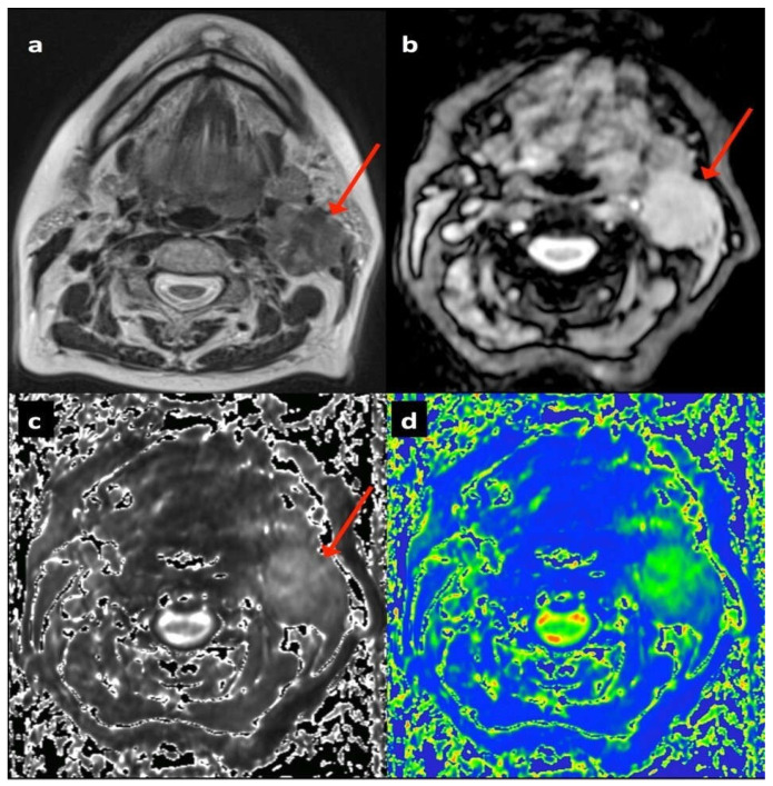 Figure 1
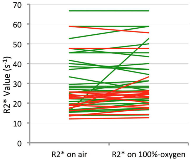 Figure 2
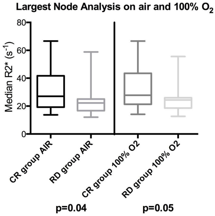 Figure 3
Peer Reviews
No public reviews on file for this paper yet. If you reviewed it on a platform where reviews are public (OpenReview, ICLR, NeurIPS, ICML), you can paste yours below so the community can read it here.
Videos
No videos yet. Explain this paper in a talk, walkthrough, or lecture? Add one.
Taxonomy
TopicsMRI in cancer diagnosis · Advanced MRI Techniques and Applications · Head and Neck Cancer Studies