Association of the Middle Accessory Cerebral Artery With Bihemispheric Anterior Cerebral Artery and Median Artery of the Corpus Callosum
Gervith Reyes Soto, Carlos Castillo-Rangel, Luis Delgado-Reyes, Danil Nurmukhametov, Carlos Ernesto López Lara, Julio Cesar Pérez Cruz, Andreina Rosario Rosario, Manuel De Jesus Encarnacion Ramirez

TL;DR
This study reports rare cerebral artery variations found in a cadaver, emphasizing their importance for neurosurgical planning.
Contribution
The paper documents a rare combination of three vascular anomalies in a single individual, enhancing anatomical understanding for neurosurgery.
Findings
A right middle accessory cerebral artery (MACA) originated from the anterior communicating artery and had two perforating branches.
The right A1 segment was dominant, while the left A1 was hypoplastic, forming a bihemispheric anterior cerebral artery.
A median artery of the corpus callosum (MACC) arose from the left A3 segment and supplied the callosal sulcus and cingulate gyrus.
Abstract
As part of a microsurgical anatomy study of the recurrent artery of Heubner (RAH), we identified three rare vascular variants in a 45-year-old female cadaver: the right middle accessory cerebral artery (MACA), bihemispheric anterior cerebral artery (Bihem-ACA), and median artery of the corpus callosum (MACC). These anomalies were documented through meticulous dissection and detailed morphometric analysis, underscoring the value of cadaveric studies in elucidating complex cerebral vascular anatomy. The specimen was obtained within 24 hours postmortem. The cerebral arteries were injected with red latex and fixed using a standard protocol: initial perfusion with formaldehyde, followed by immersion in 10% formalin for a total fixation period of two months. Dissection was performed under a Carl Zeiss OPMI™ surgical microscope (Carl Zeiss Meditec AG, Jena, Germany) at 6x-40x magnification.…
Genes, proteins, chemicals, diseases, species, mutations and cell lines named across the full text — each resolved to its canonical identifier and authoritative record.
Click any figure to enlarge with its caption.
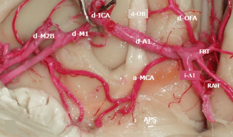 Figure 1
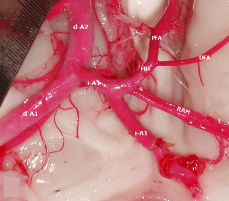 Figure 2
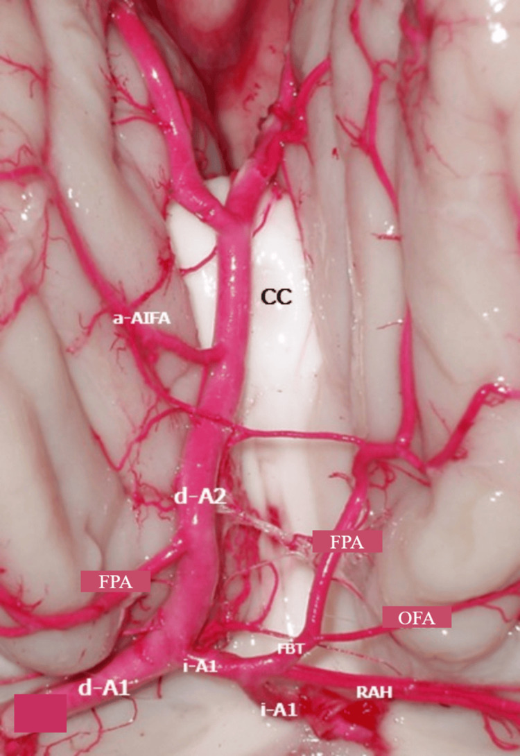 Figure 3
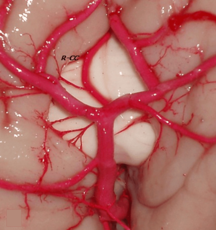 Figure 4
Peer Reviews
No public reviews on file for this paper yet. If you reviewed it on a platform where reviews are public (OpenReview, ICLR, NeurIPS, ICML), you can paste yours below so the community can read it here.
Videos
No videos yet. Explain this paper in a talk, walkthrough, or lecture? Add one.
Taxonomy
TopicsTraumatic Brain Injury and Neurovascular Disturbances · Advanced Neuroimaging Techniques and Applications · Cerebrospinal fluid and hydrocephalus