Diagnostic Accuracy and Interrater Agreement of FDG-PET/CT Lymph Node Staging in High-Risk Endometrial Cancer: The SENTIREC-Endo Study
Jorun Holm, André Henrique Dias, Oke Gerke, Annika Loft, Kirsten Bouchelouche, Mie Holm Vilstrup, Sarah Marie Bjørnholt, Sara Elisabeth Sponholtz, Kirsten Marie Jochumsen, Malene Grubbe Hildebrandt, Pernille Tine Jensen

TL;DR
FDG-PET/CT scans help detect lymph node cancer spread in high-risk endometrial cancer patients but are best used with surgical methods for accurate diagnosis.
Contribution
The study confirms FDG-PET/CT's role as a complementary tool for lymph node staging when combined with sentinel lymph node mapping in high-risk endometrial cancer.
Findings
FDG-PET/CT showed 56% sensitivity and 91% specificity for detecting lymph node metastases.
High interrater agreement (95% agreement, κ = 0.84) was observed among specialists evaluating FDG-PET/CT scans.
FDG-PET/CT is recommended for clinical use alongside sentinel node biopsy to guide targeted dissection of PET-positive nodes.
Abstract
This study looked at how well FDG-PET/CT scans detect cancer spread to lymph nodes in women with high-risk endometrial cancer. It included 227 women undergoing treatment at three Danish hospitals. The scans correctly identified lymph node spread in many patients, especially along the major vessels, but were less reliable in finding all cases where cancer had spread to the lymph nodes. Specialists from different hospitals largely agreed on which lymph nodes looked suspicious, showing that the method was consistent across hospitals. While FDG-PET/CT alone cannot replace surgical staging, we confirmed that it added value when used alongside the sentinel lymph node mapping surgical procedure. The combined approach allows doctors to better identify the nodes that need removal for investigation, while avoiding unnecessary surgery with the removal of all lymph nodes. The findings support…
Genes, proteins, chemicals, diseases, species, mutations and cell lines named across the full text — each resolved to its canonical identifier and authoritative record.
Click any figure to enlarge with its caption.
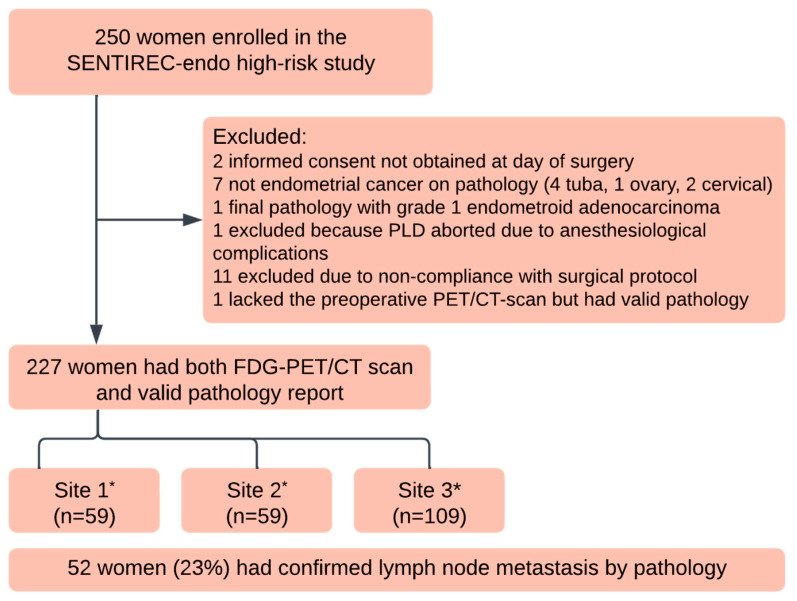 Figure 1
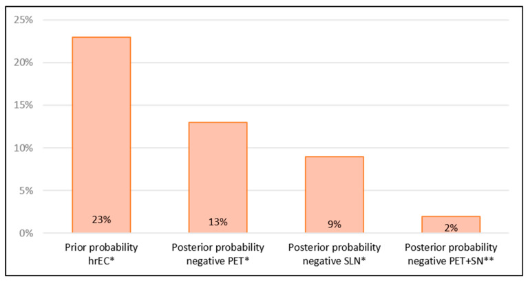 Figure 2
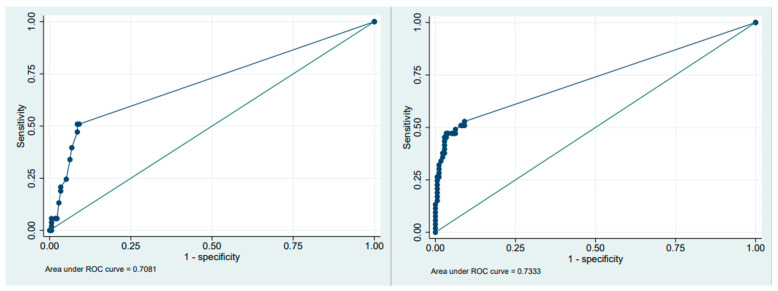 Figure 3
Peer Reviews
No public reviews on file for this paper yet. If you reviewed it on a platform where reviews are public (OpenReview, ICLR, NeurIPS, ICML), you can paste yours below so the community can read it here.
Videos
No videos yet. Explain this paper in a talk, walkthrough, or lecture? Add one.
Taxonomy
TopicsEndometrial and Cervical Cancer Treatments · Radiomics and Machine Learning in Medical Imaging · Ovarian cancer diagnosis and treatment