Association between frailty and echocardiographic findings in hospitalized older adults with preserved ejection fraction
Daniel Betancourt, Jose Zuluaga, Fernando Arango, Tatiana Murillo, Daniel Hincapié

TL;DR
Frailty in older adults is linked to heart function issues, even when heart pumping is normal, showing structural and functional changes in the heart.
Contribution
This study demonstrates that frailty independently predicts diastolic dysfunction and cardiac structural changes in older adults with preserved ejection fraction.
Findings
Frailty is strongly associated with diastolic dysfunction (adjusted odds ratio of 3.49).
Frail individuals show larger atrial volumes and smaller ventricular volumes.
Pulmonary artery systolic pressure and tricuspid regurgitation velocity are elevated in frail patients.
Abstract
This study aims to examine the association between frailty and cardiac structure and function in hospitalized older adults with preserved ejection fraction, using echocardiographic parameters and the Fried frailty index. A cross-sectional analytical study was conducted in two referral centres. A total of 269 individuals aged 60 years or older were included. The exclusion criteria were conditions that affect ventricular mechanics. Patients were categorized into non-frail, prefrail, and frail groups. Transthoracic echocardiography included 2D imaging, Doppler, and Global Longitudinal Strain (GLS) of the left ventricle. Comparative analysis was considered statistically significant if P < 0.05. Frailty was significantly associated with diastolic dysfunction, with an adjusted odds ratio of 3.49 (95% CI: 1.90–6.39, P < 0.001). After adjusting for potential confounders—including age,…
Genes, proteins, chemicals, diseases, species, mutations and cell lines named across the full text — each resolved to its canonical identifier and authoritative record.
Click any figure to enlarge with its caption.
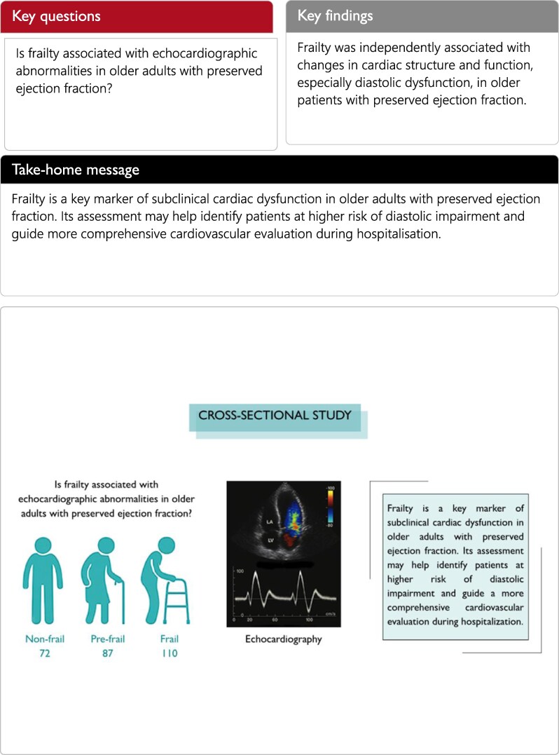 Figure 1
 Figure 2
| Non-frail ( | Prefrail ( | Frail ( |
| |
|---|---|---|---|---|
| Male | 42 (58.3%) | 43 (49.4%) | 38 (34.6%) |
|
| Age (years) | 68.4 ± 6.2 | 75.1 ± 8.3 | 78.1 ± 8.9 |
|
| Age >80 (years) | 3 (4.2%) | 24 (27.6%) | 52 (47.3%) |
|
| BMI (kg/m2) | 26.0 ± 3.7 | 24.9 ± 4.0 | 24.8 ± 4.6 | 0.088 |
| Arterial hypertension | 43 (59.7%) | 60 (69.0%) | 76 (69.1%) | 0.358 |
| Diabetes mellitus | 11 (15.3%) | 30 (34.5%) | 29 (26.4%) |
|
| Atrial fibrillation | 3 (4.2%) | 5 (5.8%) | 9 (8.2%) | 0.567 |
| CKD | 7 (9.7%) | 8 (9.2%) | 11 (10.0%) | 1.000 |
| COPD | 3 (4.2%) | 16 (18.4%) | 39 (35.5%) |
|
| OSA | 3 (4.2%) | 1 (1.2%) | 4 (3.6%) | 0.436 |
| Coronary heart disease | 9 (12.5%) | 10 (11.5%) | 9 (8.2%) | 0.597 |
| Smoking history | 5 (6.9%) | 5 (5.8%) | 8 (7.3%) | 0.909 |
| NYHA ≥ III | 3 (4.2%) | 13 (14.9%) | 53 (48.2%) |
|
| Non-frail ( | Prefrail ( | Frail ( |
| |
|---|---|---|---|---|
| LVESD (mm) | 25.7 ± 4.0 | 26.1 ± 4.8 | 25.0 ± 4.7 | 0.603 |
| LVEDD (mm) | 43.3 ± 3.9 | 41.9 ± 4.9 | 41.3 ± 6.3 |
|
| IVS (mm) | 8.3 ± 1.3 | 8.5 ± 1.4 | 8.8 ± 1.6 | 0.132 |
| LVPW (mm) | 8.1 ± 1.2 | 8.2 ± 1.2 | 8.7 ± 1.8 | 0.349 |
| LVEDV (mL/m2) | 54.6 ± 10.4 | 55.6 ± 9.5 | 53.3 ± 12.6 |
|
| LVESV (mL/m2) | 22.9 ± 6.7 | 22.5 ± 5.0 | 21.8 ± 5.8 |
|
| RWT | 0.38 ± 0.06 | 0.40 ± 0.07 | 0.42 ± 0.07 |
|
| LVMI (g/m2) | 63.1 ± 14.0 | 66.0 ± 14.5 | 73.0 ± 22.1 | 0.286 |
| RVDbasal (mm) | 35.7 ± 4.8 | 36.5 ± 5.4 | 36.7 ± 5.6 | 0.901 |
| RVDmid (mm) | 31.6 ± 4.5 | 32.0 ± 4.5 | 32.5 ± 5.0 | 0.625 |
| RVDlong (mm) | 68.9 ± 7.4 | 67.5 ± 7.6 | 67.7 ± 7.4 | 0.096 |
| LAD (mm) | 39.1 ± 5.0 | 39.5 ± 7.7 | 40.7 ± 6.9 | 0.493 |
| LAVI (mL/m2) | 30.5 ± 8.0 | 35.0 ± 8.8 | 39.6 ± 13.3 |
|
| RAVI (mL/m2) | 21.5 ± 6.7 | 23.9 ± 6.6 | 26.0 ± 9.1 |
|
| Non-frail ( | Prefrail ( | Frail ( |
| |
|---|---|---|---|---|
| LVEF (%) | 59.6 ± 3.7 | 58.8 ± 4.6 | 59.7 ± 3.9 | 0.784 |
|
| 0.83 ± 0.25 | 0.80 ± 0.26 | 0.77 ± 0.94 |
|
|
| 8.7 ± 2.4 | 10.6 ± 3.6 | 12.4 ± 6.4 |
|
|
| 6.2 ± 1.4 | 5.6 ± 1.4 | 5.0 ± 1.4 |
|
|
| 7.9 ± 1.7 | 7.0 ± 2.1 | 6.5 ± 1.9 |
|
| TRV (m/s) | 2.6 ± 0.3 | 2.9 ± 0.3 | 2.9 ± 0.5 |
|
| PASP (mmHg) | 36.7 ± 10.2 | 39.7 ± 12.8 | 41.0 ± 11.0 |
|
| TAPSE (mm) | 21.7 ± 2.5 | 21.5 ± 3.2 | 21.7 ± 3.8 | 0.759 |
| TASA (cm/s) | 12.2 ± 2.1 | 12.7 ± 2.7 | 12.3 ± 2.9 | 0.601 |
| FAC (%) | 48.2 ± 6.1 | 48.6 ± 6.9 | 48.0 ± 5.8 | 0.531 |
| LVGLS (%) | −23.0 ± 2.0 | −22.0 ± 2.6 | −21.9 ± 2.7 | 0.592 |
Peer Reviews
No public reviews on file for this paper yet. If you reviewed it on a platform where reviews are public (OpenReview, ICLR, NeurIPS, ICML), you can paste yours below so the community can read it here.
Videos
No videos yet. Explain this paper in a talk, walkthrough, or lecture? Add one.
Taxonomy
TopicsFrailty in Older Adults · Global Health Care Issues · Cardiovascular Function and Risk Factors
Introduction
Since 1980, for the first time in history, most people can expect to live beyond the age of 60 years.^1^ However, as life expectancy continues to increase worldwide, there has also been a rise in the prevalence of non-communicable chronic diseases, particularly cardiovascular diseases such as heart failure.^2^ Moreover, geriatric syndromes such as frailty have emerged, defined as a biological syndrome characterized by a progressive decline in physiological reserves and resistance to stressors due to the cumulative dysfunction of multiple physiological systems, leading to increased vulnerability to adverse outcomes.^3–5^ This condition not only has health, social, and economic implications but also represents a significant challenge in public health.^6^
The relationship between frailty and cardiovascular disease has been widely studied,^7^ but whether frailty itself leads to worse clinical outcomes by directly affecting cardiac structure and function remains controversial.^8^ Echocardiography has evolved as a key tool in cardiovascular assessment, with the quantification of chamber size and function being the cornerstone of cardiac imaging.^8^ Some studies have suggested an association between frailty and increased left ventricular mass,^9–11^ whereas others have not supported this conclusion.^12–14^ Furthermore, previous research has failed to demonstrate a significantly lower left ventricular ejection fraction in frail individuals.^9,12,15^
The aim of this study was to assess frailty using the physical phenotype derived from the Cardiovascular Health Study^16^ in an older population hospitalized in two referral centres and to analyse its association with cardiac structure and function.
Methods
Study population
A total of 269 older individuals aged 60 and above, who walk independently or with assistive devices such as a cane or walker, and who underwent a clinically indicated transthoracic echocardiogram at SES Hospital Universitario de Caldas and Hospital Universitario Santa Sofía in Manizales, Colombia, from July 2023 to June 2024, were prospectively enrolled.
The exclusion criteria were patients with congenital heart disease, cardiomyopathy, moderate or severe valvular heart disease, severe right ventricular dilation with septal displacement and dyssynchrony, segmental wall motion abnormalities, valvular prostheses or cardiac devices, immunochemotherapy, delirium, hospitalized in the intensive care unit, poor acoustic window, left ventricular ejection fraction < 50%, and acute heart failure at the time of admission. Acute heart failure was defined as the presence of typical symptoms (e.g. dyspnoea, orthopnoea, and oedema) along with elevated NT-proBNP levels (> 900 pg/mL for patients ≤ 75 years and > 1800 pg/mL for those > 75 years).
This study was conducted in accordance with the Declaration of Helsinki, and participants were required to provide written informed consent. The study protocol was approved by the ethics committees of each participating hospital (approval numbers: R-20230421-1 and DEI-054-23).
Definition of frailty
The frailty phenotype was assessed using the Fried Frailty Index.^16^ The criteria for defining frailty included weight loss, exhaustion, physical activity, gait speed, and grip strength.
Weight loss was defined as an unintentional loss of more than 10 pounds (4.5 kg) in the previous year.Exhaustion was determined based on self-reported responses to two items from the Centre for Epidemiologic Studies Depression Scale.^17^Physical activity was assessed using the exercise scale to measure function in advanced activities of daily living.Grip strength values were obtained using a Takei Smedley Hand Dynamometer III, which measures grip strength in kilograms-force (kg/f). Weakness was defined as values below 26 kg/f for men and below 16 kg/f for women, based on the cut-off points established by the Foundation for the National Institutes of Health.^18^Gait speed, measured in metres per second (m/s), is a validated parameter in the literature, typically assessed over distances of 3, 5, or 10 m.^19^ In this study, the 3-m assessment was used. Each patient underwent two gait speed measurements, and the highest recorded speed was used for analysis. Slowness was defined as a gait speed below 0.8 m/s, based on the cut-off point established in the International Mobility in Ageing Study.^20^
Patients were classified as ‘frail’ if they had impairments in ≥ 3 components, as ‘prefrail’ if they had impairments in 1 or 2 components, and as ‘non-frail’ if they had no apparent impairments.
Collection of data for clinical variables
The clinical information, including demographic data, medical history, and structural and functional echocardiographic variables, was obtained from medical records. The variables used to define frailty were assessed by two of the authors, who are experts in geriatrics.
Echocardiography
Echocardiographic images of the participants were obtained using a Philips EPIQ CVX echocardiograph, available at both institutions. Standard 2D views of three to five cardiac cycles were collected: three cycles for patients with sinus rhythm and five cycles for those with atrial fibrillation, following guideline recommendations.^21^ Cardiac structure and function were assessed using two-dimensional transthoracic echocardiography, colour Doppler, and spectral Doppler. Additionally, the Global Longitudinal Strain (GLS) of the left ventricle was analysed in all patients.
Diastolic dysfunction was defined according to current literature,^22^ using the following criteria: (i) average E/e′ ratio > 14; (ii) e′ velocity: < 7 cm/s septal or < 10 cm/s lateral; (iii) tricuspid regurgitation velocity > 2.8 m/s; and (iv) left atrial volume index > 34 mL/m². Diastolic dysfunction was diagnosed when more than 50% of the specified criteria were met.
All transthoracic echocardiograms in this study were performed and interpreted by a single echocardiography specialist with Level 3 training. Frailty was assessed independently by the study authors using Fried’s criteria, and they were blinded to the echocardiographic findings at the time of data collection.
Statistical analyses
According to the Fried phenotype score, patients were classified into three groups: non-frail, prefrail, and frail.
Continuous variables were expressed as means ± standard deviation if they followed a normal distribution, as determined by the Kolmogorov–Smirnov test. If they did not follow a normal distribution, they were expressed as median and inter-quartile range. Differences between groups were compared using one-way ANOVA. Categorical variables were expressed as counts and frequency (%), and comparisons between groups were made using the χ^2^ test or Fisher’s exact probability method, as appropriate.
Logistic regression models were used to analyse the association between clinical data, structural and functional cardiac parameters, and frailty in older adults. Odds ratios (ORs) and 95% confidence intervals (CIs) were reported. A P-value of < 0.05 was considered statistically significant. All statistical analyses were performed using Stata software, version 16.1.
Results
Baseline characteristics
A total of 269 hospitalized patients were included, with a range between 60 and 96 years and a mean of 74.5 ± 8.9 years (95% CI: 73.4–75.6). The gender distribution was 54.4% female and 45.6% male. According to the Fried phenotype, 40.9% of participants were classified as frail, 32.3% as prefrail, and 26.8% as non-frail.
Clinical characteristics showed a high prevalence of hypertension (66.5%), followed by diabetes mellitus (26.0%) and chronic obstructive pulmonary disease (COPD) (21.6%). Additionally, chronic kidney disease (9.7%), atrial fibrillation (6.3%), and coronary artery disease (10.4%) were reported. Table 1 presents the baseline characteristics of the study population.
Echocardiographic structural parameters
Frail patients had smaller left ventricular indexed volumes (LVEDVI: 53.3 vs. 55.6 mL/m^2^ in prefrail and 54.6 mL/m^2^ in non-frail, P = 0.007) and larger left atrial indexed volumes (LAVI: 39.6 vs. 35.0 mL/m^2^ in prefrail and 30.5 mL/m^2^ in non-frail, P = 0.019). A progressive increase in relative wall thickness (RWT) was also observed as frailty severity increased (0.38 in non-frail vs. 0.40 in prefrail and 0.42 in frail, P = 0.043). Table 2 lists the cardiac structural parameters.
Echocardiographic functional parameters
In terms of functional parameters, no significant differences were found in left ventricular ejection fraction (LVEF) among the groups (P = 0.784). However, frail patients exhibited significant diastolic abnormalities, reflected by a progressive decrease in the E/A ratio (0.83 in non-frail vs. 0.77 in frail, P = 0.014) and an increase in the E/e′ ratio (8.7 ± 2.4 in non-frail vs. 12.4 ± 6.4 in frail, P = 0.003).
Additionally, pulmonary artery systolic pressure (PASP) was significantly higher in frail patients (41.0 vs. 36.7 mmHg in non-frail, P = 0.002). Table 3 presents the cardiac functional parameters.
Multivariable regression model
Stepwise forward logistic regression was performed, and the only variable associated with diastolic dysfunction was frailty (OR: 3.49, 95% CI: 1.90–6.39, P < 0.001).
Discussion
The main finding of this study was the significant and independent association between frailty and diastolic dysfunction, with an adjusted OR of 3.49 (95% CI: 1.90–6.39, P < 0.001). This result supports the hypothesis that frailty is not only a marker of vulnerability in older adults but may also reflect underlying alterations in cardiac function that predispose individuals to worse outcomes.^23^ Our findings are consistent with previous studies, such as that of Xi et al., who, using a similar multidimensional definition of frailty, identified a higher prevalence of left ventricular hypertrophy and diastolic dysfunction in hospitalized frail patients.^8^
The results of our study show both similarities and differences compared with previous investigations in Anglo-Saxon populations. In the study by Gharacholou et al., conducted at the Mayo Clinic, frailty was associated with increased left atrial volume, lower stroke volume, and higher pulmonary pressures; however, no relationship was identified with left ventricular mass.^12^ Our results confirm the association between frailty and increased left atrial volume, which may reflect chronic pressure overload, but differ in terms of left ventricular mass, as our analysis found a significant difference between frail and non-frail patients. These discrepancies may be explained by differences in the study population, as our analysis focused on hospitalized patients with preserved ejection fraction, whereas Gharacholou’s study included a broader spectrum of ventricular dysfunction.
On the contrary, in the study by Topriceanu et al., based on the 1946 British birth cohort, frailty in middle age was associated with left ventricular hypertrophy, increased filling pressures, and reduced systolic function in older age.^10^ Similarly, our study also found left ventricular hypertrophy when comparing frail and non-frail patients. However, our main finding supports the notion that frailty primarily affects diastolic function. These differences may be explained by the longitudinal design of the British study, which enabled the assessment of long-term cardiac changes, whereas our study evaluated a hospitalized population at a single time point, limiting the ability to analyse progressive alterations in ventricular function.
A notable finding of this study was the significant increase in PASP in frail patients. This increase in PASP is closely associated with the presence of diastolic dysfunction, as one of the diagnostic criteria for diastolic dysfunction is a tricuspid regurgitation velocity >2.8 m/s. Therefore, the elevated PASP further supports the presence of diastolic dysfunction in frail patients, reinforcing its role in this population’s cardiovascular profile.^24^
In contrast to other studies that have reported a reduction in GLS of the left ventricle in frail patients,^8,25^ our analysis did not find significant differences in this parameter between groups. This absence of GLS differences is likely related to the selection criteria of our study, which included only patients with preserved left ventricular function and no structural cardiac damage. By focusing on a population with normal systolic function, the potential impact of frailty on myocardial contractility may have been mitigated, highlighting instead its predominant effect on diastolic function and atrial remodelling.
To avoid confounding, patients with acute heart failure at admission were excluded. Diagnosis was based on the presence of typical symptoms and elevated NT-proBNP levels (as described above), ensuring that echocardiographic findings reflected changes associated with frailty rather than acute decompensation.
We acknowledge that using the New York Heart Association (NYHA) classification to assess functional limitation due to dyspnoea may cause some confusion, as the scale was originally developed specifically for patients with heart failure. However, in our study, the NYHA classification was employed because of its simplicity and widespread use in clinical practice to describe degrees of dyspnoea, irrespective of the underlying aetiology. Therefore, the NYHA classification served as a general measure of functional status rather than as a definitive indicator of heart failure, since patients with heart failure were excluded from the study.
While reverse causality and residual confounding cannot be eliminated in an observational design, the associations observed in our study show that frailty remains independently linked to diastolic dysfunction. There is, however, biological plausibility for a shared, self-reinforcing pathway: low-grade inflammation, neurohormonal activation, and reduced physical activity are known to drive both musculoskeletal catabolism (frailty) and adverse myocardial remodelling (diastolic dysfunction),^26^ potentially creating a vicious cycle in which early frailty and sub-clinical left ventricular dysfunction amplify one another.
Importantly, the prevalence of frailty in our cohort was considerably higher compared with previous studies, with 40.9% of frail patients vs. the 29.1% reported by Xi et al.^8^ This suggests that factors such as socioeconomic differences, access to health care, and the management of cardiovascular diseases may influence the prevalence of frailty and its impact on cardiac function.^27–29^
Finally, few studies have evaluated the relationship between frailty and echocardiographic findings, and only one previous study has included Speckle Tracking technology in its analysis. Notably, this is the first study to examine this association in a hospitalized Ibero-American population with preserved ejection fraction, expanding the applicability of these findings and emphasizing the importance of considering frailty as a key determinant in the cardiac assessment of older adults.
Limitations
The present study has several limitations. First, its observational design allows exploration of associations between frailty and cardiac structure and function, but cannot establish causality. Second, lipid profiles and information on cardiovascular medications were not collected, preventing adjustment for these potential confounders. Although the echocardiography specialist was not informed of the patients’ frailty status, we acknowledge that this did not represent true blinding, as the examiner had direct contact with the patients during image acquisition. Some frailty components were self-reported, introducing possible misclassification bias. Reliance on clinical records may entail data inaccuracies and missing details, and the exclusion of intensive care patients limits applicability to critically ill cohorts. Because only hospitalized patients were evaluated, the findings may not be generalizable to the broader older population. Moreover, although all participants were acutely admitted to internal medicine, cardiology or geriatric wards, the specific reasons for admission or discharge were not recorded, which constitutes an additional potential confounder.
Conclusions
This study reveals a significant association between frailty and diastolic dysfunction in hospitalized older adults, highlighting the need for specific cardiac assessments in this population.
Lead author biography
Daniel Betancourt Zuluaga is a medical doctor specializing in Internal Medicine and Geriatrics. He is a member of the Research Group on Geriatrics and Gerontology at the Faculty of Health Sciences, Universidad de Caldas, Colombia. His clinical and academic work focuses on the health and care of older adults, with a particular interest in the intersection of cardiology and geriatrics. He has contributed to scientific publications in respected journals, including the European Journal of Medical and Health Sciences. In 2024, he completed an international clinical rotation in cardiogeriatrics at the Hospital Clínico Universitario de Valencia in Spain.
Supplementary Material
oeaf087_Supplementary_Data
The reference list from the paper itself. Each links out to its DOI / PubMed record.
- 1United Nations . Development in an Ageing World. New York: United Nations, 2007. p 1–22. https://fiapam.org/wp-content/uploads/2013/06/El-desarrollo-en-un-mundo-que-envejece.pdf
- 2Davila CA, Pardo AM. Changes in life expectancy due to chronic disease-related mortality in older adults. Mexico 2000–2013. Revista Ciencias de la Salud 2017;15:223–235.
- 3Fried LP, Tangen CM, Walston J, Newman AB, Hirsch C, Gottdiener J, Seeman T, Tracy R, Kop WJ, Burke G, Mc Burnie MA. Frailty in older adults: evidence for a phenotype. J Gerontol A Biol Sci Med Sci 2001;56:M 146–M 156.11253156 10.1093/gerona/56.3.m 146 · doi ↗ · pubmed ↗
- 4Damluji AA, Chung SE, Xue QL, Hasan RK, Moscucci M, Forman DE, Bandeen-Roche K, Batchelor W, Walston JD, Resar JR, Gerstenblith G. Frailty and cardiovascular outcomes in the National Health and Aging Trends Study. Eur Heart J 2021;42:3856–3865.34324648 10.1093/eurheartj/ehab 468PMC 8487013 · doi ↗ · pubmed ↗
- 5Morley JE, Vellas B, Van Kan GA, Anker SD, Bauer JM, Bernabei R, Cesari M, Chumlea WC, Doehner W, Evans J, Fried LP. Frailty consensus: a call to action. J Am Med Dir Assoc 2013;14:392–397.23764209 10.1016/j.jamda.2013.03.022PMC 4084863 · doi ↗ · pubmed ↗
- 6Lemus GA, Cárdenas JM, Curcio CL, Moreno GA. Effects of frailty on the adverse outcomes of cardiac surgery in the elderly. Rev Colomb Cardiol 2020;27:250–261.
- 7He D, Wang Z, Li J, Yu K, He Y, He X, Liu Y, Li Y, Fu R, Zhou D, Zhu Y. Changes in frailty and incident cardiovascular disease in three prospective cohorts. Eur Heart J 2024;45:1058–1068.38241094 10.1093/eurheartj/ehad 885 · doi ↗ · pubmed ↗
- 8Xi L, Xuemei Z, Ling Y, Changchun C, Zhuo H, Jinyang Q, Xin W. Correlation between frailty and cardiac structure and function in echocardiography in elderly patients with normal ejection fraction. Aging Clin Exp Res 2023;35:775–784.36871112 10.1007/s 40520-023-02363-5 · doi ↗ · pubmed ↗