A Crumpet, a Canine and a Cryoprobe: A Case of Tooth Aspiration
Matthew Donnan, Melanie Wong, Elina Chi, Dominic Keating

TL;DR
A 66-year-old man aspirated a canine tooth, causing pneumonia and sepsis, which was successfully treated by removing the tooth with a cryoprobe.
Contribution
Demonstrates the successful use of a cryoprobe for retrieving an aspirated tooth in a clinical case.
Findings
Tooth aspiration can lead to post-obstructive pneumonia and sepsis.
A cryoprobe was effective in removing the aspirated canine tooth.
Timely intervention is crucial for source control in such cases.
Abstract
Foreign body inhalation can lead to post‐obstructive pneumonia and sepsis, requiring timely removal to achieve source control. We report a case of tooth aspiration successfully retrieved with a cryoprobe. A 66‐year‐old man presented with sepsis secondary to post‐obstructive pneumonia following aspiration of a canine tooth. The tooth was successfully removed with a cryoprobe.
Genes, proteins, chemicals, diseases, species, mutations and cell lines named across the full text — each resolved to its canonical identifier and authoritative record.
Click any figure to enlarge with its caption.
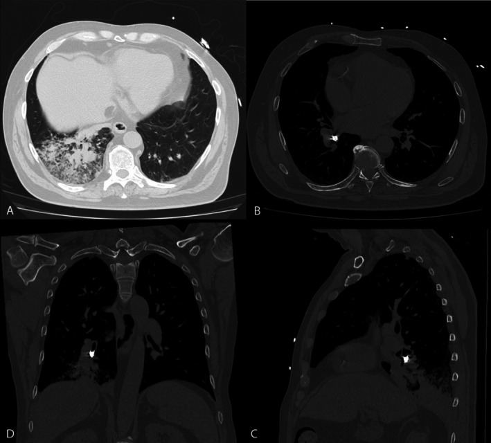 Figure 1
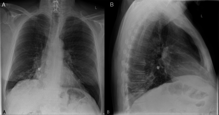 Figure 2
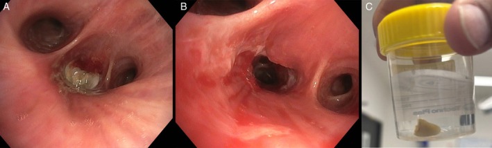 Figure 3
 Figure 4
Peer Reviews
No public reviews on file for this paper yet. If you reviewed it on a platform where reviews are public (OpenReview, ICLR, NeurIPS, ICML), you can paste yours below so the community can read it here.
Videos
No videos yet. Explain this paper in a talk, walkthrough, or lecture? Add one.
Taxonomy
TopicsForeign Body Medical Cases · Restraint-Related Deaths · Traumatic Ocular and Foreign Body Injuries
A 66‐year‐old man presented to hospital with a 1‐week history of a productive cough following inadvertent aspiration of a canine tooth while eating a crumpet. On examination, he was febrile (38.2°C), tachycardic (heart rate 125 beats per minute), hypotensive (systolic blood pressure 90 mmHg), with coarse crepitations audible at the right lung base. His white cell count was 10.6 × 10^9^/L and his CRP was 308 mg/L. Chest x‐ray (Figure 1) and subsequent computed tomography (Figure 2) demonstrated a lucency at the orifice of the right lower lobe bronchus, with post‐obstructive consolidation and collapse. He was treated with intravenous antibiotics and underwent a flexible bronchoscopy which demonstrated a tooth lodged within the right lower lobe bronchus resulting in distal obstruction. A 1.1 mm cryoprobe was used to remove the tooth, and extensive mucopurulent secretions were suctioned (Figure 3).
Foreign body aspiration is relatively uncommon in adults. Removal of the aspirated material is essential to prevent complications including airway mucosal ulceration and post‐obstructive pneumonia. While at times either rigid bronchoscopy or traditional grasping devices (forceps, basket) may be suitable to remove aspirated teeth, a cryoprobe can be an effective alternative if the tooth pulp is exposed or there is mucus adherent to it [1, 2].
Author Contributions
Matthew Donnan, Melanie Wong and Elina Chi: conceptualisation, draft writing, review, editing. Dominic Keating: supervision, writing – review and editing.
Consent
The authors declare that written informed consent was obtained for the publication of this manuscript and accompanying images using the consent form provided by the Journal.
Conflicts of Interest
The authors declare no conflicts of interest.
The reference list from the paper itself. Each links out to its DOI / PubMed record.
- 1H. Ishimoto , N. Sakamoto , S. Moriyama , et al., “Removal of an Aspirated Tooth From the Bronchus Using a Cryoprobe: A Case Report,” Respirology Case Reports 9, no. 12 (2021): e 0880.34853696 10.1002/rcr 2.880PMC 8612864 · doi ↗ · pubmed ↗
- 2H. Azam and P. Wu , “Bronchoscopic Retrieval of an Aspirated Tooth Following High‐Speed Motor Vehicle Accident,” Respirology Case Reports 12, no. 8 (2024): e 01444.39086723 10.1002/rcr 2.1444 PMC 11290951 · doi ↗ · pubmed ↗