Anterior segment optical coherence tomography in pupillary seclusion diagnosis and follow-up
Jordi Izquierdo-Serra, Juan Pablo Figueroa-Vercellino, Marina Dotti-Boada, Néstor Ventura-Abreu, Marta Pazos

Abstract
Genes, proteins, chemicals, diseases, species, mutations and cell lines named across the full text — each resolved to its canonical identifier and authoritative record.
Click any figure to enlarge with its caption.
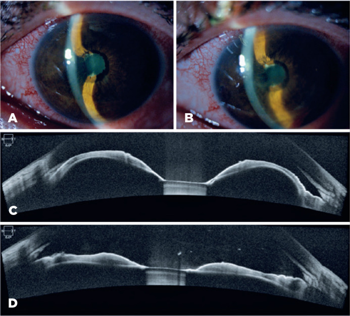 Figure 1
Peer Reviews
No public reviews on file for this paper yet. If you reviewed it on a platform where reviews are public (OpenReview, ICLR, NeurIPS, ICML), you can paste yours below so the community can read it here.
Videos
No videos yet. Explain this paper in a talk, walkthrough, or lecture? Add one.
Taxonomy
TopicsOcular Surface and Contact Lens · Glaucoma and retinal disorders · Ophthalmology and Visual Impairment Studies
Dear Editor,
Anterior segment optical coherence tomography (AS-OCT) has allowed the advancement toward a better understanding of the anatomy of the anterior segment, which is of capital importance in glaucoma care, allowing high-quality three-dimensional images of the anterior segment and dimension measurements of distances and areas^(1)^. These determinations aim for a better understanding of the dynamic changes in high-risk groups of patients, as in primary angle glaucoma disease^(2)^.
AS-OCT could also be of great utility in more straight forward, minimally invasive examinations in less frequent and clinically challenging conditions such as uveitic glaucomas. A 47-year-old man presented at the ophthalmology emergency department with eye pain and photophobia in his left eye (OS) in the previous week. He had consulted the department 2 weeks before for an episode of acute anterior uveitis in the same eye and was receiving treatment with topical corticoids and mydriatics. His visual acuity was 20/30. Slit-lamp examination revealed periciliary hyperemia, anterior chamber cells +2, 360^o^ posterior synechiae, and subsequent iris bombe appearance with peripheral iridocorneal contact (Figure 1A). Intraocular pressure (IOP) was 26 mmHg. Extensive synechiae precluded the fundus examination; therefore, ocular ultrasonography was performed, which ruled out a posterior pushing mechanism. AS-OCT detailed the angle closure caused by the anterior iris bowing secondary to seclusio pupillae (Figure 1C). After Nd:YAG laser peripheral iridotomy, AS-OCT revealed flattening of the iris with opening of the angle (Figure 1B and 1D). The IOP decreased to 18 mmHg and stabilized in low-teen values in the following visits. Iris bombe is an uncommon complication of uveitic glaucoma^(3)^ that presents with apposition of the iris to the lens, which prevents aqueous flow from the posterior to the anterior chamber. As a consequence, the pressure in the posterior chamber increases, causing anterior bowing of the peripheral iris and obstruction of the trabecular meshwork, which may result in an acute angle closure.
Figure 1. Photograph of the anterior segment showing an iris bombe due to a pupillary seclusion (A) and its resolution after laser peripheral iridotomy (LPI) (B). Wide angle-to-angle anterior segment optical coherence tomography (AS-OCT) demonstrating iris bombe with angle closure (C) and its resolution after LPI (D).
In most of these cases, iridotomy can restore aqueous humor outflow^(4)^. Although gonioscopy examination remains to be the gold standard technique for iridocorneal angle examination, recently, AS-OCT has proven to be a reliable high-resolution noninvasive imaging technique that can quantify not only the anterior chamber depth and volume^(4)^ but also the iris curvature and position, thus allowing angle closure diagnosis when irido-trabecular contact is visualized^(5)^. As illustrated in this case, when inflammation and poor collaboration hinder the gonioscopic examination, AS-OCT may be useful in performing a proper diagnosis and follow-up of this subgroup of angle closure.
The reference list from the paper itself. Each links out to its DOI / PubMed record.
- 1Angmo D Nongpiur ME Sharma R Sidhu T Sihota R Dada T. Clinical utility of anterior segment swept-source optical coherence tomography in glaucoma Oman J Ophthalmol 2016913102701382110.4103/0974-620X.176093 PMC 4785705 · doi ↗ · pubmed ↗
- 2Zhao M Sun Q Oatts J Hu G Ge L Zhu B Changes in intraocular pressure and angle structure after dilation in primary angle-closure suspects with visually significant cataract Ophthalmology 2020 S 01616420(20):30667-910.1016/j.ophtha.2020.07.00932652206 · doi ↗ · pubmed ↗
- 3Moorthy RS Mermoud A Baerveldt G Minckler DS Lee PP Rao NA. Glaucoma associated with uveitis Surv Ophthalmol 1997415361394916383510.1016/s 0039-6257(97)00006-4 · doi ↗ · pubmed ↗
- 4Ikegawa W Suzuki T Namiguchi K Mizoue S Shiraishi A Ohashi Y. Changes in anterior segment morphology of iris bombe before and after laser peripheral iridotomy in patients with uveitic secondary glaucoma J Ophthalmol 2016201684962012787275510.1155/2016/8496201 PMC 5107836 · doi ↗ · pubmed ↗
- 5Ang M Baskaran M Werkmeister RM Chua J Schmidl D Aranha dos Santos V Anterior segment optical coherence tomography Prog Retin Eye Res 2018661321562963506810.1016/j.preteyeres.2018.04.002 · doi ↗ · pubmed ↗