Feasibility of in vivo small animal imaging using a clinical total-body PET/CT system
Julia G. Mannheim, Wenhong Lan, Maurizio Conti, Franziska Siedler, Marcel A. Krueger, Kristina Herfert, Christian la Fougère, Fabian P. Schmidt

TL;DR
This study shows that a clinical total-body PET/CT scanner can be used for small animal imaging, offering good quantification despite lower spatial resolution.
Contribution
Demonstrates the feasibility of using clinical total-body PET/CT systems for preclinical rodent imaging with robust SUVmean quantification.
Findings
The Biograph Vision Quadra TB-PET/CT achieved comparable recovery coefficients and lower noise than the Inveon DPET for larger structures.
SUVmean values were consistent across organs between the clinical and preclinical scanners.
Position-dependent effects were not observed in phantom or mouse scans across the extended axial FOV.
Abstract
Clinical PET scanners have long been explored for preclinical imaging, but limited spatial resolution and sensitivity have restricted their use for preclinical studies. The recent availability of total-body (TB) PET/CT scanners with extended axial fields of view (FOVs) has largely overcome sensitivity limitations, enabling potential new opportunities for small-animal imaging. This study evaluated the feasibility and performance of the Biograph Vision Quadra TB-PET/CT for rodent imaging compared to the dedicated small-animal PET scanner Inveon DPET. Recovery coefficients (RC), image noise, and optimum image reconstruction parameters were assessed using the preclinical NEMA NU 4–2008 image quality phantom and a sub-cohort of three anesthetized mice as a proof-of-concept demonstrating the feasibility of the setup. In vivo quantification accuracy was evaluated by scanning nine frozen mice…
Genes, proteins, chemicals, diseases, species, mutations and cell lines named across the full text — each resolved to its canonical identifier and authoritative record.
Click any figure to enlarge with its caption.
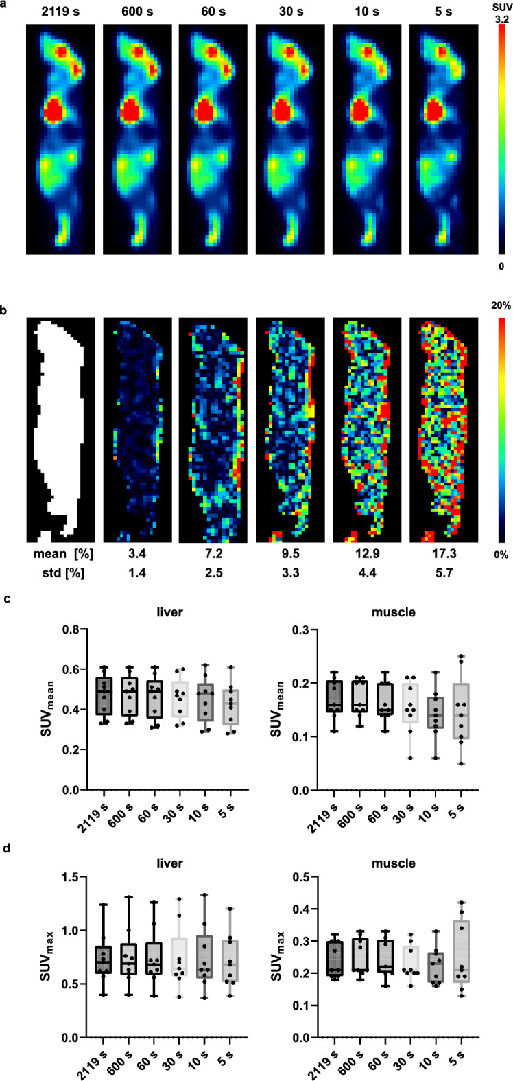 Figure 10
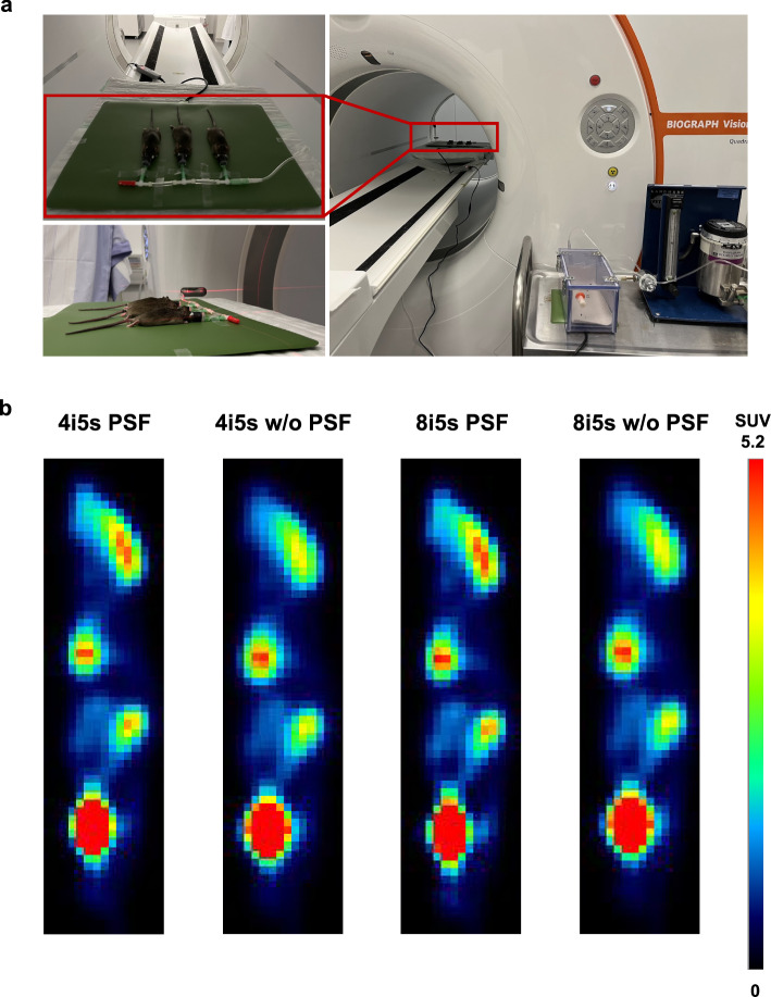 Figure 1
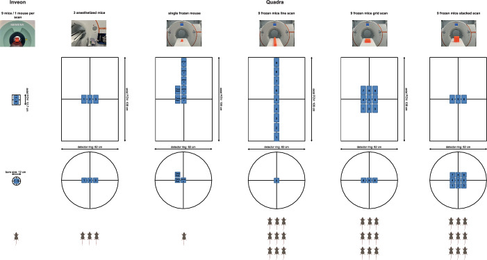 Figure 2
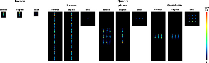 Figure 3
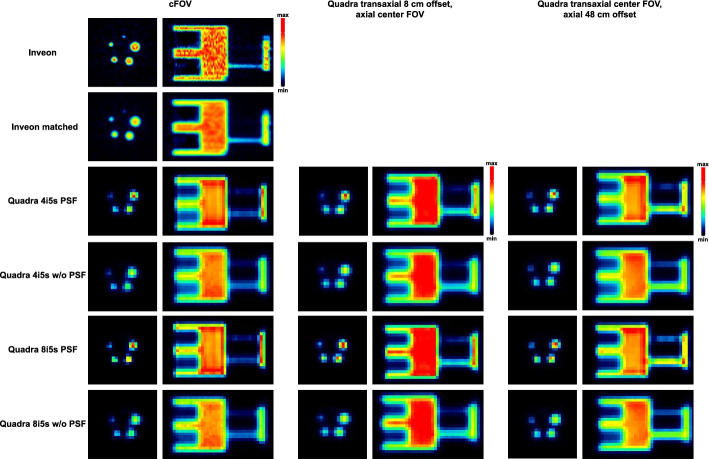 Figure 4
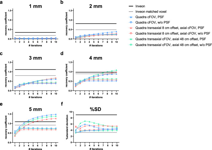 Figure 5
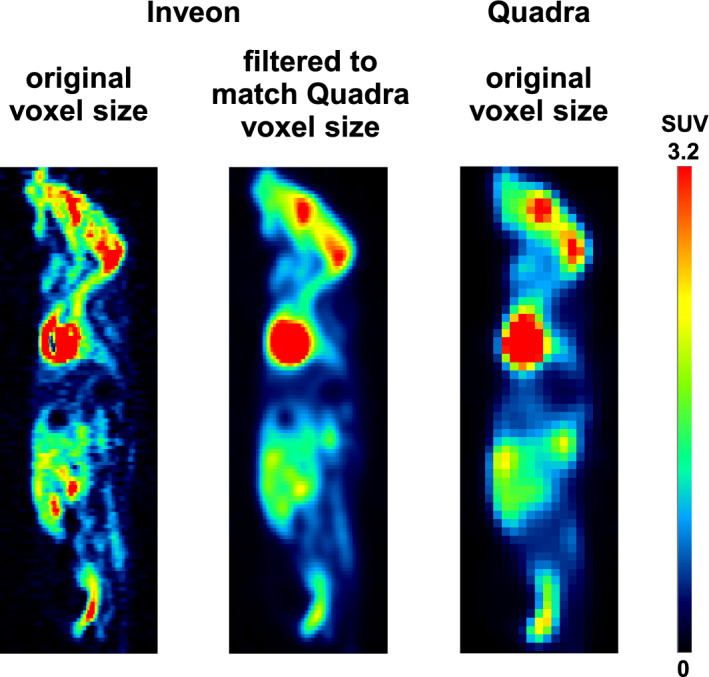 Figure 6
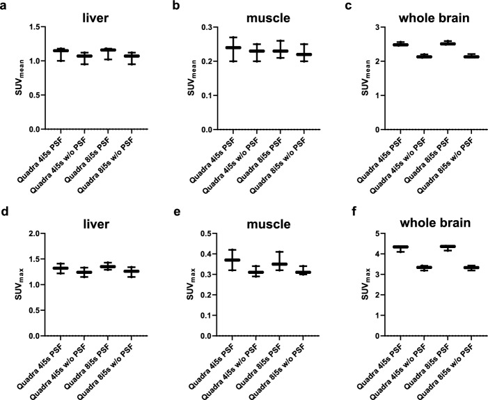 Figure 7
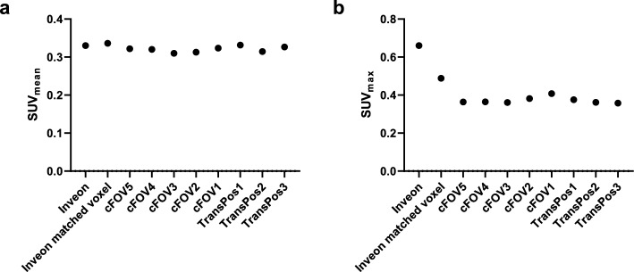 Figure 8
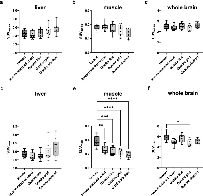 Figure 9
Peer Reviews
No public reviews on file for this paper yet. If you reviewed it on a platform where reviews are public (OpenReview, ICLR, NeurIPS, ICML), you can paste yours below so the community can read it here.
Videos
No videos yet. Explain this paper in a talk, walkthrough, or lecture? Add one.
Taxonomy
TopicsMedical Imaging Techniques and Applications · Advanced X-ray and CT Imaging · Advanced MRI Techniques and Applications