Non-Invasive Diagnostic Techniques in Penile Intraepithelial Neoplasia (PeIN): Insights from Reflectance Confocal Microscopy (RCM), Line-Field Confocal Optical Coherence Tomography (LC-OCT), and Correlation with Histopathological Features
Caterina Damiani, Cesare Ariasi, Giuseppe La Rosa, Francesca Di Lauro, Mariachiara Arisi, Vincenzo Maione, Marina Venturini, Simone Soglia

TL;DR
This paper explores non-invasive imaging techniques for diagnosing penile intraepithelial neoplasia, showing their potential to improve diagnosis and reduce invasive procedures.
Contribution
The study demonstrates the effectiveness of RCM and LC-OCT in diagnosing PeIN with features matching histopathological results.
Findings
RCM and LC-OCT revealed features like hyperkeratosis and nuclear pleomorphism matching high-grade PeIN histopathology.
Non-invasive imaging techniques can aid in the differential diagnosis of genital lesions.
The methods may reduce the need for invasive diagnostic procedures.
Abstract
Penile intraepithelial neoplasia (PeIN) is a rare but clinically significant condition that can progress to invasive squamous carcinoma. Early diagnosis is crucial but often challenging due to its heterogeneous clinical and dermoscopic presentation, which can mimic other benign or malignant lesions. In this study, we report two cases of pigmented penile lesions evaluated using non-invasive imaging techniques: reflectance confocal microscopy (RCM) and line-field confocal optical coherence tomography (LC-OCT). Both methods revealed characteristic features such as hyperkeratosis, parakeratosis, acanthosis, nuclear pleomorphism of keratinocytes, and the presence of bright intraepithelial dendritic cells, correlating closely with histopathological findings of high-grade basaloid PeIN. Our findings highlight the valuable role of RCM and LC-OCT in improving the differential diagnosis of…
Genes, proteins, chemicals, diseases, species, mutations and cell lines named across the full text — each resolved to its canonical identifier and authoritative record.
Click any figure to enlarge with its caption.
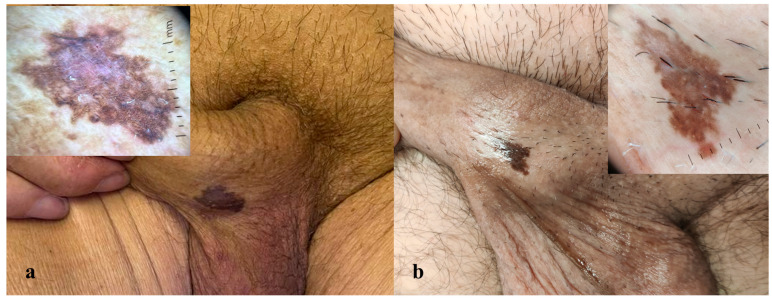 Figure 1
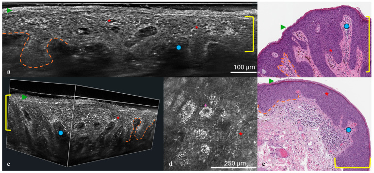 Figure 2
Peer Reviews
No public reviews on file for this paper yet. If you reviewed it on a platform where reviews are public (OpenReview, ICLR, NeurIPS, ICML), you can paste yours below so the community can read it here.
Videos
No videos yet. Explain this paper in a talk, walkthrough, or lecture? Add one.
Taxonomy
TopicsGenital Health and Disease · Cervical Cancer and HPV Research · Nonmelanoma Skin Cancer Studies