Double Myopic Choroidal Neovascular Membranes (CNVMs): A Case Report
Asli Perente, Aikaterini Giannoukaki, Doukas Dardabounis, Tryfon Rotsos, Georgios Labiris

TL;DR
A rare case of two myopic choroidal neovascularizations in one eye was diagnosed and treated successfully with anti-VEGF injections.
Contribution
The paper presents a rare case of double myopic choroidal neovascular membranes in a single eye, managed with anti-VEGF therapy.
Findings
OCT and OCT-A clearly depicted two CNVs in the same eye.
Anti-VEGF injections successfully managed the condition.
The case highlights the need for further study on this rare presentation.
Abstract
Myopic choroidal neovascularization (CNV) is the most common cause of visual morbidity in patients with pathologic myopia (PM). Early diagnosis and management are crucial to prevent permanent loss of central vision. Optical coherence tomography (OCT) and OCT angiography (OCT-A) are the key diagnostic modalities for identifying and monitoring this vision-threatening complication. We report an unusual case of a female patient with two myopic CNVs in the same eye, clearly depicted using OCT-A and successfully managed with anti-vascular endothelial growth factor (anti-VEGF) injections. This rare instance warrants further study to draw more definite conclusions regarding this common complication and its response to treatment.
Genes, proteins, chemicals, diseases, species, mutations and cell lines named across the full text — each resolved to its canonical identifier and authoritative record.
Click any figure to enlarge with its caption.
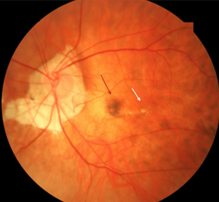 Figure 1
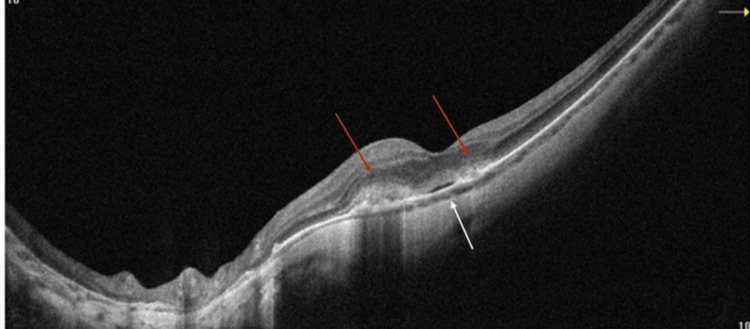 Figure 2
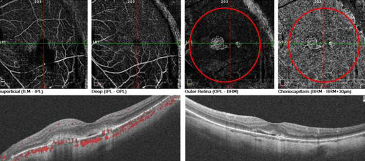 Figure 3
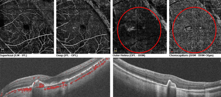 Figure 4
Peer Reviews
No public reviews on file for this paper yet. If you reviewed it on a platform where reviews are public (OpenReview, ICLR, NeurIPS, ICML), you can paste yours below so the community can read it here.
Videos
No videos yet. Explain this paper in a talk, walkthrough, or lecture? Add one.
Taxonomy
TopicsRetinal and Optic Conditions · Retinal Diseases and Treatments · Retinal Imaging and Analysis