Relationship between DTI-MRI derived metrics and radiotherapy dose range in the contralateral cerebrum in lower grade glioma
Justyna Kłos, Hildebrand Dijkstra, Hiska L. van der Weide, Jan H. Potze, Peter F. Sinnige, Kelvin Ng Wei Siang, Rudi A. J. O. Dierckx, Ronald J. H. Borra, Miranda C. A. Kramer, Anouk van der Hoorn

TL;DR
This study shows that DTI-MRI can detect radiation-induced brain damage in white matter of the cerebrum contralateral to a tumor, especially in areas receiving the highest radiation dose.
Contribution
The study demonstrates that FA from DTI-MRI is a sensitive marker for radiation-induced brain damage in white matter, specifically in high-dose regions.
Findings
FA values in white matter decreased significantly in the >50 Gy dose bin, indicating radiation-induced brain damage.
No significant changes were observed in grey matter FA or in ADC values for both white and grey matter.
FA changes were not correlated with FLAIR hyperintensity volume or ADC values, suggesting distinct mechanisms of damage detection.
Abstract
To evaluate the value of diffusion tensor imaging (DTI) MRI derived fractional anisotropy (FA) and apparent diffusion coefficient (ADC) for both white matter (WM) and grey matter (GM) of the contralateral cerebrum following radiotherapy (RT) for supratentorial lower grade glioma (LGG) as markers for radiotherapy-induced brain damage (RIBD). 14 patients were analysed. WM and GM were segmented using automated software (cNeuro) and the mean FA and ADC were extracted per RT dose bin (0-10, 10-20, 20-30, 30-40, 40-50, >50 Gy) of WM and GM. One way ANOVA with post-hoc Bonferroni’s test were used to analyse differences in FA and ADC between dose bins. Fluid-attenuated inversion recovery (FLAIR) hyperintensities were segmented in a semi-automated manner and correlated with a percentual difference in ADC and FA between dose bin ≥50 Gy and the mean of lower dose bins. Furthermore, the…
Genes, proteins, chemicals, diseases, species, mutations and cell lines named across the full text — each resolved to its canonical identifier and authoritative record.
Click any figure to enlarge with its caption.
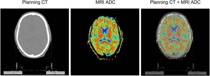 Figure 1
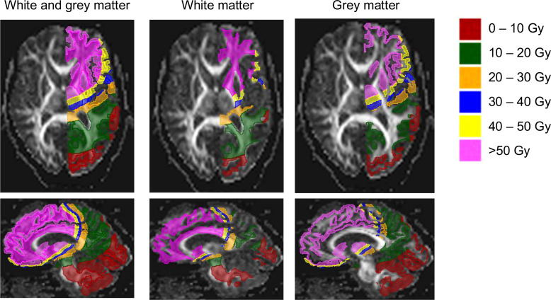 Figure 2
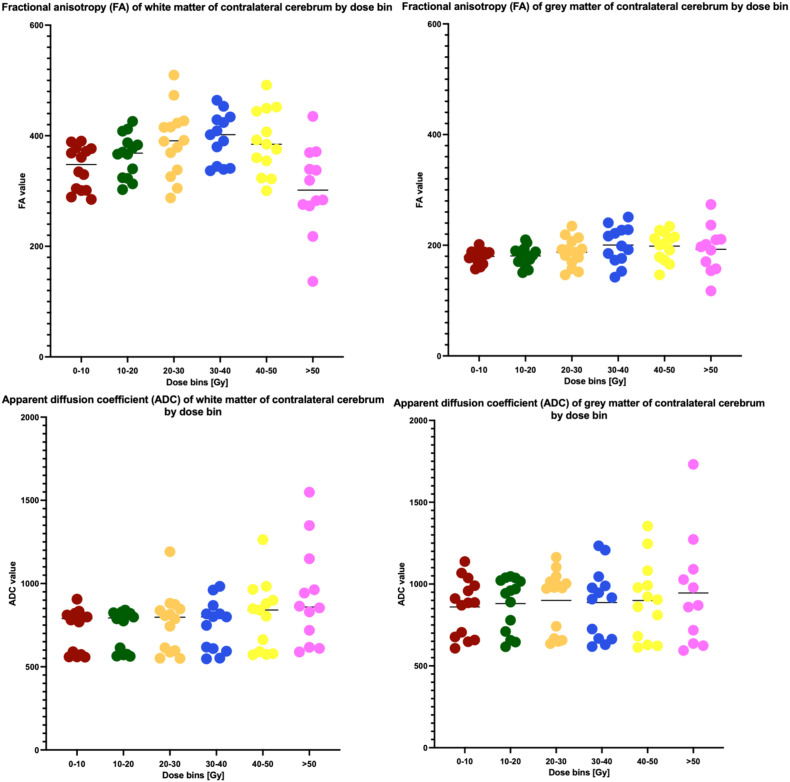 Figure 3
Peer Reviews
No public reviews on file for this paper yet. If you reviewed it on a platform where reviews are public (OpenReview, ICLR, NeurIPS, ICML), you can paste yours below so the community can read it here.
Videos
No videos yet. Explain this paper in a talk, walkthrough, or lecture? Add one.
Taxonomy
TopicsAdvanced Neuroimaging Techniques and Applications · MRI in cancer diagnosis · Glioma Diagnosis and Treatment