Transfer learning with class activation maps in compositions driving plaque classification in carotid ultrasound
Georgia D. Liapi, Christos P. Loizou, Maura Griffin, Constantinos S. Pattichis, Andrew Nicolaides, Efthyvoulos Kyriacou

TL;DR
This paper explores how CNNs classify carotid ultrasound images of plaques using class activation maps to identify which plaque features influence classification.
Contribution
The study introduces the use of class activation maps to interpret CNN-based classification of asymptomatic and symptomatic carotid plaques in ultrasound images.
Findings
Dark grayscale areas (GS ≤ 25) and juxtaluminal black areas (JBAs) were influential in both asymptomatic and symptomatic plaque classification.
Lipid cores, fibrous content, and calcified areas were more associated with asymptomatic plaques.
The model achieved 80.4% accuracy in classifying plaque images into asymptomatic and symptomatic categories.
Abstract
Carotid B-mode ultrasound (U/S) imaging provides more than the degree of stenosis in stroke risk assessment. Plaque morphology and texture have been extensively investigated in U/S images, revealing plaque components, such as juxtaluminal black areas close to lumen (JBAs), whose size is linearly related to the risk of stroke. Convolutional neural networks (CNNs) have joined the battle for the identification of high-risk plaques, although the ways they perceive asymptomatic (ASY) and symptomatic (SY) plaque features need further investigation. In this study, the objective was to assess whether class activations maps (CAMs) can reveal which U/S grayscale-(GS)-based plaque compositions (lipid cores, fibrous content, collagen, and/or calcified areas) influence the model's understanding of the ASY and SY cases. We used Xception via transfer learning, as a base for feature extraction (all…
Genes, proteins, chemicals, diseases, species, mutations and cell lines named across the full text — each resolved to its canonical identifier and authoritative record.
Click any figure to enlarge with its caption.
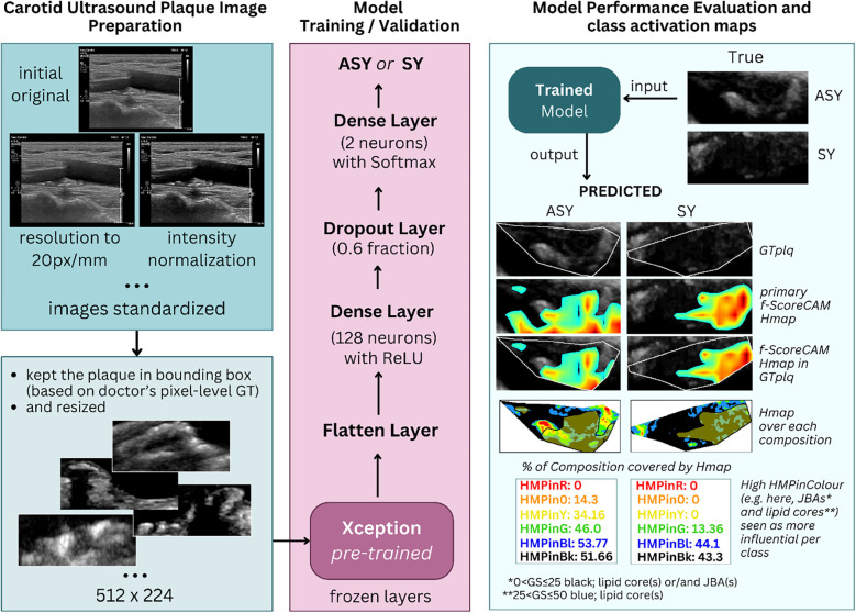 Figure 1
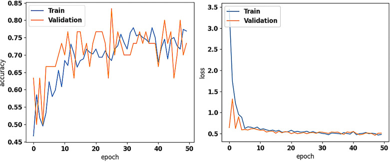 Figure 2
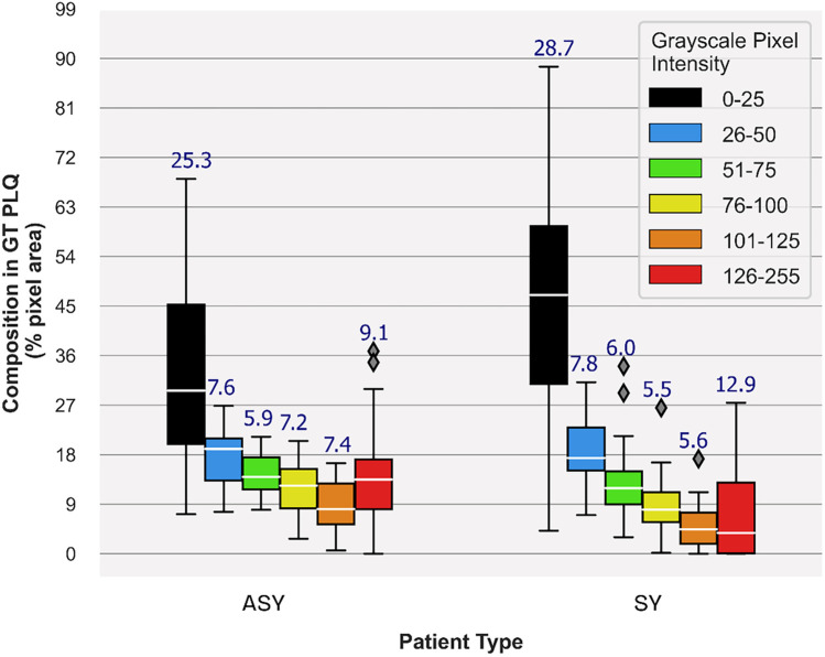 Figure 3
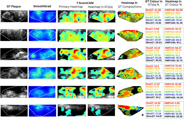 Figure 4
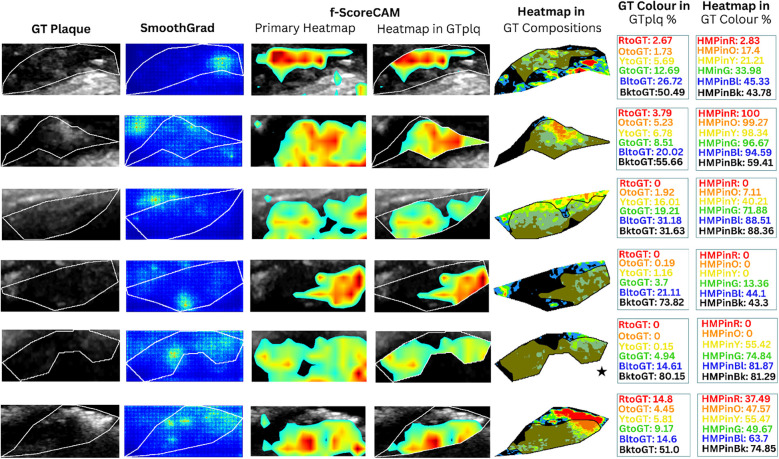 Figure 5
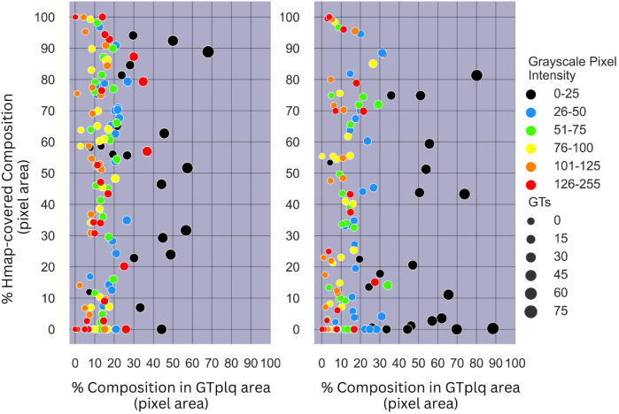 Figure 6
Peer Reviews
No public reviews on file for this paper yet. If you reviewed it on a platform where reviews are public (OpenReview, ICLR, NeurIPS, ICML), you can paste yours below so the community can read it here.
Videos
No videos yet. Explain this paper in a talk, walkthrough, or lecture? Add one.
Taxonomy
TopicsCerebrovascular and Carotid Artery Diseases · Medical Image Segmentation Techniques · Radiomics and Machine Learning in Medical Imaging