Interplay between the gut microbiome and typhoid fever: insights from endemic countries and a controlled human infection model
Philip M. Ashton, Leonardos Mageiros, James E. Meiring, Angeziwa Chunga-Chirambo, Farhana Khanam, Sabina Dongol, Happy Banda, Abhilasha Karkey, Lorena Preciado-Llanes, Helena Thomaides-Brears, Malick Gibani, Nazmul Hasan Rajib, Nazia Rahman, Prasanta Kumar Biswas

TL;DR
This study explores how the gut microbiome influences resistance to typhoid fever, identifying bacteria and metabolic functions linked to protection against the disease.
Contribution
The study identifies specific gut bacteria and SCFA-related metabolic gene clusters associated with protection against typhoid fever across multiple populations.
Findings
Four bacterial species were less abundant in typhoid patients compared to exposed individuals in Bangladesh and Malawi.
28 metabolic gene clusters, including seven involved in SCFA metabolism, were negatively associated with typhoid fever.
Participants in a controlled infection model who resisted typhoid had higher SCFA-related gene clusters.
Abstract
Typhoid fever is a systemic infection caused by Salmonella enterica serovar Typhi (S. Typhi) invasion from the gut lumen. Transmission between people occurs through ingestion of contaminated food and water, particularly in settings with poor water and sanitation infrastructure, resulting in over 10 million illnesses annually. As the pathogen invades via the gastrointestinal tract, it is plausible that the gut microbiome may influence the outcome of S. Typhi exposure. There is some evidence that bacteria producing short-chain fatty acids (SCFAs) may create an environment unfavourable to invasive Salmonella, but data from humans is limited. To investigate the association between the gut microbiome and typhoid fever, we analysed samples collected from three all-age cohorts enrolled in a prospective surveillance study conducted across three settings where typhoid fever is endemic (Dhaka,…
Genes, proteins, chemicals, diseases, species, mutations and cell lines named across the full text — each resolved to its canonical identifier and authoritative record.
Click any figure to enlarge with its caption.
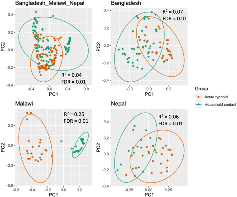 Figure 1
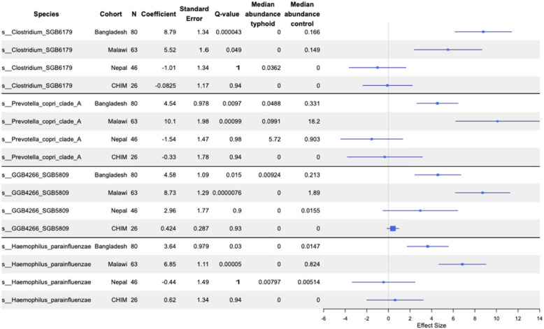 Figure 2
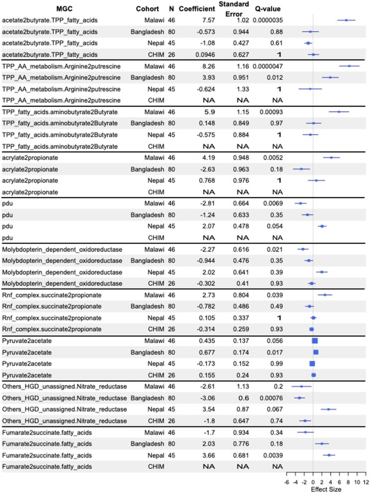 Figure 3
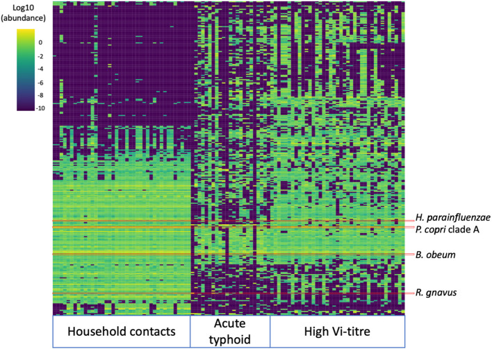 Figure 4
Peer Reviews
No public reviews on file for this paper yet. If you reviewed it on a platform where reviews are public (OpenReview, ICLR, NeurIPS, ICML), you can paste yours below so the community can read it here.
Videos
No videos yet. Explain this paper in a talk, walkthrough, or lecture? Add one.
Taxonomy
TopicsGut microbiota and health · Salmonella and Campylobacter epidemiology · Clostridium difficile and Clostridium perfringens research
Introduction
Typhoid fever, caused by invasive Salmonella enterica serovar Typhi (S. Typhi) infection, causes an estimated 10.9 million illnesses and 116,800 deaths per year, disproportionately affecting people in South and Southeast Asia and sub-Saharan Africa [42, 59]. The introduction of typhoid conjugate vaccines is expected to decrease the number of typhoid fever cases in these settings, along with improvements in water, sanitation and hygiene (WASH). However, the large burden of disease, the role of asymptomatic gallbladder carriage as a source of infection and the fact that typhoid fever often impacts the most impoverished and marginalised communities mean that eradicating this infection will be difficult [43, 48, 61].
The human gut microbiota, a complex ecosystem comprising trillions of microbial cells, plays an indispensable role in shaping our overall health and susceptibility to diseases [56]. This densely populated microbial environment is not only pivotal in the processes of digestion and nutrient absorption but also intimately connected to our immune system, determining its responses to various stimuli [8]. As the nexus between the external environment and our internal physiology, the gut microbiome is a critical determinant of the outcome of gastrointestinal infectious disease exposures, via a phenomenon termed ‘colonisation resistance’ [37, 38, 60, 65]. This protective effect arises from multifaceted interactions: direct competition for nutrients, production of antimicrobial compounds and modulation of the host’s innate and adaptive immune responses [36]. Some of the earliest work on colonisation resistance showed that suppressing the gut microbiome of mice with antibiotics dramatically reduced the dose of Salmonella required to establish infection [12]. Recent work has made great strides in understanding the mechanisms by which Salmonella and the microbiome interact [55]. Short-chain fatty acids (SCFAs) are important in the interaction between the microbiota and Salmonella, reducing the availability of oxygen in the gut [15] and acidifying the cytosol of Salmonella, thereby inhibiting growth [31]. Despite well-established knowledge about the gut microbiome’s function in preventing Salmonella infections and the established faecal-oral transmission pathway of S. Typhi, the precise influence of the gut microbiome on the outcome of S. Typhi exposure remains underexplored, complicated by the human host-specificity of S. Typhi.
While insights from animal studies of invasive infection with other S. enterica serovars can be informative, detailed studies of the interaction between the human gut microbiome and S. Typhi specifically are lacking. Controlled human infection model (CHIM) studies provide an experimental system in which to interrogate infections in humans, complementing observational studies of natural infections [23, 64]. Previous data generated from this model revealed an association between typhoid fever susceptibility and the presence of Methanobrevibacter in the gut microbiome [68]. Analysing natural infections, Haak et al. determined that typhoid fever patients in Bangladesh had reduced microbial diversity and fewer SCFA producers than their healthy counterparts [27]. A better understanding of the causal relationships between the gut microbiome and typhoid fever could lead to novel preventative mechanisms and diagnostics, as well as an improved understanding of colonisation resistance to bacteria causing serious invasive diseases in high-burden settings.
To fill this knowledge gap, we sequenced the stool microbiome of 258 participants enrolled in the STRATAA study, conducted in three diverse high typhoid fever burden settings in Asia and Africa [43]. Signals that replicated in more than one study population were further investigated in a UK-based CHIM cohort.
Materials and methods
Participants and sample collection
Detailed methods for the STRATAA study have been published previously [18, 43]. In brief, approximately 100,000 individuals were enumerated in a demographic census in three communities: Ndirande in Blantyre, Malawi; Lalitpur in Kathmandu, Nepal; and Mirpur in Dhaka, Bangladesh. Febrile patients, of any age, within the study populations were recruited via passive surveillance, and ‘acute typhoid fever’ patients were defined as those with positive blood cultures yielding S. Typhi. When participants presented with fever, stool samples were collected where possible, preferably prior to antimicrobial use. Stool specimens were transferred to − 80 °C within 6 h of collection. Data on antimicrobial use prior to enrolment was recorded [18]. Blood-culture–confirmed typhoid fever patients were then followed up, and stool samples were collected from their household contacts; asymptomatic stool culture-negative individuals were included in microbiome sequencing. The rationale for this was that having a febrile person or typhoid fever case in the household is a significant risk factor for developing typhoid fever, and therefore, asymptomatic household contacts are likely exposed to S. Typhi without becoming infected [22, 63]. It has been reported that shared household membership is positively associated with microbiota phylogenetic similarity [58, 62], so differences between household members could be responsible for, or due to, the development of typhoid fever. High Vi-titre individuals were identified from a community-based serological survey of up to 8500 age-stratified participants per site from the original demographic census. From the serological survey, samples were analysed for anti-Vi IgG antibodies, i.e. those with a twofold increase in anti-Vi IgG titres and an absolute titre of 50 EU/mL or higher at the second timepoint [43]. The participants at each site with the highest Vi responses were followed up, and stool samples were collected for microbiome analysis [32]. Stool cultures were S. Typhi-negative in all but one participant [32],however, it is known that carriers shed only intermittently and high-Vi individuals are an acceptable predictor for S. Typhi carriage [50, 53]. For the microbiome sub-study, we randomly selected for sequencing stool samples from up to 40 participants from each site, from each of three groups: acute typhoid fever cases, household contacts and serosurvey participants with high-Vi titres.
DNA extraction and sequencing
DNA extraction from stool was done using the Fast-Prep 24 FastDNA Spin Kit (MP Biomedicals, CA, USA) according to the manufacturer’s instructions. DNA was sent to the Wellcome Sanger Institute (UK) for metagenomic sequencing using Illumina HiSeq 2500 or HiSeq 4000 to generate 150 bp paired-end reads, yielding an average of 14.8 million read pairs (standard deviation 2.3 million) per sample.
Bioinformatics
Raw sequencing data was quality trimmed and adapters were removed using bbduk v38.96 with the parameters ‘ktrim = r k = 23 mink = 11 hdist = 1 tbo tpe qtrim = r trimq = 20 minlength = 50’. Sequence deriving from human DNA was removed by mapping to a human reference genome (GCF_009914755.1) and the hostile tool [17]. Taxonomic profiling was carried out with metaphlan v4.0.6 with database version mpa_vOct22_CHOCOPhlAnSGB_202212 [10].
We used the BiG-MAP program [46] (https://github.com/medema-group/BiG-MAP, commit e7b8042) to compare metagenomic readsets against a database of non-redundant metabolic gene clusters (MGCs) identified using gutSMASH [47] from a collection of reference genomes from the Culturable Genome Reference, the Human Microbiome Project and other Clostridia genomes (https://zenodo.org/records/7252625#.ZFVTrexBz0r). Each MGC is assigned to a species which is the species from which the representative sequence representing the cluster was obtained. BiG-MAP calculated the number of reads per kilobase per million reads (RPKM) for each metabolic gene cluster.
A sub-sample of 8.8 million reads was taken from each readset using the ‘sample’ command of the seqtk tool (v1.3-r106). Sub-sampled reads were then analysed with Resistance Gene Identifier (RGI) v6.0.2 bwt command against the CARD v3.2.7 reference database [3]. Due to uncertainty regarding some CARD database classifications (e.g. OXA-1 was classified as a carbapenemase, a curation error which has subsequently been resolved), we used the drug class information for each gene from the NCBI AMR Reference Gene Database (refgene) v2023-08–08.2 (https://www.ncbi.nlm.nih.gov/pathogens/refgene). Only “Core” genes from NCBI refgene were analysed; these genes are almost entirely mobilisable AMR determinants and do not include mutational resistance. Extended-spectrum beta-lactamases (ESBLs) were identified based on the gene product descriptions in NCBI refgene. Only genes with an average percent coverage of 100% in the RGI analysis were included in further analysis. The number of reads mapped to each gene (RGI completely mapped reads) was normalised by the length of the gene (RGI reference length) to generate a ‘reads per kilobase of AMR gene’ metric. There was no need to normalise for the number of reads, as all readsets had been sub-sampled to the same number of reads. RGI results were parsed and analysed using the scripts described in the Data Availability section of this manuscript. AMR genes identified in S. Typhi were obtained from [5].
Statistical analysis
Statistical analyses were done in R (v4.1.0). To quantify alpha diversity, Shannon’s index was calculated using the alpha.div function in the rbiom package (v1.0.3), and ANOVA analysis was carried out with the aov function (stats package, v4.1.0). The Bray–Curtis metric of beta diversity was calculated with the vegdist function of the R package vegan (v2.6.4). Principal coordinate analysis (PCoA) was carried out with the cmdscale function (package stats, v4.1.0), and statistics comparing beta diversity between participant groups (i.e. acute typhoid, household contacts, presumptive carrier) was carried out using a PERMANOVA approach implemented by the adonis2 function of the vegan package (v2.6.4). Associations between taxa and phenotypes of interest (e.g. participant group) were explored using multivariable linear modelling implemented in the maaslin2 function of the maaslin2 R package (v1.6.0) [40]. Depending on the analysis, co-variates such as age, sex, antibiotic exposure and sequencing run were included, as described. For maaslin2 analyses, Benjamini–Hochberg correction was applied for multiple testing. For analyses of data from endemic cohorts a q-value threshold of 0.05 was used to identify significant associations, while for CHIM data analyses, due to the small sample size, the default maaslin2 q-value threshold of 0.25 was used.
Controlled human infection model analysis
We used data from a Salmonella Typhi and Paratyphi A CHIM cohort to validate the associations identified in the endemic countries. Briefly, healthy adults aged 18–60 were recruited to be challenged with a single oral dose of either S. Typhi or S. Paratyphi A [23]. The primary endpoint for the model was a diagnosis of typhoid or paratyphoid fever, defined as a temperature of ≥ 38 °C persisting for ≥ 12 h and/or S. Typhi/Paratyphi A bacteraemia in a sample collected ≥ 72 h after oral challenge. Only data from participants who were being challenged for the first time were analysed here. Stool samples were taken from participants prior to the challenge and stored at − 80 °C. DNA extraction, sequencing and analyses were undertaken using the methods described above. The primary maaslin2 analyses for this cohort included only species or MGC classes that were significantly associated with the participant group in at least two endemic country cohorts; a secondary analysis was carried out without this restriction. Analyses were done separately for participants challenged with either S. Typhi or S. Paratyphi A, in addition to a combined analysis including both pathogens.
Results
Description of population
Stool microbiome sequences were successfully generated for 258 participants from Bangladesh (n = 80), Malawi (n = 102) and Nepal (n = 76). Across the three populations, there were three participant groups; 92 patients with acute typhoid fever, 97 household contacts of acute typhoid cases and 69 people with high anti-Vi titres. The baseline characteristics of participants are described in Table 1. Overall, acute typhoid fever patients were significantly younger than household contacts, while in Bangladesh and Malawi, high-Vi titre participants were significantly older than household contacts (Additional file 1: Fig. S1, Table 1). Overall, in Malawi, Bangladesh and Nepal, 62%, 53% and 54% of participants were female, respectively (Table 1, Additional file 1: Fig. S2). In Bangladesh and Malawi, sex distribution was similar between the participant groups, whereas in Nepal, 82% of household contacts and 67% of carriers were female, while only 24% of typhoid fever cases were female (Table 1; Additional file 1: Fig. S2). Of the acute typhoid fever cases, 37.5%, 73.9% and 44.8% from Bangladesh, Malawi and Nepal, respectively, reported antimicrobial use in the 2 weeks prior to stool sample collection. Table 1. Characteristics of participants in the microbiome sub-study of STRATAA surveillanceBangladeshMalawiNepal****Acute typhoidN402329Median age, years(range)6 (1–60)10 (3–40)17 (5–38)Female (%)48%61%24%Antibiotics in last 2 weeks (%)38%74%45%CarrierNNA^a^3930Median ageNA^a^3243.9Female (%)NA^a^56%67%Antibiotics in last 2 weeks (%)NA^a^NA^b^NA^b^Household contactN404017Median age, years(range)29 (4–65)24 (18–41)35 (15–48)Female (%)65%65%82%Antibiotics in last 2 weeks (%)NA^b^NA^b^NA^b^^a^Carriers from Bangladesh were not included in the analysis due to sample protocol differences^b^Antibiotic usage in the 2 weeks previous was an exclusion criterion for carriers and household contacts
Acute typhoid fever patients compared with household contacts
First, we compared stool metagenomic profiles of acute typhoid patients with those of household contacts, within each of the three study populations, to explore microbiome signatures associated with typhoid fever. The participant group was not significantly associated with differences in alpha diversity in any population (Additional file 1: Fig. S3; Additional file 2: Table S1). Beta diversity varied significantly between typhoid fever patients and household contacts across all sites (Fig. 1, Additional file 2: Table S2). The proportion of beta diversity variance explained by the participant group was highest in Malawi (R^2^ = 0.25, FDR = 0.01), followed by Bangladesh (R^2^ = 0.07, FDR = 0.01), Nepal (R^2^ = 0.06, FDR = 0.01 and the combined analysis (R^2^ = 0.04, FDR = 0.01) (Fig. 1; Additional file 2: Tables S2–S5). The participant group explained the greatest beta diversity variance of the factors investigated (which also included sex, age and reported antibiotic use) in a combined analysis of all three sites and for each site individually. In Malawi, prior antibiotic usage and the interaction of prior antibiotic usage with age and sex were also significantly associated with beta diversity, although these variables explained a lower proportion of variance than the participant group (R^2^ 0.02–0.05; Additional file 2: Table S4). In Nepal, sex was significantly associated with beta diversity and explained almost as much variance as the participant group (R^2^ = 0.04; Additional file 2: Table S5), which was itself associated with sex at this site. Firmicutes were the dominant phylum in typhoid fever patients and household contacts at all three sites, followed by Bacteroidetes in Malawi and Nepal and Actinobacteria in Bangladesh (Additional file 1: Fig. S4; Additional file 2: Table S6).Fig. 1. Principal coordinate analysis of the beta diversity between household contacts and typhoid fever participants across all three sites and for each site individually. The proportion of variance explained (R^2^) and the Bonferroni-corrected FDR from a PERMANOVA analysis including participant group, sex and antibiotic usage are displayed on each plot. In the combined analysis, the country of sampling was also included in the PERMANOVA analysis
Maaslin2 analysis of taxonomic profiles identified 92, 23 and 0 species significantly associated with household contact vs. typhoid fever participant groups in Malawi, Bangladesh and Nepal, respectively (Fig. 2; Additional file 2: Tables S7–S8). No taxa showed significant associations across all three sites; however, four showed consistent associations at two sites (Bangladesh and Malawi), all of which were negatively associated with typhoid fever (Fig. 2). These four species were Prevotella copri clade A, Haemophilus parainfluenzae, Clostridium SGB6179 and a Veillonellaceae spp represented by a metagenome-assembled genome, GGB4266_SGB5809. Although there were differences in age between cases and household contacts at all sites, age was included as a covariate in maaslin2, and the associations with typhoid status were also evident within age groups where data from that age group was available (Figs. 5, 6, 7 and 8). Of these four species, none was significantly associated with typhoid fever in Nepal or the CHIM; most samples from the CHIM did not contain any reads assigned to these species (Additional file 2: Table S29).Fig. 2. Forest plot of species that significantly differed between household contacts and typhoid fever participants in at least two endemic countries. Results from all three endemic countries and the CHIM cohort are shown for context. CHIM maaslin2 analysis only included species that were significantly associated in at least two of the endemic country sites. Species are labelled with their GTDB identifier, note that “s__ GGB4266_SGB5809” refers to a proposed novel genus/species of Veillonellaceae
Some species were associated with typhoid fever at a single endemic country site. In Bangladesh, there were 19 species for which the relative abundance significantly differed between typhoid fever participants and household contacts that did not differ at any other site or in the CHIM (Additional file 2: Table S7). Three species were negatively associated with typhoid fever (Prevotella copri clade C, Romboutsia timonensis and Ligilactobacillus ruminis), while 16 species including 7 species of Actinomyces were positively associated with typhoid fever (Additional file 2: Table S7). In Malawi, 70 species were negatively associated with typhoid fever, including Ruminococcus gnavus, Roseburia intestinalis, Roseburia inulinivorans and Faecalibacterium prausnitzii (Additional file 2: Table S8), while 18 species were positively associated with typhoid fever including 11 (61%) that are only in the Metaphlan4 database as metagenome-assembled genomes (Additional file 2: Table S8).
To complement our taxonomic analysis, we also carried out functional gene analysis. We explored two dimensions of variation in metabolic gene clusters (MGCs). First, we examined the distribution of specific MGCs, investigating participant group differences at a granular, sub-type level (e.g. a specific RNF complex from a strain of Bacteroides ovatus). Second, we compared the abundance of different MGC types between participant groups (e.g. all RNF complex MGCs). This dual approach enables us to capture both detailed and broad patterns of functional gene distribution within the microbiome.
We identified 264 specific MGCs that significantly differed between typhoid fever patients and household contacts in the Malawi cohort, 126 in Bangladesh and 2 in Nepal (Additional file 1: Tables S24–S26). Neither of the specific MGC associations identified in the Nepal data were replicated in the Malawi or Bangladesh cohorts. There were 28 specific MGCs significantly associated with typhoid fever in both Bangladesh and Malawi (summarised in Table 2, full information in Additional file 2: Table S9), and all were negatively associated with typhoid fever in both settings. Six of these specific MGCs were linked to SCFA metabolism (‘pyruvate2acetate.formate’) and five to anaerobic metabolism (‘Rnf complex’), associated with Prevotella and Haemophilus (see Fig. 2; Additional file 2: Table S9). None of these 28 specific MGCs showed negative associations with acute typhoid fever in Nepal. Table 2. Summary of the specific MGCs negatively associated with typhoid fever in both Bangladesh and Malawi and the species of each MGC reference sequenceMGC_classnSpecies Pyruvate2acetate.formate6Haemophilus aegyptius, Haemophilus haemolyticus, Haemophilus parainfluenzae, Prevotella copri, Prevotella sp., Prevotella sp.Rnf_complex5Haemophilus parainfluenzae, Prevotella copri, Prevotella sp., Prevotella sp., Prevotella sp.TPP_AA_metabolism4Clostridium celatum, Haemophilus pittmaniae, Haemophilus sputorum, Prevotella sp.Respiratory_glycerol2Aggregatibacter sp., Haemophilus parainfluenzaeArginine2putrescine.Putrescine2 spermidine1Romboutsia sp.Formate_dehydrogenase1Haemophilus sp.Fumarate2 succinate1Haemophilus sp.OD_eut_pdu_related.PFOR_II_pathway1Paeniclostridium sordelliiOD_fatty_acids1Dialister succinatiphilusOthers_HGD_unassigned1Prevotella copriPFOR_II_pathway1Prevotella sp.TPP_AA_metabolism.Arginine2putrescine1Haemophilus sp.TPP_fatty_acids1Prevotella copriporA1Prevotella coprisuccinate2propionate1Dialister invisus
Full information in Additional file 1: Table S9
There were 31 MGC types that significantly differed between typhoid fever patients and household contacts in the Malawi cohort, 8 in Bangladesh and 1 in Nepal (Fig. 3; Additional file 2: Table S30). There was one MGC type that was negatively associated with typhoid fever in both Malawi and Bangladesh, ‘TPP_AA_metabolism.Arginine2putrescine’. Other MGC types that were negatively associated with typhoid fever in Malawi only included ‘acetate2butyrate.TPP_fatty_acids’, ‘TPP_fatty_acids.aminobutyrate2Butyrate’, ‘acrylate2propionate,’ and ‘Rnf_complex.succinate2propionate,’ while in Bangladesh, ‘Pyruvate2acetate’ was negatively associated with typhoid fever. In Bangladesh, ‘Others_HGD_unassigned.Nitrate_reductase’ was positively associated with typhoid fever, while in Malawi, two MGC types were positively associated with typhoid fever—‘Molybdopterin_dependent_oxidoreductase’ and ‘pdu’. In Nepal, ‘Fumarate2 succinate.fatty_acids’ was significantly negatively associated with typhoid fever.Fig. 3. Forest plot of selected MGC types that differed significantly between household contacts and typhoid fever participants in at least one cohort. If an entry is NA, then there was insufficient of that MGC type identified in that cohort for maaslin analysis
We hypothesised the taxa and MGCs that were found in higher abundance in asymptomatic household contacts of typhoid fever patients may be protective against developing disease upon exposure to S. Typhi. We sought to explore this more directly by assessing their abundance in CHIM participants who did or did not develop enteric fever upon challenge with typhoidal Salmonella, in a UK-based study [23]. We sequenced the microbiomes of 13 participants who were subsequently challenged with S. Typhi and 13 with S. Paratyphi A, of which 7 and 6 were diagnosed with enteric (typhoid or paratyphoid) fever, respectively (Additional file 2: Table S18). There was no significant difference (FDR < 0.05) in alpha or beta diversity at baseline among CHIM participants by age at challenge, sex, or subsequent enteric fever diagnosis (Additional file 2: Tables S19–20; Additional file 1: Fig. S15). In an analysis including only the 28 MGC classes that were identified as negatively associated with typhoid in both Malawi and Bangladesh, two were also negatively associated with typhoid fever diagnosis in CHIM participants: an ‘Rnf_complex.Glycine_cleavage.succinate2propionate’ MGC (coefficient = − 3.4, q-value = 0.22) and an ‘Rnf_complex’ MGC (coefficient = − 6.2, q-value = 0.22) (Additional file 1: Fig. S16, full results available in Additional file 2: Table S23). None of the 28 MGC classes tested were associated with paratyphoid fever or a combination of both typhoid and paratyphoid fever. We investigated whether any of the species associated with typhoid in both Malawi and Bangladesh were associated with susceptibility to typhoid and/or paratyphoid fever in the CHIM cohort; none of them was (see Fig. 2). All four species that were associated with typhoid fever in the endemic cohorts were not detected in the majority of samples from the CHIM (Additional file 2: Table S29).
Analysis of high-Vi participants
High-Vi participants may represent asymptomatic gallbladder carriers or individuals with recent exposure or sub-clinical infection, either of whom may be a source of transmission. Detection of carriers is important for typhoid fever control and will remain so even as disease incidence reduces due to vaccination and improvements in WASH; therefore, we sought to explore the microbiome signature of this group. Samples from Bangladesh were not included in this analysis, as laboratory processing of the high Vi-titre participant samples from Bangladesh was not consistent with the other sites or participant groups. Using PERMANOVA, the participant group was significantly associated with beta diversity in both countries (age and sex were not significant; Additional file 2: Table S11–12). The participant group explained greater variance in the Malawi cohort (R^2^ 0.25, FDR = 0.01) than in Nepal (R^2^ 0.07, FDR = 0.01; Additional file 2: Tables S11–S12; Additional file 1: Fig. S10). The three participant groups have distinct beta-diversity signatures, with high-Vi individuals closer to typhoid patients than to household contacts (Fig. 4). In the Malawi cohort, 125 bacterial species significantly differed between household contacts and high Vi-titre individuals (Additional file 2: Table S13), and 41 of these also significantly differed between household contacts and typhoid fever (Additional file 2: Table S21). These 41 species include H. parainfluenzae,* Blautia obeum* and Ruminococcus gnavus (all less prevalent in acute typhoid and high-Vi individuals than household contacts; Additional file 2: Fig. S11). Amongst Nepal samples, only one species significantly differed between household controls and high-Vi individuals (the Firmicutes SGB GGB9790 SGB15413). There were no species associated with household contacts compared with typhoid patients from Nepal, and SGB15413 did not differ between these groups (Additional file 2: Fig. S12).Fig. 4. Heatmap of the abundance of the 400 most abundant species in household contacts, acute typhoid fever cases and high Vi-titre participants from Malawi. The columns are sorted by participant group, and rows are sorted by hierarchical clustering across all samples
The combined relative abundance of species only described as species genome bins (SGBs) was significantly lower in household contacts in Malawi (13.9%), compared with both typhoid fever patients (34.8%, P = 0.012) and high Vi-titre participants (47.1%, P = 2.2 × 10^−16^) in this setting (Additional file 1: Fig. S13). The 50 SGBs with the highest summed abundance across all participant types in Malawi belonged to Firmicutes (n = 41), Actinobacteria (n = 4), Bacteroidetes (n = 3) and Proteobacteria (n = 2) (Additional file 2: Table S22). The most common families of the 50 most abundant SGBs were Oscillospiraceae (n = 13), Lachnospiraceae (n = 7) and Clostridiaeceae (n = 4) (Additional file 2: Table S22). In Nepal, high Vi-titre individuals had lower SGB relative abundance than acute typhoid cases (16.8% vs. 29.9%, p = 0.03; Additional file 1: Fig. S13).
Antimicrobial resistance genes
We recently reported antimicrobial resistance (AMR) genotypes and phenotypes for S. Typhi isolated from acute typhoid fever cases in Bangladesh, Malawi and Nepal [20]. Quinolone resistance was common in Bangladesh and Nepal but rare in Malawi; here, we saw a similar pattern reflected in the gut microbiomes, with 2% quinolone resistance in Malawi, 30% in Bangladesh and 15% in Nepal (only mobile genetic element encoded quinolone determinants were analyzed in the metagenomic data; Additional file 2: Table S14). Macrolide resistance was identified in one S. Typhi isolates from Bangladesh (0.6%) but not in Nepal or Malawi; here, we found higher rates of macrolide resistance in microbiomes from individuals in Bangladesh (38%) and Nepal (35%) and lower in Malawi (11%). For the older drugs, S. Typhi isolates from Malawi had near-universal resistance to older sulfonamides and tetracyclines, and this was mirrored in high rates of resistance genes to these drugs in the microbiome samples (92% sulfonamides, 52% tetracycline). Extended-spectrum beta-lactamases (ESBLs) were not detected in any S. Typhi isolates but were common in the microbiomes in Nepal (17%) and Bangladesh (9%); less so in Malawi (3%).
The proportion of samples with macrolide and trimethoprim resistance genes that have been observed in S. Typhi was significantly higher in participants with acute typhoid fever than in household contacts or high Vi-titre participants (macrolides = 42%, 14% and 22%, respectively, chi-square test p-value = 4.4 × 10^−5^; trimethoprim = 32%, 12% and 20%, respectively; chi-square test p-value = 0.005). There was no significant difference between participant types in the proportion of samples with resistance genes to any other drug classes. There was no statistically significant association between prior reported antibiotic usage and the number of AMR genes (Wilcoxon-rank sum P-value = 0.52) or the number of AMR gene classes (Wilcoxon rank sum P-value = 0.56) in typhoid fever patients across all sites (Additional file 1: Fig. S14).
Discussion
In this study, we examined the relationship between the stool microbiome and typhoid fever in three settings with endemic disease. Our analyses identified four microbial species (Fig. 2), linked to anaerobic fermentation and SCFA metabolism (Table 2), that differentiated typhoid fever patients from household contacts in Bangladesh and Malawi. While these taxa were not found in the UK-based CHIM participants, a different MGC with a potential link to SCFA metabolism was higher in participants who were not susceptible to typhoid fever. Looking at the abundance of different MGC types (as opposed to specific MGCs), we found four types of MGC involved in SCFA metabolism that were negatively associated with typhoid fever in Malawi and one in Bangladesh. Overall, our data support a protective role for SCFA-producing microbes, although with our study design, it is not possible to deconvolute the impact of the microbiome on typhoid from the impact of typhoid on the microbiome.
The strengths of our study include a larger sample size than previous investigations of the interaction between typhoid fever and the microbiome, and a multi-site study design to increase generalisability. In contrast to previous studies of the microbiome and typhoid fever, we used shotgun metagenomics to characterise the gut flora, enabling in-depth functional analysis and investigation of AMR genes. Notably, using the same methodology to analyse participants from different countries, most of the taxa we associated with typhoid fever differed between locations. This suggests that susceptibility to disease in different human populations may be modified by different species, which could hamper the generalisability of study findings between populations. It is notable that the most consistent microbial signature for disease susceptibility, identified across multiple study populations with small sample sizes, was a functional signature (specifically gene clusters associated with SCFA metabolism) rather than any specific taxa that perform this function.
One of the key limitations of our study is the lack of age and sex matching between typhoid fever and household contact groups. The age and sex composition of the household controls reflect the occupants of the household during the daytime visits of the study workers. This discrepancy may introduce bias, as age and sex influence the composition and function of the microbiome, and the microbiota may not reach a mature state until adolescence [29, 67]. Although age has been linked to differences in alpha and beta diversity in the literature [29, 67], it was not significantly associated with these ecological variables in our study. Although these variables were included in the statistical analyses, the lack of matching weakens our findings. Our use of a case–control study design in the endemic countries generated numerous hypotheses for subsequent research, but it does not prove causal relationships. Additionally, we did not collect dietary information from participants. Diet is a major determinant of microbiome composition and function, and variations in dietary habits could confound the associations we observed [13]. Lastly, accounting for the effect of antibiotics on the microbiome in these cohorts presents a complex challenge, as it can significantly alter microbiome composition and potentially mask or mimic associations with typhoid fever. These challenges could largely be addressed using CHIM studies. Unfortunately, the only available CHIM data was from a different study population (in the UK) and with a small sample size (n = 13 exposed to S. Typhi). The use of larger high-income country CHIMs and the development of CHIMs in populations from settings with endemic disease would be helpful to better elucidate specific microbial signatures of protection. The Bangladesh and Nepal sites have considerable diversity of S. Typhi circulating [20], and we know that minor differences between Salmonella strains can result in different interactions with the microbiota [25]; however, we did not carry out an analysis including the sub-lineage of S. Typhi as a covariate, due to limited sample size. While the primary site of S. Typhi invasion is the ileum, we have sampled the colon, which may be less relevant to S. Typhi invasion. However, the colon is the primary enteric site for the production and absorption of SCFAs, which have been shown to act on the peripherally and in the ileum to modulate immune environments and intestinal barrier functions [6, 9].
Our use of shotgun metagenomics enabled us to also investigate the prevalence of AMR genes associated with resistance in S. Typhi across the three sites and participant groups. Macrolide and trimethoprim resistance genes were more common in acute typhoid cases compared with the household controls and high Vi-titre participants, highlighting that typhoid and associated antimicrobial usage impose selective pressure on the gut microbiome. This supports the idea that reducing disease via immunisation could reduce AMR beyond just S. Typhi. Higher levels of resistance to sulphonamides in Malawi likely reflect the common use of trimethoprim-sulfamethoxazole in HIV/AIDS programs [39], while higher microbiome prevalence of acquired ESBL and fluoroquinolone-resistance genes in Bangladesh than Malawi reflects the local epidemiology (e.g. recent studies of bloodstream infections show that in Bangladesh, 72% and 75% were resistant to ciprofloxacin and third-generation cephalosporins respectively, compared with 31% and 30% in Malawi) [1, 44]. The gut microbiome prevalence of resistance genes followed a similar pattern to the prevalence of resistance genes in S. Typhi observed from these sites [20] for tetracycline, sulphonamides and quinolones, which likely reflects a shared evolutionary pressure on the isolates and the microbiome. The fact that non-ESBL beta-lactamases were identified in fewer metagenomes than extended-spectrum beta-lactamases is because we only counted beta-lactamase encoding genes that are commonly found in S. Typhi.
Zhang et al. investigated the 16S community profile and metatranscriptome features associated with susceptibility to typhoid fever in a CHIM [68]. One of their primary findings was that the archaeal genus Methanobrevibacter was enriched in people who were challenged with S. Typhi but did not develop the disease. In contrast, we only identified one association with a Methanobrevibacter species (M. smithii; a positive association with typhoid fever in Malawi. Furthermore, Zhang et al. identified that Prevotella was higher in people who developed typhoid fever, in contrast to our findings that it was negatively associated with typhoid fever in natural infections in endemic settings. One source of these discrepancies could be that Zhang et al. employed 16S, while we used shotgun metagenomics. Haak et al. observed more SCFA-producing bacteria in both healthy control and febrile but non-typhoid fever participants in their study in Bangladesh compared with typhoid fever patients, and a higher faecal load of SCFAs in controls than any febrile group [27], which corresponds well with our findings. It has also been shown that higher SCFAs in an acidic environment, and a strict anaerobic environment, can inhibit the growth of Enterobacteriaceae such as Salmonella via both direct and indirect mechanisms [15, 31, 55]. One caveat is that the site of S. Typhi invasion in the gut may not be at an acidic pH. Recently, a higher relative abundance of butyrate-producing bacteria was associated with a reduced risk of hospitalisation for infections in a prospective study carried out in high-income European countries [33]. Two MGC types positively associated with typhoid fever in Malawi were molybdopterin-dependent oxidoreductase and propanediol utilisation microcompartments, both of which enable growth in the inflamed gut [21, 69], suggesting that the microbiota of typhoid fever participants in Malawi may be in a state of dysbiosis. Arginine to putrescine was the only MGC type associated with health in both Bangladesh and Malawi, polyamines such as putrescine promote gut barrier integrity [52], and can act as immunomodulators [49].
Our findings from the STRATAA cohorts can be interpreted from two perspectives: (i) the microbiota protecting against typhoid and (ii) typhoid causing microbiota changes. In contrast, the CHIM study design enables us to specifically assess whether microbiota present prior to pathogen exposure are associated with the outcome of that exposure (i.e. developing disease or not). Unfortunately, none of the taxa negatively associated with typhoid fever in the endemic settings was present in most of the CHIM participants, making it impossible to validate the association. It is well established that microbiome species composition differs between human populations, particularly between those in high-income countries and low- and middle-income countries [26]. However, the metabolic gene cluster findings identified a recurring finding across the endemic settings and the CHIM; an association between species encoding SCFA metabolising genes and non-susceptibility to typhoid fever. While it should be noted that the CHIM sample size was small, the fact that we identified a statistically significant difference supports the idea that the effect is quite strong. While our CHIM analyses lacked power, which is reflected in the high q-values obtained, the q-value for the SCFA MGC association was below the default maaslin2 threshold for significant findings.
Both P. copri and H. parainfluenzae, which were significantly lower in typhoid fever patients compared with household contacts in both Bangladesh and Malawi, are associated with increased gut inflammation [34, 57]. P. copri is thought to increase Th17 inflammation [24], while H. parainfluenzae stimulates intestinal (IFN-γ) + CD4 + T cells [57], two mechanisms that play a key role in response to Salmonella infections [7, 51]. Increased inflammation could prime the host to respond more rapidly to pathogenic exposures, leading to enhanced control of infections. It is highly plausible that the gut microbiome plays a role in shaping the response of the immune system to pathogen challenges [19]. This intriguing association needs further investigation, as the role of inflammation in enabling Salmonella to overcome colonisation resistance in mice is well established [55, 65]. Among the species negatively associated with typhoid fever in Malawi only was Ruminococcus gnavus, which could influence susceptibility to enteric infection via IgA stimulation [14], protect against enteropathogenic E. coli [41], stimulate host tryptophan catabolism [28] (S. Typhi requires tryptophan to grow in macrophages [11]) and produce secondary bile acids including chenodeoxycholic acid and iso-LCA that have anti-virulence effects on Salmonella [35, 66].
The direction of causality cannot be determined from our study design, and a murine model of Salmonella Typhimurium infection recently reported shifts in gut microbiome composition, including a reduction in Ruminococcaceae taxa associated with acetate and butyrate production, following infection [54]. It is therefore plausible that the microbiome is modified in people suffering from typhoid fever. Gut disruption is known to alter the gut microbiome, for example, colorectal cancer patients from Morocco and Kenya had reduced P. copri in their gut [4, 45]. Potential triggers for microbiome alterations in typhoid fever could include antibiotic exposure, dietary changes or anorexia due to sickness, S. Typhi-induced malabsorption in the small intestine resulting in alterations to the nutrient composition within the large intestine, and the direct impact of fever on microbiome composition as the increase in temperature could favour the growth of particular bacterial species [16, 30].
We cannot be certain about the exact cause of higher levels of anti-Vi antibodies in the high Vi-titre participants. Potential sources of immune stimulation include recent symptomatic or asymptomatic S. Typhi exposure/infection, exposure/infection with Citrobacter freundii which can be Vi-antigen positive, or chronic S. Typhi carriage. It is striking that there was a clear divergence in microbiota profile between these participants and acute cases and household contacts in Malawi, as this suggests that there is an interaction between the cause of the high Vi-titre and the gut microbiota. The 41 species that were significantly lower in both typhoid fever cases and high Vi-titre participants included the potentially immune-modulatory H. parainfluenzae [57], the bile acid modifying B. obeum (formerly Ruminococcus obeum) and R. gnavus. Notably, it has been demonstrated that bile salt hydrolases encoded by B. obeum can inhibit Vibrio cholerae virulence gene activation and colonization [2], and it is intriguing to hypothesise a similar mechanism might protect against S. Typhi gallbladder carriage and/or systemic infection. The influence of these species on susceptibility to S. Typhi infection, gallbladder carriage and immune reactivity should be investigated further.
Conclusions
We found that typhoid fever patients in Malawi and Bangladesh display distinct microbiome signatures compared to household contacts, marked by a lower abundance of SCFA producers and inflammation-related species. The negative association between SCFA-producing genes and typhoid fever susceptibility was also replicated to some extent in our UK-based challenge study. The differences in species composition in the microbiota of these distinct cohorts make direct comparison and therefore validation difficult, highlighting the importance of establishing challenge studies in endemic settings to directly address important mechanistic questions.
Supplementary Information
Additional file 1. Fig. S1: Age distribution of participants in the healthy control, acute typhoid, and presumptive carrier groups, broken down by study site. Statistical annotations are the p-values resulting from Wilcoxon rank sum tests. Fig. S2: Sex split of the different participant groups across the different sites. Fig. S3: Alpha diversity for acute typhoid fever patients and healthy household contacts for each endemic country study site. Fig. S4: Bar chart showing the prevalence of eight common phyla in acute typhoid patients and healthy household contacts, for each study site. Fig. S5: Prevalence of Prevotella copri clade A across age brackets in Malawi, Bangladesh, and Nepal. Fig. S6: Prevalence of Clostridium SGB6179 across age brackets in Malawi, Bangladesh, and Nepal. Fig. S7: Prevalence of GGB4266_SGB5809 across age brackets in Malawi, Bangladesh, and Nepal. Fig. S8: Prevalence of Haemophilus parainfluenzae across age brackets in Malawi, Bangladesh, and Nepal. Fig. S9: Alpha diversity of acute typhoid patients, high Vi-titre participants, and household contacts from Malawi and Nepal. Statistical annotations are the p-values resulting from Wilcoxon rank sum tests. A single asterisk indicates a p-value < 0.01, triple asterisk indicates a p-value < 0.0001. Fig. S10: Beta diversity of acute typhoid fever, high Vi-titre participants, and household contacts from Malawi and Nepal. Fig. S11: Prevalence of B. obeum, H. parainfluenzae, and R. gnavus across three participant groups (household contacts, typhoid cases, and high Vi-titre) in Malawi. Fig. S12: Heatmap of top 400 taxa from Nepalese participants – household contacts, acute typhoid cases and high-Vi titre participants. Fig. 13: The abundance of species only described as species genome bins in the metaphlan database from Malawian and Nepalese household contacts, acute typhoid, and high-Vi titre participants. Fig. S14: Comparison of A) the number of AMR gene and B) the number of AMR gene classes identified in participants from all three sites who did and did not report antibiotic usage prior to sampling. Fig. S15: Beta-diversity plot of 26 CHIM participants challenged with S. Typhi or S. Paratyphi, coloured by whether they were diagnosed with typhoid/paratyphoid fever following pathogen exposure. Fig. S16: Comparison of prevalence of A) succinate2priopionate and B) rnf MGCs between people who were susceptible (disease) and non-susceptible (no_disease) to challenge with S. Typhi
Additional file 2. Table S1: ANOVA analysis of alpha diversity from healthy household contacts and typhoid fever patients from all three sites. Table S2: PERMANOVA analysis of beta diversity from healthy household contacts and typhoid fever patients from all three sites. Table S3: PERMANOVA analysis of beta diversity from healthy household contacts and typhoid fever patients from all Bangladesh. Table S4: PERMANOVA analysis of beta diversity from healthy household contacts and typhoid fever patients from Malawi. Table S5: PERMANOVA analysis of beta diversity from healthy household contacts and typhoid fever patients from Nepal. Table S6: Median proportion of reads assigned to different phyla from healthy household contacts and typhoid fever patients from all three sites. Table S7: Species associated with health or disease in Bangladesh only. Positive co-efficient (coef) means the species is associated with health, negative co-efficient means it’s associated with typhoid fever. Table S8: Species associated with health or disease in Malawi only. Positive co-efficient (coef) means the species is associated with health, negative co-efficient means it’s associated with typhoid fever. Table S9: Full information about Metabolic Gene Clusters associated with health in both Bangladesh and Malawi. Table S10: ANOVA analysis of alpha diversity from healthy household contacts, typhoid fever patients, and high Vi-titre participants from Malawi and Nepal. Table S11: PERMANOVA analysis of beta diversity from healthy household contacts, typhoid fever patients, and high Vi-titre participants from Malawi. Table S12: PERMANOVA analysis of beta diversity from healthy household contacts, typhoid fever patients, and high Vi-titre participants from Nepal. Table S13: Species associated with high-Vi titre compared with household controls in Malawi only. Positive coefficient (coef) is associated with health, negative co-efficient is associated with high-Vi titre. Table S14: Metagenome identification of AMR genes commonly identified in Salmonella Typhi per country. Table 15: Metagenome identification of AMR genes commonly identified in Salmonella Typhi per participant group. Table S16: The average weighted importance of species to random forest classification of samples as being from control or presumptive carrier participants. Table S17: The correlation between the proportion of S. Typhi isolates and the proportion of microbiome samples from each site with AMR genes to quinolones, sulphonamides and tetracycline. Table S18: Demographic information on CHIM participants. Table S19: Alpha diversity ANOVA results for CHIM data. Table 20: PERMANOVA analysis of beta-diversity amongst CHIM participants challenged with S. Typhi and S. Paratyphi. Table 21: Species associated with household controls vs both typhoid fever and high Vi titre from Malawi. Table S22: SGBs identified in Malawi. Table S23: Associations between MGCs belonging to classes identified associated with household controls compared with acute typhoid cases and no disease following challenge in the CHIM. Table S24: MGCs significantly different between household contacts and typhoid cases in Nepal. Table S25: MGCs significantly different between household contacts and typhoid cases in Bangladesh. Table S26: MGCs significantly different between household contacts and typhoid cases in Malawi. Table S27: Endemic country cohort participant information. Table S28: metadata on CHIM samples. Table S29: The prevalence and abundance of species in the CHIM that were associated with typhoid fever in Malawi and Bangladesh. Table S30: Combined results of maaslin analysis of MGC types in four cohorts (Bangladesh, Malawi, Nepal, CHIM). Column headings indicate which cohort-specific results are from mal is Malawi, bgd is Bangladesh, nep is Nepal, and patch is the CHIM. If a result is NA, then there was insufficient of the MGC type identified in that cohort for maaslin analysis
The reference list from the paper itself. Each links out to its DOI / PubMed record.
- 1Bunker, Jeffrey J., Christoph Drees, Andrea R. Watson, Catherine H. Plunkett, Cathryn R. Nagler, Olaf Schneewind, A. Murat Eren, and Albert Bendelac. 2019. B cell superantigens in the human intestinal microbiota. Sci Translat Med. 11 (507): eaau 9356. 10.1126/scitranslmed.aau 9356.10.1126/scitranslmed.aau 9356 PMC 675855031462512 · doi ↗ · pubmed ↗
- 2Constantinides, Bede, Martin Hunt, and Derrick W Crook. 2023. Hostile: accurate decontamination of microbial host sequences. Bioinformatics. 39 (12): btad 728. 10.1093/bioinformatics/btad 728.10.1093/bioinformatics/btad 728PMC 1074977138039142 · doi ↗ · pubmed ↗
- 3Dyson, Zoe A., Philip M. Ashton, Farhana Khanam, Angeziwa Chunga Chirambo, Mila Shakya, James E. Meiring, Susan Tonks, et al. 2024. Pathogen diversity and antimicrobial resistance transmission of Salmonella Enterica Serovars Typhi and Paratyphi A in Bangladesh, Nepal, and Malawi: a genomic epidemiological study. The Lancet Microbe. 0 (0). 10.1016/S 2666-5247(24)00047-8.10.1016/S 2666-5247(24)00047-8PMC 1130042438996496 · doi ↗ · pubmed ↗
- 4Haak, Bastiaan W, Hanna K de Jong, Sarantos Kostidis, Martin Giera, Rapeephan R Maude, Rasheda Samad, Lalith Wijedoru, et al. 2020. Altered patterns of compositional and functional disruption of the gut microbiota in typhoid fever and nontyphoidal febrile illness. Open Forum Infectious Diseases. 7 (7). 10.1093/ofid/ofaa 251.10.1093/ofid/ofaa 251PMC 737141632715018 · doi ↗ · pubmed ↗
- 5Huus, Kelsey E., and Ruth E. Ley. 2021. Blowing hot and cold: body temperature and the microbiome. m Systems 6 (5): 10.1128/msystems.00707-21. 10.1128/msystems.00707-21.10.1128/m Systems.00707-21PMC 855295634581596 · doi ↗ · pubmed ↗
- 6Lopez, Christopher A., Brittany M. Miller, Fabian Rivera-Chávez, Eric M. Velazquez, Mariana X. Byndloss, Alfredo Chávez-Arroyo, Kristen L. Lokken, Renée M. Tsolis, Sebastian E. Winter, and Andreas J. Bäumler. 2016. Virulence factors enhance Citrobacter Rodentium expansion through aerobic respiration. Science (New York, N.Y.) 353 (6305): 1249–53. 10.1126/science.aag 3042.10.1126/science.aag 3042 PMC 512791927634526 · doi ↗ · pubmed ↗
- 7Mc Grath, Conor J., Edgaras Laveckis, Andrew Bell, Emmanuelle Crost, Nathalie Juge, and Stephanie Schüller. 2022. Development of a novel human intestinal model to elucidate the effect of anaerobic commensals on Escherichia coli infection. Disease Models & Mechanisms. 15 (4): dmm 049365. 10.1242/dmm.049365.10.1242/dmm.049365 PMC 906649035302159 · doi ↗ · pubmed ↗
- 8Obuya, Sarah, Amr Elkholy, Nagavardhini Avuthu, Michael Behring, Prachi Bajpai, Sumit Agarwal, Hyung-Gyoon Kim, et al. 2022. A signature of Prevotella Copri and Faecalibacterium Prausnitzii depletion, and a link with bacterial glutamate degradation in the Kenyan colorectal cancer patients. J Gastro Oncol. 13 (5): 2282–92. 10.21037/jgo-22-116.10.21037/jgo-22-116PMC 966006236388691 · doi ↗ · pubmed ↗