An Immunocompromised Man With a Chest Wall Mass
Patrick M. Lin, Julia Wickman, Jonathon Haverty

Abstract
Genes, proteins, chemicals, diseases, species, mutations and cell lines named across the full text — each resolved to its canonical identifier and authoritative record.
Click any figure to enlarge with its caption.
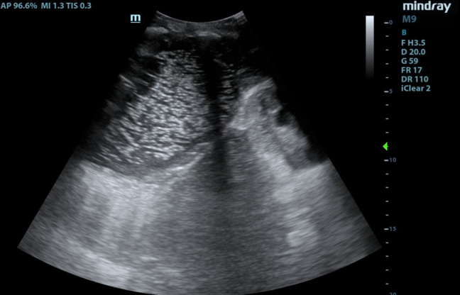 Figure 1
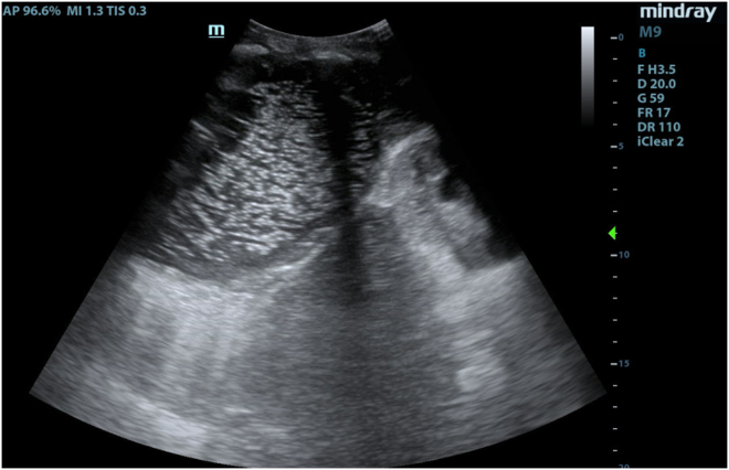 Figure 2
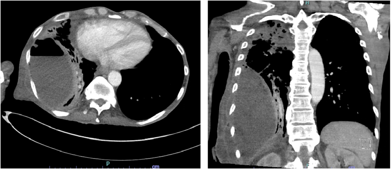 Figure 3
Peer Reviews
No public reviews on file for this paper yet. If you reviewed it on a platform where reviews are public (OpenReview, ICLR, NeurIPS, ICML), you can paste yours below so the community can read it here.
Videos
No videos yet. Explain this paper in a talk, walkthrough, or lecture? Add one.
Taxonomy
TopicsUltrasound in Clinical Applications · Pleural and Pulmonary Diseases · Trauma Management and Diagnosis
Patient Presentation
1
A 57-year-old male with a history of Crohn’s disease on infliximab presented to the emergency department with 1 month of dyspnea and a progressively expanding chest wall “mass.” Examination showed an erythematous, fluctuant area over the patient’s right lateral chest wall. His vitals were normal, other than tachycardia at 107 bpm. Subcutaneous incision and drainage yielded 400 mL of brisk, purulent drainage. Point-of-care ultrasound (POCUS) of the right posterolateral lung zone demonstrated a large echogenic punctiform collection in the pleural space, communicating with the skin. The collection demonstrated positive “spine sign,” and B-lines were visualized emanating from the posterior pleura (Fig 1, Video 1).Figure 1A large echogenic, punctiform pleural collection is noted cranial to the diaphragm, with the right kidney seen caudal to the diaphragm.Video 1The spine sign is noted as a continuous hyperechoic line far-field to the pleural space. Confluent B-lines are seen emanating from the posterior pleura.
Diagnosis: Empyema Necessitans
2
Empyema necessitans (EN) describes a rare complication of empyema where the purulent infection dissects beyond the pleural space into neighboring chest wall musculature and subcutaneous tissue.1^,^2 Our case initially appeared to be a skin abscess, but ultrasonographic signs suggested an exudative pleural effusion, which was confirmed to be EN on computed tomography (Fig 2).3^,^4 The collection required extensive operations, including video-assisted thoracoscopic surgery, open thoracotomy, and lung decortication. The patient was discharged home ambulatory on postoperative day 7.Figure 2. Computed tomography imaging demonstrated a 1-L collection lateral to the right lower lobe containing an air-fluid level. Multifocal consolidation and scarring to the right lung were noted.
EN is typically found in immunocompromised and postoperative patients, although nowadays, it is rare due to the prevalence of antibiotics. Diagnosis of EN is often difficult due to its masquerading as a subcutaneous abscess. Thus, our report reinforces the use of POCUS as a quick, accurate tool for the early detection of EN. POCUS changed management, guiding further thoracic imaging and involvement of a multidisciplinary surgical team, ultimately leading to the resolution of the patient’s condition.
Funding and Support
By JACEP Open policy, all authors are required to disclose any and all commercial, financial, and other relationships in any way related to the subject of this article as per ICMJE conflict of interest guidelines (see www.icmje.org). The authors have stated that no such relationships exist.
The reference list from the paper itself. Each links out to its DOI / PubMed record.
- 1Zeinali Nezhad N.Gholami Shahrebabak A.Shahpar A.Empyema necessitans as a rare manifestation of Staphylococcus aureus Clin Case Rep 1242024 e 869710.1002/ccr 3.8697 PMC 1098370638562573 · doi ↗ · pubmed ↗
- 2White-Dzuro C.G.Assi P.E.Thomas H.C.Thayer W.P.Unusual presentation of empyema necessitans: case report and review of the literature Gen Thorac Cardiovasc Surg 6962021102610303355904410.1007/s 11748-021-01601-9 · doi ↗ · pubmed ↗
- 3Bhoil R.Ahluwalia A.Chopra R.Surya M.Bhoil S.Signs and lines in lung ultrasound J Ultrason 21862021 e 225e 2333454027710.15557/Jo U.2021.0036 PMC 8439137 · doi ↗ · pubmed ↗
- 4Ahmed A.A.Martin J.A.Saul T.Lewiss R.E.The thoracic spine sign in bedside ultrasound. Three cases report Med Ultrason 16220141791812479185210.11152/mu.201.3.2066.162.aara 1 · doi ↗ · pubmed ↗