GSK3β phosphorylated tau filaments are probably not like those from AD brains
Sjors H. W. Scheres

Abstract
Genes, proteins, chemicals, diseases, species, mutations and cell lines named across the full text — each resolved to its canonical identifier and authoritative record.
Click any figure to enlarge with its caption.
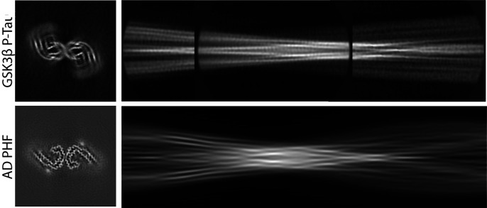 Figure 1
Peer Reviews
No public reviews on file for this paper yet. If you reviewed it on a platform where reviews are public (OpenReview, ICLR, NeurIPS, ICML), you can paste yours below so the community can read it here.
Videos
No videos yet. Explain this paper in a talk, walkthrough, or lecture? Add one.
Taxonomy
TopicsGenetic Neurodegenerative Diseases · Alzheimer's disease research and treatments · Cellular transport and secretion
The filamentous assembly of tau is a hallmark of multiple neurodegenerative diseases, including Alzheimer’s disease (AD). In 2017, we introduced software for helical reconstruction of electron cryomicroscopy (cryo-EM) images (1), and we used this software to determine the structures of tau paired helical and straight filaments (PHFs and SFs) extracted from the brain of an individual with AD (2). Since then, we and others have used this software to determine more than five hundred amyloid structures (3). These combined efforts have shown that a given protein can adopt multiple amyloid structures and that, for tau and other proteins, specific folds characterize different diseases (4).
Recently, Chakraborty et al. reported that phosphorylation with the serine/threonine kinase GSK3β catalyzes the assembly of recombinant tau (5). They used our software to calculate a cryo-EM reconstruction of the resulting filaments. The authors posit that our software overestimated the resolution (3.8 Å), and claim that the correct resolution is ~5 Å. However, rather than suffering from overestimated resolution, the reconstruction most likely represents an incorrect local minimum of the helical reconstruction process. This well-documented problem (6, 7) is the reason why users of our software are advised to remain critical of unexpected features in the reconstruction (8). The reconstruction of the GSK3β phosphorylated tau filaments shows many unexpected features: Densities are disconnected and without recognizable protein-like features (Fig. 1A). For comparison, a reconstruction of an AD PHF, at 5 Å resolution, shows fully connected amino acid main chains, with side chain densities depending on their size and the resolution (Fig. 1B).
We published detailed protocols on how to deal with local minima in amyloid structure determination (7, 8). These instructions were not followed for the GSK3β phosphorylated tau filaments. Because the resulting reconstruction is likely incorrect, it cannot distinguish between the many different structures that have been reported for tau filaments to date. Comparison of a projected cross-over of an AD PHF (Fig. 1D) and an assembly of reference-free 2D class averages of GSK3β phosphorylated tau filaments (Fig. 1C) suggests that they are different. Therefore, the conclusions that these filaments are “AD-like,” and that they are “more similar to” filaments from extracellular vesicles than from brain extracts, are not justified by the data presented by Chakraborty et al. Cryo-EM structure determination, when done correctly, leaves no ambiguity as to whether two structures are the same or not, and incorrect structures should not become part of the scientific discourse. Submission of cryo-EM reconstructions to the Electron Microscopy Data Base (EMDB) (9), and of unprocessed images to EMPIAR (10), allows scrutiny by the scientific community; the former should be mandated by the journal.
The reference list from the paper itself. Each links out to its DOI / PubMed record.
- 1S. He, S. H. W. Scheres, Helical reconstruction in RELION. J. Struct. Biol. 198, 163–176 (2017).28193500 10.1016/j.jsb.2017.02.003PMC 5479445 · doi ↗ · pubmed ↗
- 2A. W. P. Fitzpatrick , Cryo-EM structures of tau filaments from Alzheimer’s disease. Nature 547, 185–190 (2017).28678775 10.1038/nature 23002 PMC 5552202 · doi ↗ · pubmed ↗
- 3M. R. Sawaya, M. P. Hughes, J. A. Rodriguez, R. Riek, D. S. Eisenberg, The expanding amyloid family: Structure, stability, function, and pathogenesis. Cell 184, 4857–4873 (2021).34534463 10.1016/j.cell.2021.08.013PMC 8772536 · doi ↗ · pubmed ↗
- 4S. H. W. Scheres, B. Ryskeldi-Falcon, M. Goedert, Molecular pathology of neurodegenerative diseases by cryo-EM of amyloids. Nature 621, 701–710 (2023).37758888 10.1038/s 41586-023-06437-2 · doi ↗ · pubmed ↗
- 5P. Chakraborty , GSK 3β phosphorylation catalyzes the aggregation of tau into Alzheimer’s disease-like filaments. Proc. Natl. Acad. Sci. U.S.A. 121, e 2414176121 (2024).39693350 10.1073/pnas.2414176121 PMC 11670061 · doi ↗ · pubmed ↗
- 6E. H. Egelman, Ambiguities in helical reconstruction. Elife 3, e 04969 (2014).25486515 10.7554/e Life.04969 PMC 4371874 · doi ↗ · pubmed ↗
- 7S. H. W. Scheres, Amyloid structure determination in RELION-3.1. Acta Crystallogr. D. Struct. Biol. 76, 94–101 (2020).32038040 10.1107/S 2059798319016577 PMC 7008511 · doi ↗ · pubmed ↗
- 8S. Lövestam, S. H. W. Scheres, High-throughput cryo-EM structure determination of amyloids. Faraday Discuss. 240, 243–260 (2022).35913272 10.1039/d 2fd 00034 b PMC 9642048 · doi ↗ · pubmed ↗