High prevalence of vector-borne pathogens in the blood of clinically healthy dogs in Hong Kong
Thamali Manathunga, Mariaelisa Carbonara, Omid Nekouei, Jairo Alfonso Mendoza-Roldan, Wing Yan Jacqueline Tam, Frederic Beugnet, Domenico Otranto, Vanessa R. Barrs

TL;DR
This study found that many healthy dogs in Hong Kong are infected with vector-borne pathogens, highlighting the need for better surveillance and prevention.
Contribution
The first epidemiological survey of Leishmania spp. in Hong Kong dogs, revealing high prevalence of vector-borne diseases.
Findings
45.6% of dogs tested positive for at least one vector-borne pathogen.
Dirofilaria spp. had the highest prevalence at 20.9%, with some identified as Dirofilaria asiatica.
Leishmania spp. was detected in 11.4% of dogs, suggesting possible endemicity in Hong Kong.
Abstract
Leishmaniosis and other canine vector-borne diseases (CVBDs) pose a major risk for veterinary and public health globally, especially where humans and dogs live in close proximity. Although mosquito and tick vectors are abundant in Hong Kong, surveillance for CVBDs has been limited. A serological and molecular survey of 158 healthy owned (n = 64) and free-roaming unowned (n = 94) dogs with outdoor access in Hong Kong was performed to determine CVBD prevalence. Point-of-care (POC) immunoassays were used to detect (i) antibodies to Leishmania spp., Ehrlichia spp., and Anaplasma spp., and (ii) Dirofilaria immitis and Angiostrongylus vasorum antigens, in canine sera. Conventional polymerase chain reaction (PCR) was also carried out to detect the molecular prevalence of all five pathogens as well as Hepatazoon canis, Babesia gibsoni, and Trypanosoma evansi. In addition, for Leishmania spp.…
Genes, proteins, chemicals, diseases, species, mutations and cell lines named across the full text — each resolved to its canonical identifier and authoritative record.
Click any figure to enlarge with its caption.
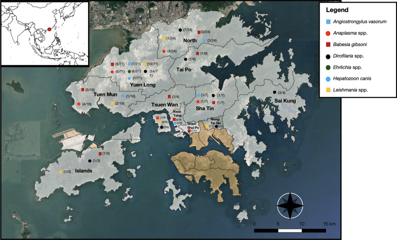 Figure 1
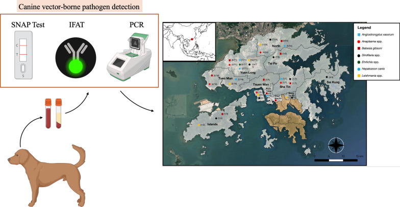 Figure 2
Peer Reviews
No public reviews on file for this paper yet. If you reviewed it on a platform where reviews are public (OpenReview, ICLR, NeurIPS, ICML), you can paste yours below so the community can read it here.
Videos
No videos yet. Explain this paper in a talk, walkthrough, or lecture? Add one.
Taxonomy
TopicsVector-borne infectious diseases · Mosquito-borne diseases and control · Viral Infections and Vectors
Background
A plethora of biotic (e.g., the introduction of arthropod vectors, dog movements, closer proximity of wildlife populations due to habit loss) and abiotic factors (e.g., global warming and urbanization of rural areas) may represent a risk for increasing the abundance and geographical distribution of canine vector-borne diseases (CVBDs), including those of zoonotic concern [1]. Although the Hong Kong Special Administrative Region (SAR), located at the southeastern tip of China, is a densely populated cosmopolitan city of 7.4 million people [2], the proportion of dog ownership is relatively low compared to many other countries, with only 5.7% of total households owning at least one dog [3].
Free-roaming “village” dogs represent the most common carnivores in Hong Kong SAR and pose a risk for the transmission of CVBDs, many of which are zoonotic [4]. Indeed, Hong Kong has a subtropical monsoon climate, providing favorable environmental conditions for vector proliferation. For example, Aedes albopictus mosquitoes, the primary vectors of canine filariosis, are highly abundant in Hong Kong [5], with Dirofilaria immitis causing canine heartworm disease [6]. This filarioid is the agent of human dirofilariasis worldwide, along with other species such as Dirofilaria repens and Dirofilaria asiatica (formerly Dirofilaria sp. “hongkongensis”), the latter causing subcutaneous nodules in cats and dogs [7] as well as in humans [8, 9].
Despite limited tick surveillance, four genera (i.e., Rhipicephalus, Haemaphysalis, Ixodes, and Hyalomma) have been recorded as being present in Hong Kong by the Food and Environmental Hygiene Department (FEHD) [10]. In particular, Rhipicephalus sanguineus sensu lato (s.l.) and Haemaphysalis longicornis are adapted to the subtropical climate of Hong Kong and may transmit Babesia vogeli, Ehrlichia canis (vectored by R. sanguineus s.l.), and Babesia gibsoni (vectored by H. longicornis) [11, 12].
From the limited investigations that have been performed so far, babesiosis, due to B. gibsoni, is the most common CVBD in Hong Kong [6, 13, 14], with a molecular prevalence ranging from 3.8% to 31% in owned dogs, and of up to 44% in free-roaming unowned dogs [6, 13, 14]. By contrast, B. vogeli has seldom been reported in dogs from Hong Kong (molecular prevalence < 5%) [13, 14]. Babesia hongkongensis has been detected at a low prevalence (1.7%) in free-roaming cats but not dogs from Hong Kong [15, 16]. Other vector-borne pathogens (VBPs), including E. canis, Hepatozoon canis, Anaplasma platys, and the potentially zoonotic Anaplasma phagocytophilum, have also been reported with low occurrence among dogs from Hong Kong [6, 13, 14].
So far, canine leishmaniosis has only been reported in two purebred dogs (Belgian Malinois) living in the same household in Hong Kong [17]. The first case was a dog imported from the USA, which presented with chronic cutaneous leishmaniosis, while the second case was autochthonous, presenting with systemic leishmaniosis [17]. Both cases were due to Leishmania infantum, the most important zoonotic Leishmania species, causing cutaneous and/or visceral signs in dogs and humans, depending on the host’s immune response. However, the primary vector (i.e., phlebotomine sand flies) has not yet been reported in Hong Kong, and non-vectorial, transplacental, or transmission by direct contact was suspected in both cases [17].
Given the paucity of information about CVBDs in this region of China, this study aimed to determine the prevalence of leishmaniosis and other CVBDs in healthy dogs with outdoor access in Hong Kong, using a comprehensive range of molecular and serological tests.
Methods
Sample collection
From December 2022 to March 2023, blood samples were collected from 158 dogs presented to a neutering clinic in the New Territories of Hong Kong (Fig. 1). All dogs were assessed by the attending veterinarians as healthy. Age, sex, breed, neuter status, outdoor walks per day, outdoor environment, and primary residential district were recorded for each dog. Whole blood was collected into ethylenediaminetetraacetic acid (EDTA) and plain tubes. The serum was separated after centrifugation at 10,000×g for 10 min. Sera and EDTA blood samples were stored at – 80 ºC.Fig. 1. Map of the study areas indicated by district, showing the location of dogs enrolled, according to their positivity to vector-borne pathogens. Tan shaded regions indicate locations where only negative samples were detected, while gray-shaded regions represent areas where positive samples were identified
Serological antibody and antigen tests
An immunofluorescence antibody test (IFAT) was used to detect anti-Leishmania immunoglobulin G (IgG) antibodies, as reported previously [18]. Samples that showed clear promastigote fluorescence at a cut-off dilution of 1:80 were considered positive and then titrated until no fluorescence was observed. Leishmania antibodies were also detected using a commercially available rapid enzyme-linked immunosorbent assay (ELISA)-based test kit (SNAP^®^ Leish 4Dx^®^ test; IDEXX Laboratories, Westbrook, ME, USA).
Serum samples were tested for D. immitis antigen using two point-of-care (POC) immunoassays (SNAP^®^ Heartworm RT test kit and SNAP^®^ Leish 4Dx^®^ test; IDEXX Laboratories, Westbrook, ME, USA). Angiostrongylus vasorum antigen was detected using a commercial rapid immunochromatographic test kit (Angio Detect™ Test, IDEXX Laboratories, Westbrook, ME, USA). Antibodies against Anaplasma spp. (i.e., A. phagocytophilum, A. platys) and Ehrlichia spp. (i.e., E. canis, E. ewingii, E. chaffeensis) were detected using the SNAP^®^ Leish 4Dx^®^ test (IDEXX Laboratories, Westbrook, ME, USA). The SNAP^®^ Leish 4Dx^®^ test has been validated for use in dogs [19], as has the Angio Detect™ test [20] and the SNAP^®^ Heartworm RT test [21].
Molecular analysis
Genomic DNA was extracted from EDTA blood using the QIAamp DNA Blood and Tissue Kit (Qiagen GmbH, Hilden, Germany), according to the manufacturer’s instructions. For all samples, DNA of flagellated protozoa (i.e., Leishmania spp., Trypanosoma evansi), Rickettsiales (Anaplasma spp., Ehrlichia spp., and Wolbachia spp.), tick-borne apicomplexan protozoa (Babesia spp. and Hepatozoon spp.), and nematodes (i.e., Dirofilaria spp., A. vasorum) was searched for using conventional polymerase chain reaction (PCR) protocols listed in Table 1. All PCR products were analyzed by electrophoresis on 2% agarose gels, purified, and subjected to Sanger sequencing. Sequences were edited using Geneious Prime^®^ 2024.0.3, and the Basic Local Alignment Search Tool (BLAST) on the National Center for Biotechnology Information (NCBI) GenBank was used to determine species identity.Table 1. Pathogen targeted in blood samples, and list of primers used in the conventional PCR protocolsPathogenTarget genePrimerAmplicon length (base pairs, bp)ReferencesLeishmania spp.Kinetoplast DNA (kDNA) minicircleMC1 (5′ GTTAGCCGATGGTGGTCTTG 3′)MC2 (5′ CACCCATTTTTCCGATTTTG 3′)447[64]Dirofilaria spp.Cytochrome c oxidase subunit-1 (cox1)Diro_COX1_F (5′ GCTTTGTCTTTTTGGTTTACTTTT 3′)Diro_COX1_R (5′ TCAAACCTCCAATAGTAAAAAGAA 3′)880[65]Angiostrongylus vasorum**18S rRNANC18SF1 (5′ AAAGATTAAGCCATGCA 3′)NC5BR (5′ GCAGGTTCACCTACAGAT 3′)1700[66]Anaplasma spp./Ehrlichia spp./Wolbachia pipientis^a^16S rRNAEHR-16SD (5′ GGTACCYACAGAAGAAGTCC 3′)EHR-16SR (5′TAGCACTCATCGTTTACAGC 3′)345[67]Babesia spp./Hepatozoon spp./18S rRNAHepF300 (5′ GTTTCTGACCTATCAGCTTTCGACG 3′)HepR900 (5′ CAAATCTAAGAATTTCACCTCTGAC 3′)581[68]Trypanosoma evansiVariant surface glycoprotein gene (vsg gene)TevF (5′ TGCAGACGACCTGACGCTACT 3′)TevR (5′ CTCCTAGAAGCTTCGGTGTCCT 3′)227[69]^a^Endosymbiont of filarioids
For samples that were seropositive for Leishmania spp. antibodies by IFAT and/or commercial test kit, a real-time PCR (qPCR) was performed to detect L. infantum kinetoplast DNA (kDNA) minicircle fragments. A duplex real-time PCR assay (dqPCR) was also performed to detect both L. infantum and Leishmania tarentolae DNA. Details of the primers, probes, and protocols used for qPCR assays are provided in Table 2.Table 2. Primers and probes used for quantitative polymerase chain reaction (qPCR) of Leishmania-seropositive samplesqPCR targetPrimers/probesAmplicon length (base pairs, bp)ReferenceKinetoplast minicircle DNA from L. infantumLEISH-1 (5′ AACTTTTCTGGTCCTCCGGGTAG 3′)LEISH-2 (5′ ACCCCCAGTTTCCCGCC 3′)TaqMan-minor groove binder probe:(FAM-5′AAAAATGGGTGCAGAAAT 3′ non-fluorescent quencher-MGB)120[70]Duplex PCR for detection of L. infantum and L. tarentolae internal transcribed spacer 1 (ITS1) DNAITS1-F (5′ GCAGTAAAAAAAAGGCCG 3′)ITS1-R (5′ CGGCTCACATAACGTGTCGCG 3′)150[71]
Data analysis
To calculate the overall prevalence of Dirofilaria spp., A. vasorum, Ehrlichia spp., Anaplasma spp., and Leishmania spp., a sample was considered positive when it tested positive on at least one serological assay and/or by PCR. The overall prevalence of Babesia spp. and Hepatozoon spp. was calculated based only on molecular results.
To measure the level of agreement between different tests for each pathogen, Cohen’s kappa statistics were calculated [22]. The level of agreement was defined as follows: no agreement (κ = 0), slight agreement (κ = 0.01–0.2), fair agreement (κ = 0.21–0.4), moderate agreement (κ = 0.41–0.6), substantial agreement (κ = 0.61–0.8), or almost perfect agreement (κ = 0.81–1). The potential associations between exposure to tick-borne pathogens and independent variables (e.g., sex, age, environment, being stray, and outdoor walk frequency) were evaluated using simple logistic regressions with a 0.05 significance level. All statistical analyses were conducted in Stata v18 (StataCorp LLC, College Station, TX, USA).
Results
Of the 158 dogs included in this study, 94 (59.5%) were free-roaming and unowned, while 64 (40.5%) were owned. All dogs were mongrels and had outdoor access. There were 53 sexually intact male dogs (33.5%) and 105 intact female dogs (66.5%), ranging in age from 5 months to 6.5 years (median 1.4 years). One hundred dogs (63.3%) were older than 1 year.
Overall, 72 (45.6%; 95% confidence interval [CI] 37.8–53.3) dogs tested positive on molecular and/or serological tests for at least one pathogen (Fig. 1), with the highest prevalence recorded for Dirofilaria spp. (20.9%; 95% CI 14.6–27.2). Other pathogens detected in decreasing order of prevalence were B. gibsoni (15.2%; 95% CI 9.6–20.8), Leishmania spp. (11.4%; 95% CI 6.4–16.4), Anaplasma spp. (7.6%; 95% CI 3.5–11.7), H. canis (4.4%; 95% CI 1.2–7.6), Ehrlichia spp. (3.8%; 95% CI 0.8–6.8), and A. vasorum (0.63%; 95% CI 0.0–3.5) (Table 3). No T. evansi DNA was detected.Table 3. Serological and molecular results of canine vector-borne pathogens detected in 158 dogs from Hong KongPathogenPositive Ag or Ab testsNo. (%; 95% CI)PCR-positiveNo. (%; 95% CI)Species identified by sequencing(no.)Ag- or Ab-positive/PCR-positiveAg- or Ab-positive/PCR-negativeAg- or Ab-negative/PCR-positiveTotal prevalence: no. of positive on Ag, Ab, and/or molecular tests (%; 95% CI)Dirofilaria spp.30 (19; 12.9–25.1)19 (12; 7.0–17.1)Dirofilaria immitis (16)Dirofilaria asiatica (2)Wolbachia pipientis (15)1614333 (20.9, 14.6–27.2)Angiostrongylus vasorum1 (0.6; 0.02–3.5)0 (0)N/AN/AN/AN/A1 (0.6; 0–3.5)Anaplasma spp.12 (7.6; 3.5–11.7)4 (2.5; 0.1–5.0)Anaplasma platys (4)48012 (7.6; 3.5–11.7)Leishmania spp.18 (11.4; 6.4–16.4)0 (0)N/AN/A18N/A18 (11.4; 6.4–16.4)Ehrlichia spp.5 (3.2; 0.4–5.9)3 (1.9; 0.4–5.5)Ehrlichia canis2316 (3.8; 0.8–6.8)Babesia spp.N/A24 (15.2; 9.6- 20.8)Babesia gibsoni (24)N/AN/AN/A24 (15.2; 9.6–20.8)Hepatozoon spp.N/A7 (4.4; 1.2–7.6)Hepatozoon canis (7)N/AN/AN/A7 (4.4; 1.2–7.6)Trypanosoma evansiN/A0 (0)N/AN/AN/AN/A0 (0)Ab antibody, Ag antigen, CI confidence interval, N/A not applicable
Of the 18 (11.4%; 95% CI 6.4–16.4) dogs seropositive for Leishmania spp., 17 were positive on IFAT alone and one on the POC immunoassay. IFAT titers ranged from 1:80 to 1:320, including 10 dogs with a titer of 1:80, five with a titer of 1:160, and two with a titer of 1:320. Leishmania spp. DNA was not detected in any blood sample.
Among the 33 (20.9%; 95% CI 14.6–27.2) samples positive for Dirofilaria spp., 19 tested positive on the cox1 gene PCR for Dirofilaria spp. and/or had 16S ribosomal RNA (rRNA) Wolbachia sequences with 100% nucleotide identity to Wolbachia pipientis (GenBank Accession no: OR939250.1). Cohen’s kappa statistic, κ = 0.83, indicated perfect agreement between Dirofilaria spp. results by cox1 PCR and the Wolbachia results by 16S rRNA PCR. Similarly, both SNAP^®^ Leish 4Dx^®^ test (IDEXX Laboratories, Westbrook, ME, USA) and SNAP^®^ Heartworm RT test kit (IDEXX Laboratories, Westbrook, ME, USA) had almost perfect agreement in detecting D. immitis antigens (κ = 0.84). No agreement between IFAT and SNAP^®^ Leish 4Dx^®^ test (IDEXX Laboratories, Westbrook, ME, USA) was observed for Leishmania antibody detection (κ = 0). Discordant results were observed between molecular and serological tests for Dirofilaria, Ehrlichia, and Anaplasma (Table 3). In particular, two samples identified molecularly as D. asiatica showed different results, with one testing antigen-positive on SNAP^®^ Heartworm RT test kit (IDEXX Laboratories, Westbrook, ME, USA) and the other negative. The W. pipientis sequence was also amplified from the 16S rRNA PCR assay of the antigen-positive D. asiatica sample.
Based on both serological and molecular results, 16.5% (26/158; 95% CI 10.7–22.2) of dogs were infected or exposed to more than one VBP (Table 4). Among the Leishmania-seropositive samples, 88.9% (16/18; 95% CI 65.3–98.6) had co-infections or were exposed to at least one other pathogen. In addition, 29.1% (46/158; 95% CI 22.0–36.2) of dogs tested positive by serology or PCR for at least one tick-borne pathogen. Three dogs were co-infected with more than one tick-borne pathogen (Table 4).Table 4. Number of dogs with co-pathogen infections or exposure based on all test results (serological and molecular)Pathogen combinationsNo. co-infected casesDirofilaria spp., Leishmania spp.2Dirofilaria spp., Anaplasma spp.1Dirofilaria spp., Babesia gibsoni3Dirofilaria spp., Hepatozoon canis2Leishmania spp., Ehrlichia spp.2Leishmania spp., B. gibsoni10Ehrlichia spp., Anaplasma spp.1Ehrlichia spp., H. canis1Angiostrongylus vasorum, Dirofilaria spp.1Dirofilaria spp., Ehrlichia spp., Leishmania spp.1Dirofilaria spp., Anaplasma spp., B. gibsoni1Dirofilaria spp., Leishmania spp., B. gibsoni1Total26
In the risk factor analysis, none of the tested independent variables were associated with tick-borne infection (Table 5); therefore, multivariable modeling was not performed.Table 5. Univariable associations between tick-borne infection status and the independent variables in 158 study dogs using simple logistic regressionCategoryNo. positive (%)No. negative (%)Odds ratio (95% CI)Statistical analysisSexMale12 (22.6)41 (77.4)–χ^2^ = 1.66, df = 1, P = 0.205Female34 (32.4)71 (67.6)1.64 (0.8–3.5)Age (years) < 112 (23.5)39 (76.5)–χ^2^ = 1.38, df = 2, P = 0.501 1 to < 220 (32.3)42 (67.7)1.55 (0.7–3.6) ≥ 212 (33.3)24 (66.7)1.63 (0.6–4.2)Outdoor walks (per day) < 15 (19.2)21 (80.8)–χ^2^ = 5.08, df = 3, P = 0.166 17 (19.4)29 (80.6)1.01 (0.3–3.6) 210 (32.3)21 (67.7)1.99 (0.6–6.9) ≥ 324 (37)41 (63)2.46 (0.8–7.4)Stray Yes29 (30.8)65 (69.2)–χ^2^ = 0.34, df = 1, P = 0.561 No17 (26.6)47 (73.4)1.23 (0.6–2.5)Environment (access to vegetation) Limited8 (28.6)20 (71.4)–χ^2^ = 0.001, df = 1, P = 0.944 Yes38 (29.2)92 (70.8)1.03 (0.4–2.6)χ^2^ Chi-square value, df degrees of freedom, CI confidence interval
Discussion
Almost half of the dogs in this study tested positive for at least one VBP, suggesting that CVBDs are highly prevalent among owned and stray dogs with outdoor access in Hong Kong. Notably, 11.4% of dogs were seropositive for Leishmania spp., yet no Leishmania DNA was detected in any blood sample. In naturally occurring Leishmania spp. infections, due to their tropism for lymphoid tissues, protozoa are more abundant in lymph node and bone marrow aspirates, as well as in conjunctival swab samples, than in whole blood [23]. However, as none of these samples were collected, Leishmania infection could not be confirmed.
Although no competent sand fly vectors have yet been reported in Hong Kong, Phlebotomus chinensis, a known vector of visceral leishmaniasis in China, has been detected in the nearby provinces of Hainan and Guangdong in mainland China, suggesting that an expansion of the geographical distribution of these vectors may have occurred [24–26]. In 2023, the first case of human visceral leishmaniasis in nearby Shenzhen city (Guangdong province) was reported, although it was thought to be an imported case from an endemic area in China [27]. Sand flies belonging to the genus Sergentomyia, which prefer to feed on cold-blooded vertebrates, have been found in Hong Kong [24] and are widely distributed in the Old World [28]. The role of the herpetophilic Sergentomyia in the transmission of zoonotic Leishmania spp. is controversial, as L. infantum [29–31] and Leishmania martiniquensis [32] have been molecularly detected in these sand flies.
In the present study, samples were tested for antibodies to Leishmania spp. using a POC immunoassay and IFAT, with the latter being considered the reference test among serological tests [33]. The lack of agreement between the POC immunoassay and IFAT could be due to the high sensitivity of IFAT for the detection of subclinically infected dogs [19, 34, 35]. Low IFAT titers, which were most common in our study, are difficult to interpret, especially in the absence of molecular confirmation of the infection. Although infections by T. evansi were ruled out by PCR, the IFAT seropositivity obtained is unlikely to be due to cross-reactivity with trypanosomatids [35, 36]. Nonetheless, it should be acknowledged that in China, other Leishmania spp. (e.g., Leishmania gerbilli, Leishmania tropica, Leishmania turanica, and an undescribed Leishmania sp. closely related to Sauroleishmania) have been identified [37], including in dogs [38]. Previous studies have suggested that cross-reactivity between Leishmania spp. and E. canis may occur [39–41]. In our study, three of the 17 Leishmania-seropositive dogs were infected with E. canis, while overall, 15 (88.2%) of Leishmania-seropositive samples were co-infected with VBPs (Table 4); thus, the occurrence of cross-reactivity with other VBPs cannot be ruled out.
The proportion of dogs testing positive for D. immitis antigens by POC assay (19%; 95% CI 12.9–25.1) is higher than that previously recorded in Hong Kong (i.e., 6% and 5.6%) [6, 14], suggesting an increasing distribution of this mosquito-borne infection and the importance of adopting preventive chemoprophylaxis control measures in dog populations. While the prevalence in this study was similar among stray (19.1%) and owned dogs (23.4%), most infected animals (69.7%) were older than 1 year, reflecting increased opportunities for vector exposure when compared to younger animals. The finding of dogs PCR-positive for D. immitis but antigen-negative by POC assays may be due to a low burden of adult female worms [42–44]. However, as no heat pretreatment of sera was performed before the use of the POC assay, immune complex formation resulting in antigen-negative results on the POC assay cannot be excluded [45]. Conversely, the antigen positivity detected in a sample that was molecularly positive for D. asiatica might suggest a simultaneous infection with D. immitis, or might be due to a cross-reaction between the two Dirofilaria spp.
Although W. pipientis DNA can originate from several filarioids (e.g., Acanthocheilonema spp., Dirofilaria spp., Brugia spp.) [46], we only detected DNA of Dirofilaria spp., supporting Wolbachia spp. as an indicative marker for D. immitis infection in this study [47, 48].
The finding of one healthy dog seropositive for A. vasorum is of interest, since it has not been previously detected in East or Southeast Asia, and infected dogs usually present with severe respiratory signs [49]. Although A. vasorum has been detected in Turkey, which lies mainly in West Asia, its presence in other parts of Asia has not been documented [50]. However, as other Angiostrongylus spp., such as A. cantonensis and A. malaysiensis, have been reported in East and Southeast Asia [51, 52], cross-reactivity [53] might have occurred. The A. vasorum-seropositive dog in this study also tested positive for D. immitis on both POC immunoassays, as well as by PCR. However, cross-reactivity with A. vasorum on the Angio Detect™ Test is highly unlikely, as this event was excluded in purpose-designed studies [20, 54]. Accordingly, the A. vasorum antigen assay employed was reported to have an overall sensitivity of 85% (combined for experimentally infected dogs and field infections), and a specificity of 100% [20, 55]. Other investigators found cross-reactivity of the A. vasorum POC assay used in this study with other Angiostrongylus spp. infecting wildlife in Europe [56] or with a close relative from the family Angiostrongylidae, Gurltia paralysans, in cats from Chile [57]. Thus, further research is needed to confirm the presence of A. vasorum in Hong Kong (e.g., Baermann sedimentation with molecular confirmation).
Overall, nearly one-third (29.1%) of the dogs in this study were infected with at least one tick-borne pathogen, indirectly illustrating the tick abundance in Hong Kong, with R. sanguineus s.l. and* H. longicornis* being common in East Asia [58–60]. The employment of PCR protocols amplifying more than one pathogen, such as in the case of piroplasms and rickettsias, represents a limitation of this study, as it may have underestimated the occurrence of co-infections.
The higher prevalence of A. platys than E. canis detected in this study (i.e., 7.6% vs. 3.8%) differs from previous studies conducted on owned dogs in Hong Kong, which found a mean of 10.8% for Ehrlichia versus 2.7% for Anaplasma [6, 14], and might be related to the sampled population of dogs. Specifically, previous studies focused predominantly on sick dogs for which veterinary care was sought, compared to the healthy dog population in this study. Infection by E. canis often presents as a severe clinical illness, in contrast to A. platys infections, which are typically subclinical [61].
The discrepancies between PCR and serology results for Ehrlichia and Anaplasma are likely related to the stage of infection, since PCR-positive results are expected in acute-stage infection [62] while seropositivity may reflect past or chronic exposure [63]. Similar to other investigations in Hong Kong [6, 13, 14], E. canis was the only Ehrlichia sp. detected in our study, reflecting the distribution of R. sanguineus s.l. vectors [12].
The results of this study suggest that after dirofilariosis, babesiosis caused by B. gibsoni is the second most common CVBD in Hong Kong, with a decreasing prevalence in the last 20 years from 41% overall in stray and owned dogs [14] to 15.2% in the present study.
Conclusions
Overall, this study highlights the circulation of several zoonotic VBPs in dogs from Hong Kong, with D. immitis being the most common among both owned and stray animals. In addition, Leishmania spp. seropositivity was detected in dogs, highlighting the need for prospective studies to confirm Leishmania infection and to perform surveillance for competent vectors. Given the high exposure to arthropod-borne pathogens, year-round parasiticide treatment of animals in combination with insecticides and repellents is advocated to mitigate the risk of CVBD in dogs and in humans.
Supplementary Information
Supplementary Material 1.
The reference list from the paper itself. Each links out to its DOI / PubMed record.
- 1Census and Statistics Department. Press release. 2024. https://www.censtatd.gov.hk/en/press_release_detail.html?id=5386. Accessed 9 Feb 2025.
- 2Census and Statistics Department. Thematic household survey report No.66. 2019. https://www.info.gov.hk/gia/general/201906/21/P 2019062100361.htm. Accessed 9 Feb 2025.
- 3Food and Environmental Hygiene Department (FEHD). 2011. https://www.fehd.gov.hk/english/index.html. Accessed 9 Feb 2025.
- 4American Heartworm Society. Canine guidelines for the prevention, diagnosis, and management of heartworm (Dirofilaria immitis) infection in dogs. 2024. https://d 3ft 8sckhnqim 2.cloudfront.net/images/AHS_Canine_Guidelinesweb 22NOV 2024.pdf?1732318144. Accessed 9 Feb 2025.