Towards a quantitative theory for transmission X-ray microscopy
James G McNally, Christoph Pratsch, Stephan Werner, Stefan Rehbein, Andrew Gibbs, Jihao Wang, Thomas Lunkenbein, Peter Guttmann, Gerd Schneider

TL;DR
This paper develops a model for X-ray microscopes to better understand how they capture images of tiny objects like gold nanospheres.
Contribution
A new experimental and theoretical framework using Mie theory to evaluate TXMs with quantitative accuracy.
Findings
Including both absorption and phase properties of gold is necessary for accurate TXM image modeling.
A small tilt in condenser illumination improves qualitative agreement with measurements.
The microscope underestimates absorption by 10–20% compared to the model predictions.
Abstract
Transmission X-ray microscopes (TXMs) are now increasingly used for quantitative analysis of samples, most notably in the spectral analysis of materials. Validating such measurements requires quantitatively accurate models for these microscopes, but current TXM models have only been tested qualitatively. Here we develop an experimental and theoretical framework for evaluation of TXMs that uses Mie theory to compute the electric field emerging from a nanosphere. We approximate the microscope’s condenser illumination by plane waves at the mean illumination angle and the zone plate by a thin lens. We find that this model produces good qualitative agreement with our 3D measurements of 60 nm gold nanospheres, but only if both β and δ for the complex refractive index n = 1 – δ + iβ of gold are included in the model. This shows that both absorption and phase properties of the specimen…
Genes, proteins, chemicals, diseases, species, mutations and cell lines named across the full text — each resolved to its canonical identifier and authoritative record.
Click any figure to enlarge with its caption.
 Figure 1
 Figure 2
 Figure 3
 Figure 4
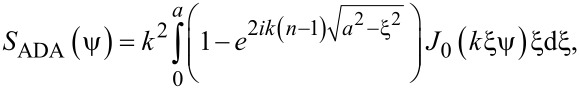 Figure 5
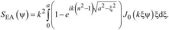 Figure 6
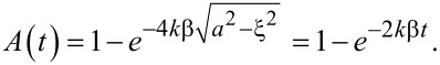 Figure 7
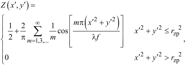 Figure 8
 Figure 9
 Figure 10
 Figure 11
 Figure 12
 Figure 13
 Figure 14
 Figure 15
 Figure 16
 Figure 17
 Figure 18
 Figure 19
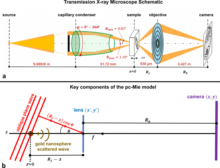 Figure 20
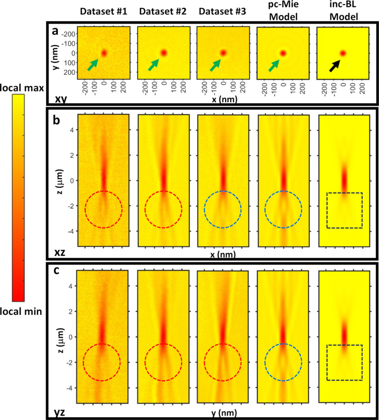 Figure 21
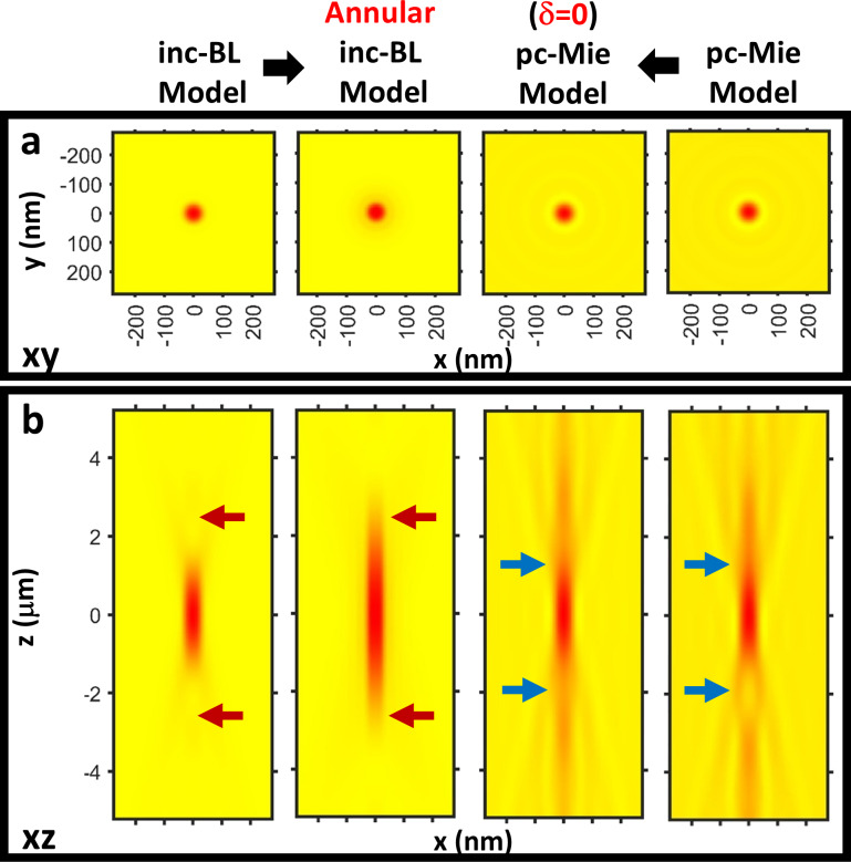 Figure 22
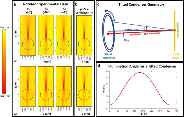 Figure 23
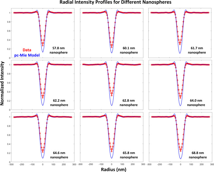 Figure 24
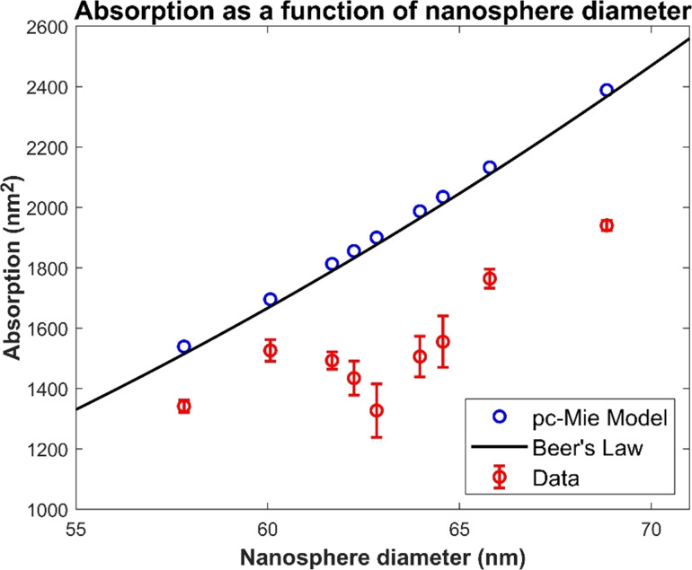 Figure 25
 Figure 26
 Figure 27
 Figure 28
 Figure 29
 Figure 30
 Figure 31
 Figure 32
 Figure 33
 Figure 34
 Figure 35
Peer Reviews
No public reviews on file for this paper yet. If you reviewed it on a platform where reviews are public (OpenReview, ICLR, NeurIPS, ICML), you can paste yours below so the community can read it here.
Videos
No videos yet. Explain this paper in a talk, walkthrough, or lecture? Add one.
Taxonomy
TopicsAdvanced X-ray Imaging Techniques · Advanced Electron Microscopy Techniques and Applications · Medical Imaging Techniques and Applications