Non-contrast photon counting computed tomography of the head: optimized modeling of off-focal radiation to reduce calvarium-related tissue inhomogeneity
Arwed Elias Michael, Martin Petersilka, Denise Schoenbeck, Matthias Michael Woeltjen, Julius Henning Niehoff, Christoph Moenninghoff, Tanja Kurzendorfer, Jan Borggrefe, Lukas Goertz, Jan Robert Kroeger

TL;DR
A new CT software reduces brain tissue inhomogeneity near the skull in non-contrast photon counting CT scans, improving image quality.
Contribution
A new software approach optimizes off-focal radiation modeling to reduce artifacts in head CT scans.
Findings
The new software significantly reduced signal differences between gray and white matter near the calvarium (p < 0.001).
Qualitative analysis showed improved gray-white differentiation in virtual monoenergetic images at 65 keV (p < 0.001).
Mathematical correction of off-focal radiation effectively reduced cortical brain tissue inhomogeneity.
Abstract
Photon counting CT (PCCT) is a promising technique for neuroradiological CT examinations. In initial studies on non-contrast PCCT of the head (NCCT), however, artifacts close to the calvarium were noticed, which lead to an inhomogeneous representation of the brain tissue. In this study, a new software for image reconstruction to reduce artifacts is evaluated. In the new CT software developed by the manufacturer, off-focal radiation was remodeled and is mathematically corrected in the NCCT in data processing during image formation. For the evaluation, 60 patients with an NCCT in the currently used software and 44 patients in the new software were included retrospectively. A detailed quantitative analysis using multiple regions of interest and a qualitative analysis with a reading by experienced radiologists was performed to evaluate image quality and tissue homogeneity below the…
Genes, proteins, chemicals, diseases, species, mutations and cell lines named across the full text — each resolved to its canonical identifier and authoritative record.
Click any figure to enlarge with its caption.
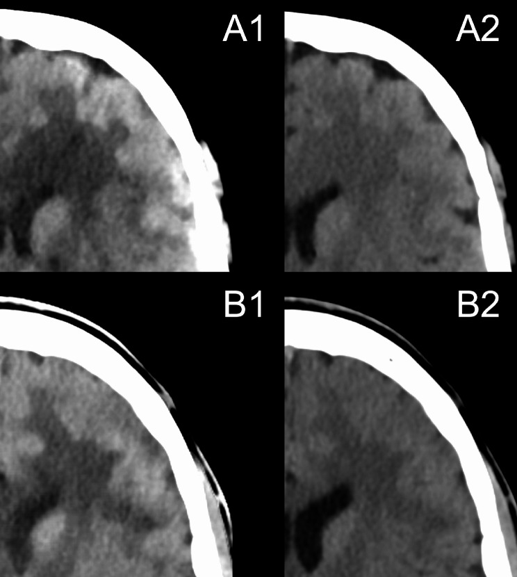 Figure 1
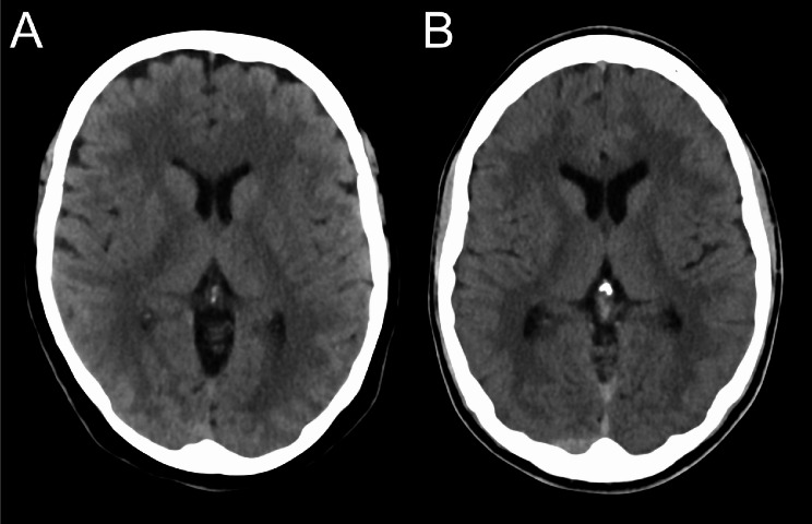 Figure 2
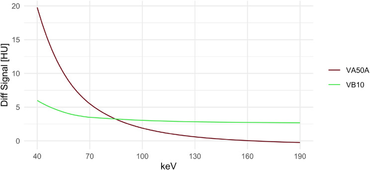 Figure 3
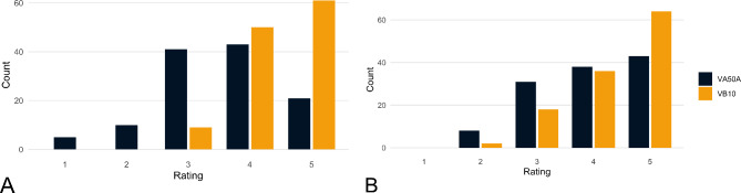 Figure 4
Peer Reviews
No public reviews on file for this paper yet. If you reviewed it on a platform where reviews are public (OpenReview, ICLR, NeurIPS, ICML), you can paste yours below so the community can read it here.
Videos
No videos yet. Explain this paper in a talk, walkthrough, or lecture? Add one.
Taxonomy
TopicsAdvanced X-ray and CT Imaging · Radiation Dose and Imaging · Medical Imaging Techniques and Applications