A deep learning model for classifying left ventricular enlargement for both transthoracic echocardiograms and handheld cardiac ultrasound
Jeffrey G Malins, D M Anisuzzaman, John I Jackson, Eunjung Lee, Jwan A Naser, Jared G Bird, Paul A Friedman, Christie C Ngo, Jae K Oh, Gal Tsaban, Patricia A Pellikka, Jeremy J Thaden, Francisco Lopez-Jimenez, Zachi I Attia, Sorin V Pislaru, Garvan C Kane

TL;DR
A deep learning model accurately detects left ventricular enlargement from heart ultrasound images, working for both standard and handheld devices.
Contribution
The model detects LV enlargement without needing patient sex or body size data and works across different ultrasound devices.
Findings
The model achieved high accuracy (AUC 0.925–0.971) in detecting LV enlargement from TTE images.
It also performed well on handheld cardiac ultrasound images (AUC 0.874–0.902).
Performance was consistent across multiple geographic locations and patient groups.
Abstract
To develop a deep learning model that: (i) utilizes transthoracic echocardiography (TTE) clips to detect left ventricular (LV) enlargement without being provided information regarding a patient’s sex and body size; and (ii) can be accurately applied to clips acquired using either standard comprehensive TTE or handheld cardiac ultrasound (HCU). Using retrospective TTE data (training: 8722 patients; internal validation: 468 patients), we developed a deep learning model that estimates a patient’s end-diastolic LV volume (indexed to body surface area and normalized across the sexes), and then thresholds this estimate to perform the following classifications: (1) normally sized LV vs. ≥ mild LV enlargement; (2) normal/mildly enlarged LV vs. ≥ moderate LV enlargement. For retrospective datasets, the model showed strong performance in TTE across three geographically distinct locations…
Genes, proteins, chemicals, diseases, species, mutations and cell lines named across the full text — each resolved to its canonical identifier and authoritative record.
Click any figure to enlarge with its caption.
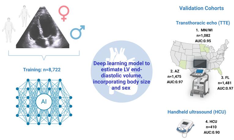 Figure 1
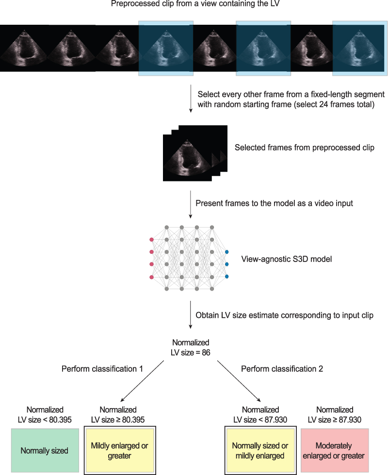 Figure 2
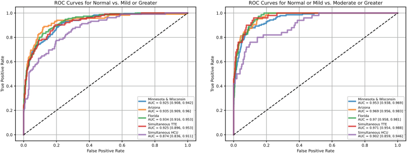 Figure 3
 Figure 4
 Figure 5
Peer Reviews
No public reviews on file for this paper yet. If you reviewed it on a platform where reviews are public (OpenReview, ICLR, NeurIPS, ICML), you can paste yours below so the community can read it here.
Videos
No videos yet. Explain this paper in a talk, walkthrough, or lecture? Add one.
Taxonomy
TopicsCardiovascular Function and Risk Factors · Cardiac Imaging and Diagnostics · Cardiac Valve Diseases and Treatments