Immunosenescence-related T cell phenotypes, structural brain imaging, and cognitive impairment in patients with schizophrenia: a moderated mediation analysis
Na Li, Yanli Li, Ting Yu, Wenjin Chen, Mengzhuang Gou, Wenkai Zheng, Zhaofan Liu, Xiaoying Wang, Jiao Fang, Jinghui Tong, Song Chen, Baopeng Tian, Chiang-Shan R. Li, Li Tian, Yunlong Tan

TL;DR
This study links immune aging in T cells and brain structure changes to cognitive issues in schizophrenia patients.
Contribution
It identifies specific T cell markers and brain regions as potential biomarkers for cognitive deficits in schizophrenia.
Findings
Schizophrenia patients show immunosenescence with fewer naïve and more memory T cells compared to healthy controls.
Higher IL-1β levels in specific T cells correlate with working memory deficits in schizophrenia patients.
Brain structure (IPL_R thickness and CP volume) mediates and moderates the link between IL-1β and cognitive impairment.
Abstract
Cognitive impairment is a core characteristic of schizophrenia. Immunosenescence has been consistently implicated in the cognitive dysfunction observed in neurodegenerative diseases, but how it may relate to cognitive deficits in schizophrenia is still unclear. We explored the associations between immunosenescence and cognitive impairment in patients with schizophrenia (SCZ, n = 65) and healthy controls (HCs, n = 39). Immunosenescence markers were assessed by flow cytometry and included the percentage of naïve or memory T cell subsets labeled by CD4+/CD8+, CD45RA+(naïve)/CD45RO (memory), or CD95+(memory), as well as the intracellular levels of selected cytokines (IL-1β, IL-6, TNF-α, and IFN-γ) in T cell subsets. T1-weighted magnetic resonance imaging was performed to assess the subcortical volume and cortical thickness. Participants were evaluated using the Positive and Negative…
Genes, proteins, chemicals, diseases, species, mutations and cell lines named across the full text — each resolved to its canonical identifier and authoritative record.
Click any figure to enlarge with its caption.
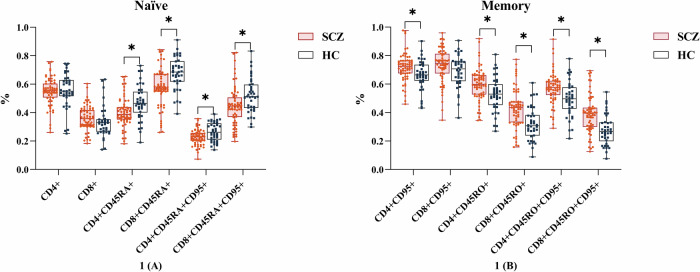 Figure 1
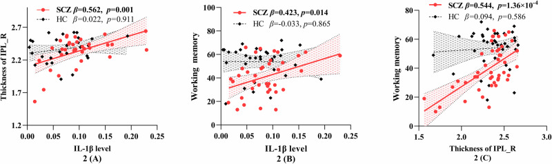 Figure 2
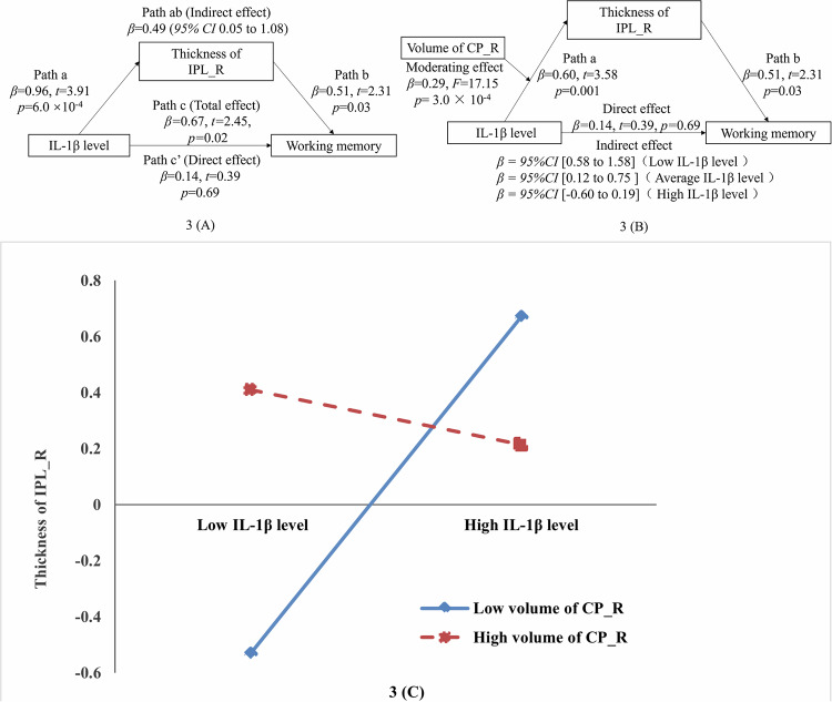 Figure 3
 Figure 4
Peer Reviews
No public reviews on file for this paper yet. If you reviewed it on a platform where reviews are public (OpenReview, ICLR, NeurIPS, ICML), you can paste yours below so the community can read it here.
Videos
No videos yet. Explain this paper in a talk, walkthrough, or lecture? Add one.
Taxonomy
TopicsNeuroinflammation and Neurodegeneration Mechanisms · Tryptophan and brain disorders · Stress Responses and Cortisol