Extravasation of Intravenous Acyclovir in a Patient With Human Herpesvirus 7 Encephalitis: A Case Report
Weeratian Tawanwongsri, Sasipaka Sindhusen, Pitchaya Jaruvijitrattana, Thanapon Sutharaphan, Taptim Stavorn

TL;DR
A patient with HHV-7 encephalitis developed skin lesions from acyclovir extravasation, emphasizing the need for prompt recognition and treatment.
Contribution
This case report presents a rare instance of acyclovir extravasation in HHV-7 encephalitis, highlighting management strategies.
Findings
Immediate vesicular skin lesions occurred at the acyclovir infusion site in a patient with HHV-7 encephalitis.
Extravasation was confirmed by rapid improvement after drug withdrawal and cold compression.
Early detection and management, including saline infiltration and hyaluronidase, minimized injury.
Abstract
Acyclovir is a widely used antiviral agent, but intravenous administration can cause cutaneous adverse reactions, particularly extravasation. We report a rare case of acyclovir extravasation in a 20-year-old female patient with human herpesvirus 7 (HHV-7) encephalitis, who developed immediate vesicular skin lesions at the infusion site. The rapid improvement after drug withdrawal and cold compression favored extravasation over herpes infection progression or drug eruption. Acyclovir’s high osmolality and alkalinity contribute to tissue damage. While no standard protocol exists, early detection, infusion discontinuation, aspiration, saline infiltration, cold compression, and hyaluronidase can help minimize injury. This case highlights the importance of prompt recognition and management to prevent complications.
Genes, proteins, chemicals, diseases, species, mutations and cell lines named across the full text — each resolved to its canonical identifier and authoritative record.
Click any figure to enlarge with its caption.
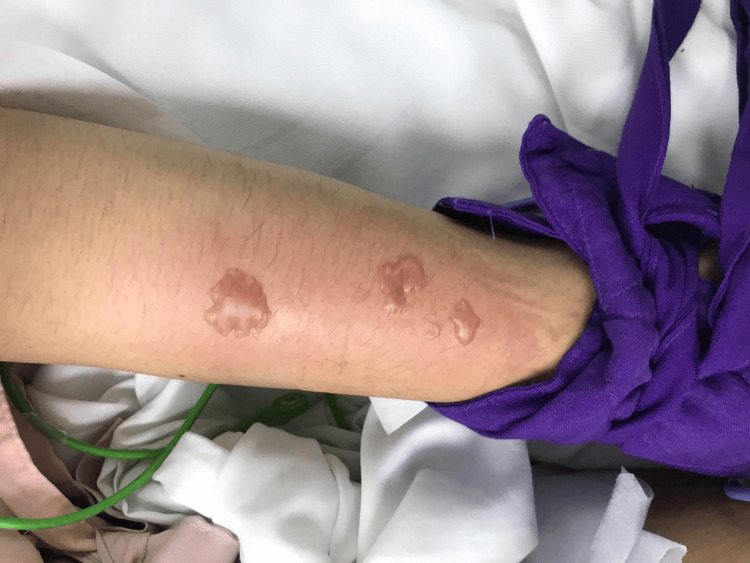 Figure 1
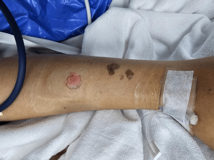 Figure 2
| Step | Action | Rationale |
| 1. Discontinue infusion | Immediately stop the IV infusion while leaving the cannula in place | Prevents further drug leakage into surrounding tissues |
| 2. Attempt aspiration | Gently aspirate any residual drug through the existing cannula | Reduces the volume of extravasated agent and potential tissue injury |
| 3. Remove cannula | Remove the IV cannula after aspiration | Minimizes additional local trauma |
| 4. Mark and assess the site | Outline the affected area and assess for swelling, erythema, blistering, or induration | Facilitates monitoring of lesion progression and clinical response |
| 5. Apply local cold compression | Apply cold compresses intermittently for 15–20 minutes several times per day (unless warm compresses are preferred based on institutional practice) | Helps reduce inflammation and local tissue reaction; approach may vary depending on drug characteristics |
| 6. Elevate the limb | Elevate the affected extremity | Promotes venous return and reduces edema |
| 7. Close observation | Monitor the site regularly, especially in high-risk patients (e.g., comatose or critically ill individuals) | Enables early detection of evolving complications |
| 8. Specialist referral | Seek consultation with dermatology, surgery, or wound care if signs of necrosis or extensive injury develop | Supports comprehensive management and potential need for debridement or reconstructive care |
| 9. Documentation and education | Document the event thoroughly and inform clinical staff | Enhances patient safety and promotes preventive measures in future practice |
Peer Reviews
No public reviews on file for this paper yet. If you reviewed it on a platform where reviews are public (OpenReview, ICLR, NeurIPS, ICML), you can paste yours below so the community can read it here.
Videos
No videos yet. Explain this paper in a talk, walkthrough, or lecture? Add one.
Taxonomy
TopicsChemotherapy-related skin toxicity · Cytomegalovirus and herpesvirus research · Herpesvirus Infections and Treatments
Introduction
Acyclovir is a nucleoside analog and serves as the first-line therapy for infections caused by herpes simplex virus (HSV) types 1 and 2 (HSV-1 and HSV-2) as well as varicella-zoster virus (VZV) [1]. It selectively targets herpesviruses through activation by the viral thymidine kinase, which phosphorylates it to acycloguanosine monophosphate. Host cellular kinases then convert it to the active triphosphate form.
Acyclovir triphosphate inhibits viral DNA polymerase and is incorporated into viral DNA, causing chain termination and blocking viral DNA synthesis and replication [2]. While generally well tolerated, intravenous (IV) acyclovir can cause cutaneous adverse reactions, particularly when extravasation occurs. Extravasation, the unintended leakage of a drug into surrounding tissues, can lead to chemical inflammation, tissue necrosis, and delayed healing, especially with vesicant or irritant drugs [3]. Despite its clinical significance, acyclovir extravasation remains rare, with limited case reports documenting its presentation and management, and cutaneous drug reactions due to extravasation are estimated to occur in approximately 1.7% of cases [4].
Extravasation refers to the inadvertent leakage of IV medication into the surrounding tissue. Depending on the properties of the infused agent, this can lead to complications ranging from mild irritation to severe tissue injury. For vesicant or irritant drugs such as acyclovir, extravasation may result in chemical inflammation, tissue necrosis, and potential long-term morbidity if not promptly identified and managed. Extravasation injuries are often driven by the osmotic characteristics and pH of the infused solution. Acyclovir has an osmolality of 278 mOsm/kg and a high alkalinity (pH = 11), both of which can induce chemical inflammation and significant tissue damage, ultimately resulting in cell death and necrosis [5,6]. The high osmolality of acyclovir solutions can exacerbate tissue injury by causing osmotic shifts, cellular dehydration, and inflammatory responses. Furthermore, its high alkalinity facilitates the breakdown of proteins, degradation of collagen, and disruption of cellular membranes, enabling hydrogen ions to penetrate deeper tissues and resulting in liquefactive necrosis [7].
Extravasation is a significant clinical concern due to its potential to cause tissue injury; however, it is largely preventable. Early detection and close monitoring during medication administration are key strategies to minimize further tissue damage and improve patient safety. As acyclovir is the drug of choice for patients with suspected or confirmed herpes encephalitis, many of whom may be unable to report early signs of extravasation due to a comatose state. In such cases, the risk of delayed detection and subsequent tissue injury is significantly increased.
Case presentation
A 20-year-old obese female patient presented to the outpatient clinic with acute fever, headache, malaise, nasal discharge, and nasal congestion. She denied any history of skin rashes. She was discharged home with supportive medications, including paracetamol and cetirizine. Five days after this, she was brought to the emergency department after experiencing a generalized tonic-clonic seizure lasting five minutes, followed by an altered level of consciousness, as well as urinary and fecal incontinence. The patient was admitted to the intensive care unit (ICU). A computed tomography (CT) scan of the brain revealed no evidence of hemorrhage, ischemia, or leptomeningeal enhancement.
Following endotracheal intubation, stabilization of vital signs, and seizure control with phenytoin, levetiracetam, and diazepam, a lumbar puncture was performed. Cerebrospinal fluid (CSF) analysis revealed clear fluid with normal opening pressure, normal glucose and protein concentrations, a white blood cell count of 15 cells/µL composed entirely of mononuclear cells (reference range: <5 cells/µL), and a red blood cell count of 3 cells/µL (reference range: 0 cells/µL). Intravenous acyclovir (10 mg/kg) was initiated, with the dose calculated based on adjusted body weight to optimize efficacy and safety. The drug was diluted in 5% dextrose to achieve a final concentration of 5 mg/mL. CSF Gram stain showed no organisms, and aerobic culture subsequently demonstrated no growth. Polymerase chain reaction (PCR) testing for the meningitis/encephalitis panel later confirmed the diagnosis of HHV-7 encephalitis.
Acyclovir was administered via a peripheral vein in the left forearm over one hour. Within five minutes after completion of the infusion, skin lesions were observed at the injection site (Figure 1). On examination, the affected area of the left dorsal forearm measured approximately 9 × 4 cm and exhibited ill-defined erythematous, edematous, and warm plaques. Within this area, four discrete, tense, yellowish vesicles were noted, each measuring approximately 2-3 cm in diameter. Cold compression was applied, and the needle was withdrawn. The patient remained in a comatose state due to postictal status; therefore, pain sensation at the lesion site could not be assessed. The immediate appearance of lesions post infusion, and their rapid improvement with cold compression, supported a diagnosis of acyclovir extravasation rather than viral progression or a delayed-type hypersensitivity reaction. Clobetasol cream was prescribed to be applied to the erythematous area twice daily to reduce inflammation.
Clinical photograph of the right forearm taken within five minutes after completion of intravenous acyclovir infusion, showing ill-defined erythematous, edematous, and warm plaques with a few small (2–3 cm), yellowish, fluid-filled vesicles.
The second dose was administered via a peripheral vein in the lower leg at the same concentration and infusion rate. Approximately 15 minutes into the infusion, a mild, warm, erythematous, and edematous plaque developed, prompting the discontinuation of the drug. Cold compression was applied immediately, and no overlying blebs were observed. Subsequently, the drug was administered via a peripheral vein in the right forearm with close monitoring. No skin lesions were observed thereafter. On the third day of ICU admission, the lesion on the left forearm was reassessed. The previously noted blebs had evolved into shallow erosions, with some becoming flaccid, while erythema and edema had resolved. A photograph taken two days later (Figure 2) documents these changes.
Clinical photograph two days after the initial lesion appearance show that previously noted blebs evolved into shallow erosions, with some becoming flaccid, while erythema and edema resolved.
Additionally, the lesions on the left lower leg had completely returned to a normal skin appearance. The erosion was treated with topical fusidic acid to prevent bacterial colonization. Although histopathological confirmation was not obtained, the diagnosis was based on the temporal association with drug administration and consistent clinical findings, supporting the likelihood of acyclovir extravasation. She regained consciousness, and acyclovir was administered for 14 days to treat encephalitis. She was subsequently extubated safely without additional cutaneous lesions and discharged home. However, during outpatient follow-up, she reported experiencing focal seizures involving the face and limbs, although these had decreased in severity by the two-month follow-up.
At the two-month follow-up, the lesion site had healed completely, with only mild post-inflammatory hyperpigmentation remaining. The patient reported no visible scarring or functional limitations, and no additional vesicles or blebs were observed during this period. Neurological status and antiepileptic therapy were regularly monitored by the neurologist.
Discussion
This was a rare case of cutaneous adverse reaction due to acyclovir extravasation, manifesting as a vesicular eruption. The differential diagnosis included acyclovir extravasation, herpes infection progression, and drug eruption secondary to acyclovir with recall phenomenon. Key clinical signs of acyclovir extravasation include swelling and induration as early indicators, followed by localized skin discoloration at the infusion site [5]. Patients may also present with pain and erythema, signaling an inflammatory response [3]. Additionally, a lack of blood return from the cannula, though not definitive, is a common finding that may suggest extravasation [8].
The progression of HSV and VZV is a key consideration in the differential diagnosis of vesicular eruptions. HSV infections typically present with multiple small, painful grouped vesicles, often accompanied by fever and lymphadenopathy [9]. VZV infections, particularly disseminated herpes zoster, can similarly manifest as vesicular eruptions, especially in immunocompromised patients. Lau et al. reported a case involving a 55-year-old male patient with HIV who developed herpes encephalitis [10]. The patient presented with diffuse, firm, yet compressible swelling of the right hand, without evidence of skin breakdown or necrosis. Management included wrapping of the affected hand, application of cold compresses, and strict elevation. Similarly, our patient developed skin lesions immediately at the infusion site following drug administration, unlike the delayed onset seen in classic herpes infections. Additionally, the lesions showed rapid improvement after infusion withdrawal and cold compression, further supporting acyclovir extravasation as the more likely cause rather than herpes infection progression. In a drug eruption secondary to acyclovir with recall phenomenon, vesicular lesions may develop. The key characteristic is confluent linear erythema along dermatomes previously affected by a herpes zoster episode, reflecting a hypersensitivity reaction triggered by drug exposure [11]. However, our patient denied any history of skin rashes or prior herpes infection, making this diagnosis less likely in this case.
Several key risk factors contribute to its occurrence, categorized into patient-related, drug-related, procedural, and healthcare provider-related factors. Both very young (younger than six years) and elderly patients (over 65 years) are more vulnerable due to fragile veins and thinner skin [12]. Cancer patients face a higher risk due to repeated venipunctures and compromised venous access from chemotherapy [13]. Neurological, cardiac, and digestive diseases further increase susceptibility [14]. Additionally, patients who are restless or unable to cooperate, such as those with cognitive impairment or severe illness, may inadvertently dislodge IV catheters, increasing the risk of leakage [12]. According to drug-related factors, vesicant and irritant drugs, such as vincristine and doxorubicin, can cause severe tissue damage upon extravasation [15]. Hypotonic or hypertonic solutions can disrupt fluid balance and cause local injury. Similarly, drugs with extreme pH values are more likely to cause chemical irritation and necrosis [7]. According to procedure-related factors, infusion tools and site selection influence extravasation risk. Indwelling and steel needles are linked to higher rates, while infusions near joints are more prone to complications due to movement [12]. Repeated venipunctures weaken veins, increasing the likelihood of leakage [8]. In addition, the experience and training of healthcare providers significantly impact extravasation outcomes. Less experienced nurses may struggle with identifying high-risk veins or recognizing early signs of infiltration. Proper training in IV placement, securement techniques, and early detection can reduce the risk and severity of complications [8]. Our patient was in a comatose state with ongoing seizures, which limited their ability to communicate discomfort or react to early signs of extravasation. This inability to cooperate increased the risk of catheter dislodgement and unrecognized IV infiltration. Additionally, the physicochemical properties of acyclovir contributed to the severity of the reaction.
The de facto prevalence of acyclovir extravasation-related skin injury remains unclear and may be underestimated in the literature, as such cases are likely underreported despite being encountered in routine clinical practice. Future multicenter cohort studies are needed to better define the incidence, contributing factors, and clinical outcomes of this complication. While there is no universally accepted standard protocol specifically for acyclovir extravasation, general guidelines for managing extravasation injuries can be applied. The most critical step is early detection and immediate discontinuation of the infusion to prevent further leakage and minimize tissue damage [16]. The next step is to aspirate any remaining drug from the cannula without applying pressure to prevent further tissue infiltration. Saline infiltration can be used locally to dilute the extravasated drug and reduce its toxic effects [16]. Cold compresses should be applied to the affected area for 15-20 minutes every four hours during the first 24-48 hours to limit drug dispersion and reduce inflammation [17]. Hyaluronidase, an enzyme that facilitates drug absorption, can be injected subcutaneously around the affected area, particularly for hyperosmolar solutions [5]. Topical corticosteroids may also be applied to reduce inflammation and prevent ulceration [18].
Continuous monitoring is essential to detect worsening symptoms such as increased pain, swelling, or necrosis, with early surgical consultation recommended if symptoms progress. Patient education plays a crucial role in preventing complications, emphasizing the importance of reporting any new symptoms immediately. Staff training is vital to ensure healthcare professionals are well-equipped to prevent, recognize, and manage extravasation effectively [19]. Table 1 outlines suggested steps for managing acyclovir extravasation; these steps should be adapted based on the severity of injury, patient-specific factors, and institutional protocols. In addition, there is a need for close observation of the infusion site during drug administration, particularly in comatose patients, to compensate for their inability to report early symptoms such as pain or discomfort.
Conclusions
This case highlights the rare occurrence of acyclovir extravasation, which can cause chemical inflammation, tissue necrosis, and delayed healing due to its high osmolality and alkalinity. The patient's immediate skin reaction, rapid improvement after drug withdrawal and cold compression, and absence of herpes infection history support extravasation over herpes progression or drug eruption. While no standard protocol exists, early detection, infusion discontinuation, aspiration, saline infiltration, cold compression, and hyaluronidase are crucial to minimizing tissue damage. Topical corticosteroids, antibiotics, close monitoring, patient education, and staff training further aid in preventing complications. This case underscores the importance of early recognition and prompt intervention to mitigate the adverse effects of acyclovir extravasation.
The reference list from the paper itself. Each links out to its DOI / PubMed record.
- 140 years after the registration of acyclovir: do we need new anti-herpetic drugs?Int J Mol Sci Majewska A Mlynarczyk-Bonikowska B 3431232022 https://pmc.ncbi.nlm.nih.gov/articles/PMC 8998721/3540878810.3390/ijms 23073431 PMC 8998721 · doi ↗ · pubmed ↗
- 2Nucleosides and nucleoside analogues as emerging antiviral drugs Mini Rev Org Chem Elzagheid MI 672679182021
- 3Cutaneous necrosis secondary to subcutaneous extravasation of acyclovir Our Dermatol Online Couissi I Douhi Z El Fid K Soughi M El Loudi S Bay Bay H Mernissi FZ 0142023 https://www.odermatol.com/odermatology/2023 e/E 477.Cutaneous-Couissi I.pdf
- 4Parenteral aciclovir for suspected herpes simplex virus infection in children: 0-18 years J Antimicrob Chemother Berkhout A Clark JE Jones CA 3543628020253955620010.1093/jac/dkae 411 · doi ↗ · pubmed ↗
- 5Acyclovir extravasation in a newborn: a case report J Med Case Rep Shamel S Zarkesh MR 2711820243884503010.1186/s 13256-024-04585-1PMC 11157697 · doi ↗ · pubmed ↗
- 6Extravasation of noncytotoxic agents: skin injury and risk classification Biol Pharm Bull Shibata Y Taogoshi T Matsuo H 7467554620233725813910.1248/bpb.b 22-00850 · doi ↗ · pubmed ↗
- 7Management of noncytotoxic extravasation injuries: a focused update on medications, treatment strategies, and peripheral administration of vasopressors and hypertonic saline Pharmacotherapy Stefanos SS Kiser TH Mac Laren R Mueller SW Reynolds PM 3213374320233693877510.1002/phar.2794 · doi ↗ · pubmed ↗
- 8Omadacycline extravasation: a general overview Int J Pharma Res Aishwarya MB Anusha J Moudgil K 11081111122020