The role of post-translational modifications in parvovirus life cycle
Peng Liu, Liqin Yang

TL;DR
This paper reviews how post-translational modifications affect the life cycle of parvoviruses, focusing on how these changes influence viral replication and infection.
Contribution
The paper provides a comprehensive review of post-translational modifications in parvovirus proteins and their roles in the viral life cycle.
Findings
Phosphorylation of NS and capsid proteins modulates NS function and capsid assembly.
Ubiquitination and SUMOylation influence intracellular trafficking of capsid proteins.
Glycosylation affects virion stability and infectivity.
Abstract
Parvoviruses are a group of single-stranded DNA viruses that lack an envelope and are widely distributed in both vertebrates and invertebrates. When they infect a host cell, parvoviruses take over the cell’s translational machinery to support the viral genome replication and proteins synthesis, following which viral proteins undergo various post-translational modifications (PTMs). Parvovirus non-structural (NS) and capsid proteins are modified by PTMs, including phosphorylation, ubiquitination, SUMOylation, and glycosylation. Phosphorylation of parvovirus mainly occurs on NS and capsid proteins, modulating the functions and activities of the NS protein and the assembly of the capsid protein. Ubiquitination and SUMOylation of parvoviral capsid proteins mainly affect intracellular trafficking during viral infection. Glycosylation of parvoviral capsid proteins is involved in the regulation…
Genes, proteins, chemicals, diseases, species, mutations and cell lines named across the full text — each resolved to its canonical identifier and authoritative record.
Click any figure to enlarge with its caption.
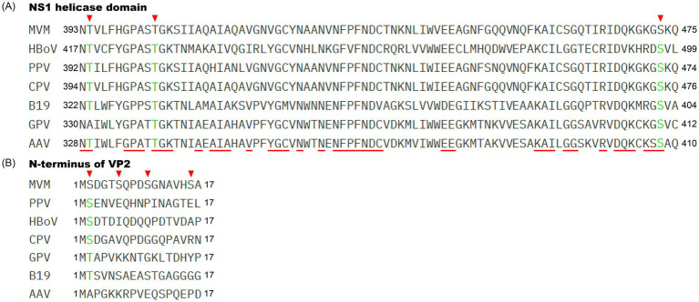 Figure 1
| Modification | Related protein | Impacts on the virus | Reference |
|---|---|---|---|
| Phosphorylation | PPV NS1 | Unknown |
|
| MVM NS1 | Regulates the NS protein’s enzymatic activity, cytotoxicity, and attachment to viral DNA | ||
| AAV Rep | Promotes viral DNA synthesis |
| |
| MVM VP1 and VP2 | Facilitates viral maturation and release | ||
| AAV capsid proteins | Inhibits the intracellular trafficking process | ||
| Ubiquitination | AAV capsid proteins | Inhibits the intracellular trafficking process | |
| MVM NS2 | Degrades viral NS2 |
| |
| AAV Reps | Degrades viral Rep proteins | ||
| SUMOylation | AAV2 Rep78 | Regulates the Rep78 protein’s stability and half-life |
|
| AAV2 capsid proteins | Inhibits the intracellular trafficking process | ||
| Glycosylation | AAV capsid proteins | May regulate virion stability and infectivity. |
Peer Reviews
No public reviews on file for this paper yet. If you reviewed it on a platform where reviews are public (OpenReview, ICLR, NeurIPS, ICML), you can paste yours below so the community can read it here.
Videos
No videos yet. Explain this paper in a talk, walkthrough, or lecture? Add one.
Taxonomy
TopicsVirus-based gene therapy research · Parvovirus B19 Infection Studies · Animal Virus Infections Studies
Introduction
1
Parvoviruses are a group of non-enveloped, single-stranded DNA viruses characterized by an icosahedral capsid measuring 18–26 nm in diameter. They exhibit a broad host range, infecting both vertebrates and invertebrates (1). Taxonomically, the family Parvoviridae is divided into three subfamilies: Parvovirinae, Densovirinae, and Hamaparvovirinae, as well as the genus Metalloincertoparvovirus (2). The viral genome, which is approximately 5 kb in length, contains left and right open reading frames (ORFs) that encode viral nonstructural (NS) proteins [called Rep proteins in adeno-associated virus (AAV) and goose parvovirus (GPV)] and capsid proteins, respectively (3). The ORFs are flanked by 5′ and 3′ terminal hairpin structures, which are essential for the regulation of viral gene expression (4). However, owing to the restricted coding capacity of the parvovirus genome, parvoviruses heavily depend on host factors and cellular machinery to perform the viral replication process after invading host cells.
In parvoviruses, the capsid is responsible for the adsorption of receptors and enters cells via the clathrin-dependent endocytic pathway (5). Under the navigation of nuclear localization signals, the capsid is transported to the nucleus through the endosomal pathway to release the viral genome (6–8). Without a viral DNA polymerase, parvoviruses rely solely on DNA replication machinery within the nucleus (9). With the aid of host factors (10, 11), parvoviral precursor mRNA is transcribed to generate matured mRNA transcripts that encode NS and capsid proteins in the cytoplasm, where they undergo post-translational modifications (PTMs).
PTMs are crucial biochemical processes in eukaryotic cells, wherein specific enzymes catalyze the formation of covalent bonds on one or more amino acid residues of target proteins. Common PTMs, including acylation, glycosylation, methylation, phosphorylation, ubiquitination, and SUMOylation, play a pivotal role in regulating protein conformation, stability, activity, subcellular localization, and interactions with other proteins (12, 13). As intracellular parasites, viruses are modified by various PTMs during the viral infection process, which are involved in viral replication, assembly, release, and immune evasion. For instance, phosphorylation of VP8 in bovine herpesvirus 1 promotes viral DNA encapsidation in cells (14); glycosylation of the SARS-CoV-2 S protein shields its epitopes and enhances viral immune evasion (15). Similar to other viruses, PTMs also play an important role in the parvovirus life cycle. In this review, based on recent advances in PTM research on parvoviruses, we summarize the current knowledge of the PTMs of parvoviral proteins, including NS and capsid proteins. We also discuss the impact of PTMs on the parvoviral life cycle, which may help us better understand parvoviral replication and pathogenesis mechanisms.
Phosphorylation
2
NS protein
2.1
The large NS protein (NS1) of parvovirus is a multifunctional protein that possesses endonuclease, helicase, ATPase, and transcription-activating activities. These enzymatic activities are vital for initiating viral genome replication (16, 17) and regulating viral gene expression with the aid of host factors, which include the transcription factor Sp1/Sp3 and the TATA-binding protein (18, 19). However, its activities are heavily regulated by phosphorylation.
Phosphorylation of the parvoviral NS1 protein was first described in porcine parvovirus (PPV) in 1985 (20). Subsequent studies demonstrated that the Rep proteins of AAV, as well as the NS proteins of MVM and canine parvovirus (CPV), are also modified by phosphorylation (21–23). Biochemical activity analysis of MVM NS1 revealed that phosphorylation increased its viral helicase, ATPase, and nickase activities (24), while dephosphorylation led to a dramatic reduction in these enzymatic activities, suggesting that the replication functions of MVM NS1 are regulated by phosphorylation. Further analysis of phosphorylation site localization revealed that the helicase domain of MVM NS1 was phosphorylated, and the T363, T394, T403, T435, and S473 were identified as phosphorylation sites (25). The rat-origin protein kinase C λ was found to target T435 and S473 of MVM NS1 to phosphorylate it, resulting in the activation of its helicase function for initiating viral DNA unwinding and replication (26). In addition, PKC η could accelerate the phosphorylation of MVM NS1, which is necessary for the production of viral double-stranded concatemeric DNA intermediates during the early stage of virus replication (27).
The cytotoxicity of MVM NS1 is also regulated through phosphorylation. The parvovirus NS1 protein is the major effector that induces cytotoxicity in host cells (28–30). The C-terminus of MVM NS1 is identified as a major regulator of NS1 cytotoxicity (31). Further mutation analysis indicated that phosphorylation at the T585 site located at the C-terminus of MVM NS1 showed higher toxicity to A9 cells (28). In addition, the phosphorylation sites T598 and T601, located at the C-terminus of CPV NS1, were found to determine CPV pathogenicity (23). Phosphorylated MVM NS1 may exert its cytotoxic by controlling host gene expression and binding host proteins. Specifically, it can induce cytotoxicity by significantly activating thyroid hormone signaling pathways in FREJ4 cells (32), promoting oncogene expression in FR3T3 rat cells (33), and altering the synthesis of intracellular phosphorylated proteins (29). At the protein level, it also can interact with endogenous casein kinase II (CKII) to alter CKII’s activity, resulting in morphological and physiological alterations in host cells (34) and even cell lysis.
Capsid protein
2.2
Phosphorylation of viral structural proteins plays a crucial role at various stages of the viral life cycle, including recognition between intracellular host proteins and viruses, transport of viral proteins, capsid assembly, and genome packaging. For example, phosphorylation of the influenza virus nucleoprotein is vital for the assembly of ribonucleoprotein complexes (35). Phosphorylation of the core protein of hepatitis B virus is essential for the nuclear import of viral RNA and the stability of viral capsid proteins (36). Current research has found that the structural proteins of parvoviruses also undergo phosphorylation, which primarily regulates the assembly of capsid proteins.
In parvoviruses, the structural proteins include two or three capsid proteins (VP1-2 or VP1-3). Phosphorylation of parvoviral capsid proteins has been identified in MVM, PPV, and AAV (20, 37–39). MVM VP1 and VP2 are phosphorylated at serine and threonine residues located at the N-terminus in NB324K cells (37). The VP2 N-terminal sequence has been identified as the major phosphorylation domain harboring four phosphorylated residues including S2, S6, S10, and S16. Mutation analysis revealed that the major phosphorylation site S2 significantly affects the viral plaque-forming ability and plaque size, suggesting that it may primarily influence the viral budding or release process (37). Further research has indicated that the phosphorylated N-terminus of VP2 acts as a nuclear export signal, promoting the nuclear export of progeny viral particles during the replication process (40). In addition, phosphorylated residues on the viral particle surface are also required for this process (41). The progeny particles exported into the cytoplasm are further modulated through sequential processing in the endoplasmic reticulum and Golgi apparatus, where full infectivity of the viral particles is conferred (42).
Similar to nuclear export, the nuclear import of MVM capsid proteins also requires phosphorylation by the Raf-1 kinase in the cytoplasm (43, 44). The capsid proteins phosphorylated by the Raf-1 kinase enhance nuclear import and can be transported into the nucleus for completing progeny particle assembly (43). However, parental AAV capsids with tyrosine phosphorylation in the cytoplasm exhibit inefficient nuclear import ability (39), suggesting that this modification may trigger host defense mechanisms.
The NS1 proteins of parvoviruses share relatively high sequence homology across different genera, especially within the helicase domain, and perform similar functions in viral genome replication. Protein sequence alignment based on the helicase domain of MVM, CPV, PPV, parvovirus B19, AAV, human bocavirus (HBoV), and GPV (Figure 1A) showed that the phosphorylation sites of T394, T403, and S473 on MVM NS1 are conserved among these parvoviruses, suggesting that phosphorylation plays a critical role in the helicase activity of parvovirus NS1 and viral genome replication. Notably, the ser in the second site of VP2 is conserved among MVM, CPV, PPV, and HBoV (Figure 1B), meanwhile the tyr in the second site of GPV and B19 VP2 may serve as a potential phosphorylation site, indicating that phosphorylation also governs the nuclear import and export dynamics of viral progeny proteins, facilitating viral maturation and release.
Schematic diagram showing the localization of potential phosphorylation sites in parvoviral NS1 helicase domain and the N-terminus of VP2. Alignment of the parvoviral helicase domain of the NS1 sequence (A) and the N-terminus of the VP2 sequence (B) The numbers on the left and right represent amino acid positions. The conserved residues in the helicase domain of parvovirus NS1 are highlighted with red lines. The identified phosphorylation sites in the MVM NS1 helicase domain and N-terminus of VP2 are indicated by the inverted red triangle. The potential phosphorylation sites are indicated in green font. The GenBank accession numbers for the NS1 or Rep and VP2 proteins of parvoviruses are as follows: the GenBank accession number for AAV2 Rep78 and VP2, CPV NS1 and VP2, HBoV NS1 and VP2, GPV Rep1 and VP2, PPV NS1 and VP2, and B19 NS1 and VP2 are DQ180605, DQ180605, KP710213, KC996729, AY684872 and AB030673, respectively; the GenBank accession number for MVM NS1 and VP2 are NP_041242 and J02275, respectively.
Ubiquitination and SUMOylation
3
Ubiquitination of parvoviral capsid proteins was first identified in AAV type 2 (AAV2) in 2000 (45). Using liquid chromatography and mass spectrometry analysis, some conserved ubiquitination sites on the AAV capsid surface were found in AAV4, AAV5, AAV9, and AAVrh10 (38, 46). These findings suggest that ubiquitination of viral capsid proteins may affect the parvoviral life cycle. In AAV2-infected cells, the viral intracellular trafficking is enhanced by treating cells with proteasome inhibitor MG132 (46, 47). Furthermore, mutation of ubiquitination sites in the AAV2 capsid protein, such as K490, K544, K549, and K55, significantly promoted the transduction efficiency of AAV2 both in vitro and in vivo (48–50). In addition, inhibition of the EGFR-PTK signaling pathway mediating ubiquitination of the AAV2 capsid protein also increased the trafficking efficiency of AAV2 (51). In contrast, the ubiquitin–proteasome pathway is required for the replication of MVM and CPV. Inhibiting the ubiquitin–proteasome pathway using MG132 in MVM- and CPV-infected cell abolished MVM and CPV replication (52), which is similar to what has been observed with human immunodeficiency virus and Rous sarcoma virus (53, 54). In addition to parvoviral capsid proteins, MVM NS2 and AAV Rep proteins also undergo ubiquitination modification and subsequent proteasomal degradation (55–57). However, the specific mechanism between the degradation of NS2 or Rep proteins via the proteasomal degradation pathway and viral replication remains to be elucidated.
In addition to ubiquitination, both the Rep and capsid proteins of AAV2 undergo SUMOylation (38, 58). SUMOylation of AAV2 Rep78 has been demonstrated to regulate its stability and extend its half-life (58). Similar to ubiquitination, SUMOylation of AAV capsid proteins has been shown to influence the efficiency of intracellular transduction (38). Inhibition of the SUMOylation pathway in AAV2-infected cells resulted in increased transduction efficiency within the cells (59). Furthermore, a systematic screening method using an siRNA library was employed to identify host factors regulating viral replication. This screening revealed that multiple host factors involved in the SUMOylation signaling pathway regulate AAV infection (60). Further knockdown of the SUMOylation components Ubc9 and Sae2 significantly enhanced AAV2 transduction. Mechanistic studies have revealed that AAV infection activates the intracellular SUMOylation machinery, which subsequently restricts AAV transduction efficiency (61).
As a delivery vehicle for gene therapy, the AAV vector cannot encode viral proteins to antagonize host restriction factors inherent to cellular defense mechanisms, since the AAV vector maintains a simplified structure composed solely of an exogenous therapeutic genome packaged within capsid proteins. Therefore, further optimization of the vector’s ubiquitination and SUMOylation target sites could improve delivery efficiency.
Glycosylation
4
Glycosylation is one of the most crucial post-translational modifications and regulates enveloped virus infection by mediating receptor binding, facilitating protein folding and trafficking, and participating in immune evasion (62, 63). However, there is very little information on parvoviral protein glycosylation. To date, glycosylation has been detected only in the capsid proteins of AAV2, AAV3, AAV5, AAV7, AAV8, AAV9, and AAVrh10using liquid chromatography and mass spectrometry analysis (38, 64, 65). Specifically, residue N499 of the AAV8 capsid protein and residue N253, N518, S537, and N551 of the AAV2 capsid protein have been identified as N-linked glycosylation sites (38, 64). In addition, both N- and O-linked glycosylation sites have been detected in the AAV9 capsid protein (65). Mutation analysis revealed that the AAV2 capsid protein with the N253Q mutation resulted in a considerable reduction in vector yield compared to AAV2 wild-type vectors, suggesting that glycosylation of the capsid is critical for virion assembly and stability (64). On the other hand, a number of potential glycosylation sites in the capsid protein of GPV, Muscovy duck parvovirus, and Aleutian mink disease virus have been predicted based on bioinformatic analyses of the capsid protein sequences (66, 67). These studies suggest that capsid protein glycosylation may be a conserved feature among all parvoviruses. However, the molecular mechanism by which parvoviral capsid glycosylation modulates viral tissue tropism, intracellular trafficking, infectivity, and immunogenicity remains to be further studied.
To date, glycosylation of NS proteins in parvovirus has not been reported. Whether the NS proteins of parvovirus possess glycosylation sites and whether these modifications affect NS proteins’ function and activity are yet to be uncovered.
Discussion
5
As an intracellular parasite, parvovirus relies heavily on a variety of cellular mechanisms to accomplish its life cycle. During parvoviral infection, parvovirus proteins undergo phosphorylation, ubiquitination, SUMOylation, and glycosylation (Table 1). Glycosylation has been detected in parvovirus capsid proteins and may regulate virion stability, receptor binding, immunogenicity, and other functions. Meanwhile, phosphorylation not only regulates the NS1 protein’s function activity and cytotoxicity but also promotes the assembly and maturation of progeny virions. Ubiquitination and SUMOylation restrict parvovirus infection, resulting in inefficient intracellular trafficking of viral capsids within cells. Nevertheless, how other PTMs—including, but not limited to, lipidation and methylation—modulate parvoviral replication dynamics remains to be fully characterized. Parvoviruses are promising vectors for gene therapy and oncolytic virotherapy. Further studies on parvoviral PTMs will contribute to the development of novel bioengineered gene therapy vectors and novel inhibitors.
To counteract viral infection, host cells deploy a broad spectrum of antiviral defenses, including interferon-induced antiviral factors and cell-intrinsic restriction factors, which directly target viral proteins or genomes. However, type I interferon signaling fails to effectively suppress infections by autonomous parvoviruses and AAV2 in both normal human and cancer cells (68). Cell-intrinsic restriction factors likely play an important role in the host’s defense against parvoviruses. Systematic screening of these antiviral factors, coupled with mechanistic studies of their viral targets, will provide a solid foundation for the development of gene therapy vectors with improved safety and efficacy.
The reference list from the paper itself. Each links out to its DOI / PubMed record.
- 1Cotmore SF Tattersall P. Parvoviral host range and cell entry mechanisms. Adv Virus Res. (2007) 70:183–232. doi: 10.1016/S 0065-3527(07)70005-2, PMID: 17765706 · doi ↗ · pubmed ↗
- 2Kibenge F Kibenge M Montes De Oca M Godoy M. Parvoviruses of aquatic animals. Pathogens. (2024) 13:625. doi: 10.3390/pathogens 13080625, PMID: 39204226 PMC 11357303 · doi ↗ · pubmed ↗
- 3Cotmore SF Tattersall P. The autonomously replicating parvoviruses of vertebrates. Adv Virus Res. (1987) 33:91–174. doi: 10.1016/s 0065-3527(08)60317-63296697 · doi ↗ · pubmed ↗
- 4Wang XS Ponnazhagan S Srivastava A. Rescue and replication of adeno-associated virus type 2 as well as vector DNA sequences from recombinant plasmids containing deletions in the viral inverted terminal repeats: selective encapsidation of viral genomes in progeny virions. J Virol. (1996) 70:1668–77. doi: 10.1128/JVI.70.3.1668-1677.1996, PMID: 8627687 PMC 189990 · doi ↗ · pubmed ↗
- 5Parker JSL Parrish CR. Cellular uptake and infection by canine parvovirus involves rapid dynamin-regulated clathrin-mediated endocytosis, followed by slower intracellular trafficking. J Virol. (2000) 74:1919–30. doi: 10.1128/jvi.74.4.1919-1930.2000, PMID: 10644365 PMC 111670 · doi ↗ · pubmed ↗
- 6Harbison CE Chiorini JA Parrish CR. The parvovirus capsid odyssey: from the cell surface to the nucleus. Trends Microbiol. (2008) 16:208–14. doi: 10.1016/j.tim.2008.01.012, PMID: 18406140 · doi ↗ · pubmed ↗
- 7Sonntag F Bleker S Leuchs B Fischer R Kleinschmidt JA. Adeno-associated virus type 2 capsids with externalized VP 1/VP 2 trafficking domains are generated prior to passage through the cytoplasm and are maintained until uncoating occurs in the nucleus. J Virol. (2006) 80:11040–54. doi: 10.1128/JVI.01056-06, PMID: 16956943 PMC 1642181 · doi ↗ · pubmed ↗
- 8Liu P Chen S Wang M Cheng A. The role of nuclear localization signal in parvovirus life cycle. Virol J. (2017) 14:80. doi: 10.1186/s 12985-017-0745-1, PMID: 28410597 PMC 5391597 · doi ↗ · pubmed ↗