Optical super-high magnification dermoscopy versus standard dermoscopy in basal cell carcinoma
Izadora Moreira do Amaral, Elisa Scandiuzzi Maciel, Daniela Surjan Milheti, Camila Arai Seque, Milvia Maria Simões e Silva Enokihara, Sérgio Henrique Hirata

Abstract
Genes, proteins, chemicals, diseases, species, mutations and cell lines named across the full text — each resolved to its canonical identifier and authoritative record.
Click any figure to enlarge with its caption.
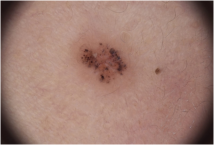 Figure 1
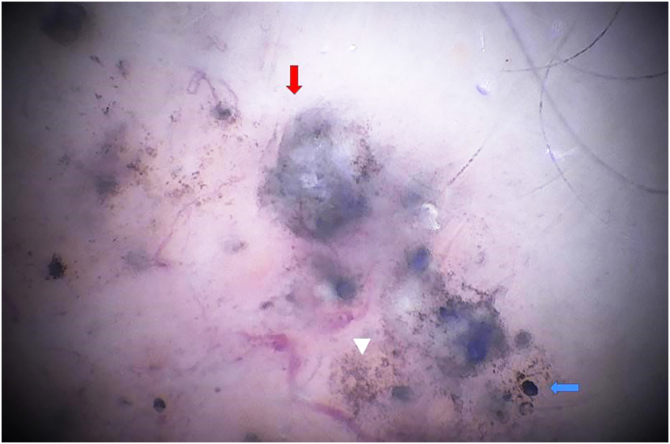 Figure 2
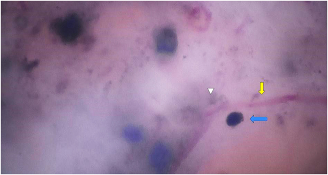 Figure 3
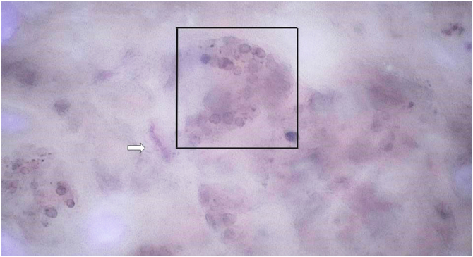 Figure 4
Peer Reviews
No public reviews on file for this paper yet. If you reviewed it on a platform where reviews are public (OpenReview, ICLR, NeurIPS, ICML), you can paste yours below so the community can read it here.
Videos
No videos yet. Explain this paper in a talk, walkthrough, or lecture? Add one.
Taxonomy
TopicsNonmelanoma Skin Cancer Studies · Cutaneous Melanoma Detection and Management · Nail Diseases and Treatments
Dear Editor,
The recent emergence of super high dermoscopy (SHD) allows magnifications of up to 400 times, which brings new perspectives to the interpretation of dermoscopic images. This technology is available with non-polarized light and the images are obtained using the Fotofinder Medicam 1000 device (Fotofinder System, Bad Birnbach, Germany) replacing the conventional terminal lens with the super high dermoscopy (SHD) lens.
To demonstrate the potential of using SHD, this report describes the case of a 78-year-old female patient with Fitzpatrick skin phototype III, with a brownish papule in the left preauricular region with progressive growth. The patient was photographed using conventional dermoscopy and super high dermoscopy with ultrasound gel immersion (Figure 1, Figure 2, Figure 3). The patient was subsequently referred for tumor excision and the material was sent for histopathological examination, with a report compatible with pigmented solid basal cell carcinoma with an adenoid component.Figure 1. Digital dermoscopy with x20 magnification. Fotofinder System, Bad Birnbach, Germany.Figure 1. Figure 2SHD with x180 magnification. The image shows blue ovoid nests at higher magnification (red arrow), rounded blue globules (blue arrow) and irregular pigmented structures (triangle).Figure 2. Figure 3SHD of the same region with x400 magnification. Telangiectasias can be seen at higher magnification, with focus (yellow arrow), surrounded by irregular pigmented structures (triangle) and rounded blue globules (blue arrow).Figure 3
SHD allows the visualization of structures that are not perceptible through conventional dermoscopy.1 In the literature, there are reports on the use of SHD in the identification and differentiation between melanomas and atypical nevi in melanocytic lesions,2 as well as in the differential diagnosis between benign facial lesions and lentigo maligna.3 Regarding basal cell carcinomas, irregularly pigmented structures, corresponding to melanocyte deposits containing melanin,4 linear vessels with peripheral dots and globules,5 vessels with a pattern similar to oak leaves,6 and hairpin vessels,4 have already been described exclusively through SHD.
The case described herein illustrates the easier identification of dermoscopic structures observed by SHD when compared to conventional dermoscopy. In Fig. 1, conventional dermoscopic examination (×20 magnification) allows the visualization of structures that less experienced examiners may mistake for globules of melanocytic lesions. Figure 2, Figure 3 show the same structures observed at SHD (x180 and x400 magnification). It is clear that these are bluish-gray globules, structures that are characteristic of basal cell carcinomas. The morphological characteristic of telangiectasias is also more easily observed at SHD, with focus and showing the characteristic peripheral dots previously described for SHD.5 Fig. 4, for comparison purposes, shows structures in an intradermal nevus with a tendency to an annular arrangement (globule), corresponding to nests formed by the union of nevus cells.Figure 4SHD at x400 magnification demonstrates a globule of an intradermal melanocytic nevus. Telangiectasias without focus (white arrow) can be observed.Figure 4
The nomenclature and description of the different structures observed at SHD are not yet standardized and the use of this technique is still in the experimental phase. It is not a substitute for conventional dermoscopy, but rather a new tool capable of aiding in diagnoses through additional information.
Financial support
None declared.
Authors' contributions
Izadora Moreira do Amaral: Collection, analysis and interpretation of data; intellectual participation in the propaedeutic and/or therapeutic conduct of the studied cases; drafting and editing of the manuscript or critical review of important intellectual content; critical review of the literature; approval of the final version of the manuscript.
Elisa Scandiuzzi Maciel: Collection, analysis and interpretation of data; intellectual participation in the propaedeutic and/or therapeutic conduct of the studied cases; approval of the final version of the manuscript.
Daniela Surjan Milheti: Collection, analysis and interpretation of data; intellectual participation in the propaedeutic and/or therapeutic conduct of the studied cases; approval of the final version of the manuscript.
Camila Arai Seque: Collection, analysis and interpretation of data; intellectual participation in the propaedeutic and/or therapeutic conduct of the studied cases; approval of the final version of the manuscript.
Milvia Maria Simões e Silva Enokihara: Collection, analysis and interpretation of data; drafting and editing of the manuscript or critical review of important intellectual content; effective participation in research orientation; intellectual participation in the propaedeutic and/or therapeutic conduct of the studied cases; approval of the final version of the manuscript.
Sérgio Henrique Hirata: Collection, analysis and interpretation of data; drafting and editing of the manuscript or critical review of important intellectual content; effective participation in research orientation; intellectual participation in the propaedeutic and/or therapeutic conduct of the studied cases; approval of the final version of the manuscript.
Conflicts of interest
None declared.
The reference list from the paper itself. Each links out to its DOI / PubMed record.
- 1Cinotti E.Rossi R.Ferrara G.Tognetti L.Rubegni P.Perrot J.L.Image gallery: super-high magnification dermoscopy can identify pigmented cells: correlation with reflectance confocal microscopy Br J Dermatol 1812019 e 13125940310.1111/bjd.17781 · doi ↗ · pubmed ↗
- 2Cinotti E.Cioppa V.Tognetti L.Perrot J.L.Rossi R.Gnone M.Super-high magnification dermoscopy in 190 clinically atypical pigmented lesions Diagnostics (Basel)13202322383744363210.3390/diagnostics 13132238 PMC 10340569 · doi ↗ · pubmed ↗
- 3Cinotti E.Cartocci A.Liso F.G.Cioppa V.Falcinelli F.Tognetti L.Super-high magnification dermoscopy can help for the diagnosis of lentigo maligna: a pilot study on 61 cases Dermatol Pract Concept 132023 e 202310110.5826/dpc.1302 a 101PMC 1018813237196274 · doi ↗ · pubmed ↗
- 4Pogorzelska-DyrbuśJ.Lallas A.Szepietowski J.C.Morphology of vessels in basal cell carcinoma in optical super-high magnification dermoscopy Acta Derm Venereol 1032023 adv 1196610.2340/actadv.v 103.11966 PMC 1041350837529846 · doi ↗ · pubmed ↗
- 5Pogorzelska-DyrbuśJ Szepietowski JC Optical super-high magnification dermoscopy of pigmented and nonpigmented nodular basal cell carcinoma J Cosmet Dermatol 212022645864603556750810.1111/jocd.15082 · doi ↗ · pubmed ↗
- 6Pogorzelska-DyrbuśJ."Oak-leaf-like" loop vessels in super-high magnification dermoscopy of basal cell carcinoma Dermatol Pract Concept 122022 e 202214710.5826/dpc.1203 a 147PMC 946451836159132 · doi ↗ · pubmed ↗