Molecular Basis of Sodium Channel Inactivation
Francisco Bezanilla, Yichen Liu, Jason Galpin, Christopher Ahern

TL;DR
This paper explains how sodium channels inactivate quickly using a new 'lock and key' model based on structural and functional experiments.
Contribution
The study proposes a novel 'lock and key' model for sodium channel inactivation, replacing the outdated 'ball and chain' model.
Findings
Fast inactivation occurs in less than 2 milliseconds through a structural rearrangement involving the IFM motif and pore-forming helices.
The IFM motif interacts with S6 segments of DIV and DIIII via hydrophobic and aromatic interactions after voltage-sensor activation.
The proposed 'lock and key' model explains how the hydrophobic gate in the pore is exposed and closed during inactivation.
Abstract
Voltage-gated sodium channels initiate action potentials and control electrical signaling throughout the animal kingdom. Fast inactivation is an essential auto-inhibitory mechanism and requisite component of sodium channel physiology. Recent structural and electrophysiological results are inconsistent with the canonical “ball and chain” model of fast inactivation thus necessitating an updated theoretical framework. Here, we use encoded fluorescence spectroscopy and high-resolution electrophysiology to capture key steps in the fast inactivation mechanism, from voltage-sensor activation to pore occlusion, an ultra-fast process which occurs in less than 2 milliseconds. Upon depolarization, activation of the domain IV voltage sensor initiates cytoplasmic DIII_DIV linker movement and quickly repositions the IFM motif into a hydrophobic pocket adjacent to the pore. This triggers a structural…
Genes, proteins, chemicals, diseases, species, mutations and cell lines named across the full text — each resolved to its canonical identifier and authoritative record.
Click any figure to enlarge with its caption.
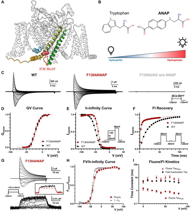 Figure 1
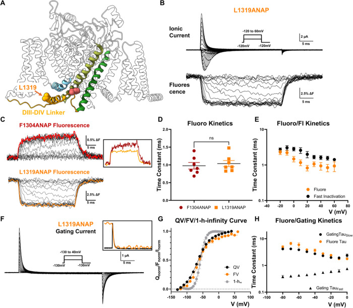 Figure 2
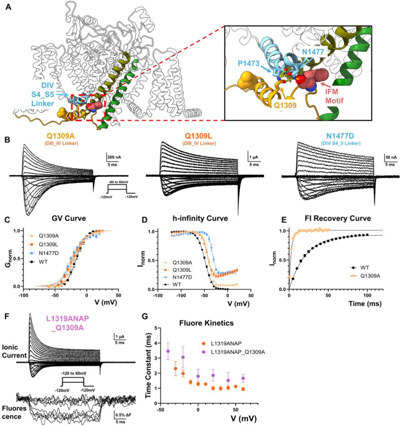 Figure 3
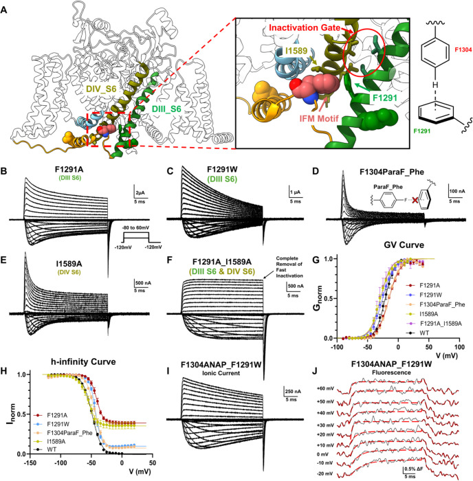 Figure 4
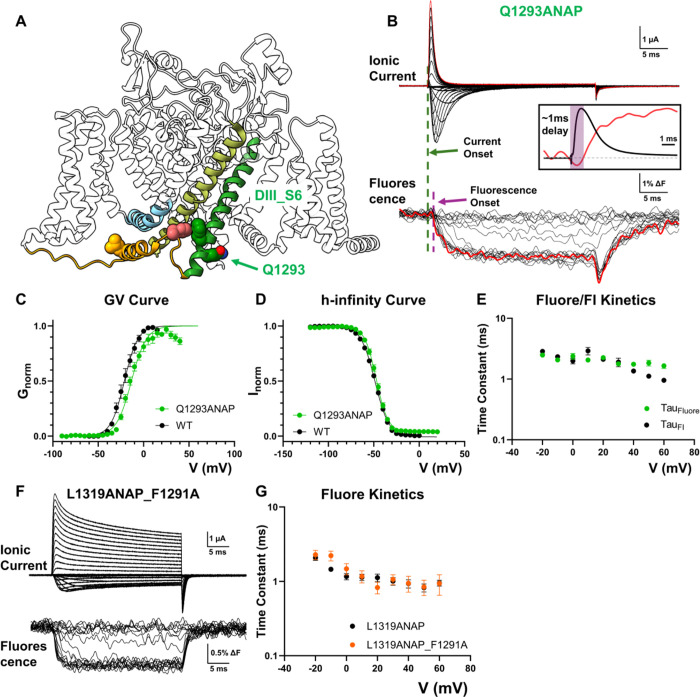 Figure 5
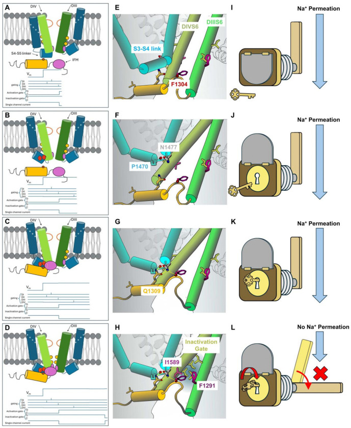 Figure 6
Peer Reviews
No public reviews on file for this paper yet. If you reviewed it on a platform where reviews are public (OpenReview, ICLR, NeurIPS, ICML), you can paste yours below so the community can read it here.
Videos
No videos yet. Explain this paper in a talk, walkthrough, or lecture? Add one.
Taxonomy
TopicsIon channel regulation and function · Neuroscience and Neuropharmacology Research · Electrochemical Analysis and Applications
Introduction
The action potential is the elementary unit of bio-excitability. At the molecular level, voltage-gated sodium (Nav) channels are responsible for the rapid upstroke of the action potential. These channels are gated to an open, sodium conducting conformation by positive changes in transmembrane potential ^1–3^. During the rising phase of the action potential, Nav channels operate in a classical positive-feedback manner: opened by membrane depolarization, the Nav channels allow further Na^+^ entry with consequent membrane depolarization, thus resulting in a cascading, all-or-nothing action potential. In this positive feedback loop, fast inactivation is an essential built-in auto-inhibitory mechanism to terminate the cascade and serves as a molecular “kill switch”. Milliseconds after the channels are open during activation, fast inactivation drives the channels into the nonconductive, fast inactivated state, facilitating the repolarization by voltage gated potassium channels. Acquired or inherited abnormalities in fast inactivation can result in severe pathological states ^4^. Additionally, due to the prevalence and diversity of Nav channels, these pathological states can manifest in numerous tissue types with a wide range of symptoms ^5–9^. Hence, a molecular understanding of the fast inactivation mechanism is of fundamental importance.
Originally proposed almost 50 years ago, the “ball and chain” model predicts that an intracellular located “inactivation particle” would bind to the open channel at the pore and physically block the ionic conduction ^10^. Later, the identification of the amino acid triad, Isoleucine-Phenylalanine-Methionine (the IFM motif) in the cytoplasmic domain III and domain IV (DIII_DIV) linker as the inactivation particle materialized the abstract concept and provided a molecular framework for fast inactivation ^11^. Numerous studies have since provided strong evidence for this model and consequently, the “ball and chain” model has been largely accepted as the textbook explanation for fast inactivation in Nav channels^12–16^.
However, upon the advent of the cryogenic electron microscopy mediated “resolution revolution” in structural biology, discrepancies emerged between the old models and new experimental results ^17^. As of today, essentially all of the available eukaryotic Nav channel structures, likely in the inactivated state due to the lack of external electric field, consistently demonstrate that the IFM motif, the inactivation particle, does not block the permeation pathway as expected ^18–21^. Instead, the motif docks in an adjacent hydrophobic pocket between the pore and voltage-sensor domains, a stark contradiction to the canonical pore-blocking “ball and chain” model. Our recent work began to address this inconsistency and identified the inactivation gate, a pore-lining double-layered hydrophobic ring at the intracellular mouth of the pore that blocks Na^+^ ion permeation in the inactivated state, instead of the IFM motif ^22^. However, as this is only the final step, additional modifications are necessary for the “ball and chain” model to accommodate the current structural and physiological results. The inactivating particle (IFM motif) is necessary for inactivation but the mechanistic linkage between voltage-sensor activation, the docked IFM motif and the newly described gate remains poorly resolved. Thus, the lack of molecular description and theoretical framework of the fast inactivation process once again represents a major conceptual gap in Nav channel biophysics and physiology.
Here, we present the full sequence of conformational changes during the fast inactivation process in mammalian Nav channels. The dynamic nature of the molecular rearrangements between the IFM binding and the gate closing requires techniques that can detect these changes while observing the actual function of the channel. To this end we have used site-directed fluorescence in voltage clamped conditions. With our improved incorporation protocol and optical recording setup, we introduced an environmentally sensitive fluorescent unnatural amino acid, ANAP, into various regions of the Nav channel and monitored the local molecular motions during fast inactivation by rapid fluorescence measurements. ANAP fluorescence recordings reported two types of movements at the IFM location during fast inactivation. A fast movement, triggered by the activation of voltage sensing domain (VSD) in domain IV (DIV), reflects the movement of the DIII_DIV linker region and posits the IFM motif into the appropriate location, conceptually like putting a key into a keyhole, that was exposed by the movement VSD of DIV. Coupled by hydrogen bonds between DIII_DIV linker and DIV S4_S5 linker, this fast component follows faithfully the movements of DIV VSD, via simultaneously measured gating currents, and proceeds prior to the closure of the fast inactivation gate. The other slower conformational change at the IFM motif triggers the fast inactivation. The aromatic side chain of the phenylalanine residue in the IFM motif forms interactions with two residues in DIII S6 and DIV S6, through T-shaped π-π and hydrophobic interactions. In tandem, these interactions propagate the conformational change from the linker to the pore and lead to the closure of the double-layered fast inactivation gate, similar to the movement of the bolt of the lock. Contrary to our expectations, modification to the interaction between the IFM motif and the hydrophobic pocket does not impede the binding of the motif itself, rather it appears to influence the effectiveness of the IFM motif at triggering fast inactivation, like creating mismatches between the key and the lock. With these current observations, we propose a “lock and key” model to describe the sequential conformational changes during fast inactivation in mammalian sodium channels.
Results
IFM Motif Undergoes Two Distinct Movements During Fast Inactivation
The IFM motif is evolutionarily conserved in Nav channels across the animal kingdom (Fig. 1A). Its importance for fast inactivation has been demonstrated repeatedly in the literature ^14,23^. However, recent structural results have cast uncertainty as to the functional role IFM motif plays in the mechanism of fast inactivation. To describe the conformational changes at the IFM position and to understand its role in fast inactivation, we replaced the phenylalanine in the IFM motif with a fluorescent, environmentally sensitive unnatural amino acid, ANAP (F1304ANAP, numbering based on rat Nav 1.4)^24,25^. ANAP (3-(6-acetylnaphthalen-2-ylamino)-2-aminopropanoic acid) has a comparable size as tryptophan which allows minimum disturbance upon incorporation (Fig. 1B). More importantly, the emission spectrum of ANAP is modulated by the hydrophobicity of its immediate environment, making ANAP an ideal reporter of molecular movement ^25^ (Fig. 1B). Robust currents were seen from F1304ANAP, and no discernable current was seen in cells where no ANAP was supplied, thus demonstrating high fidelity of encoding (Fig. 1C). F1304ANAP channels showed minor slowing effects on fast inactivation, similar to tryptophan replacement^14^, and minimal impact in activation (Fig. 1D–F). Utilizing site-directed fluorimetry with cut-open voltage clamp technique, ionic current and fluorescence signal were resolved simultaneously^26,27^. Fluorescence signals increased upon depolarization, indicating an increase in the environmental hydrophobicity seen by the side chain of ANAP (Fig. 1G). This is consistent with the notion that during fast inactivation the IFM motif transitions from a hydrophilic to more hydrophobic environment. The overlap of the steady-state fluorescence (FV) curve and fast inactivation, tracked via 1-(h-infinity) curve (Fig. 1H), indicates that the movement of the IFM motif directly reports the fast inactivation state of the Nav channels. Additionally, the off-fluorescence signal closely tracks the recovery from inactivation (Supple. Figure 1). Interestingly, the fluorescence signals that originate from F1304ANAP channels exhibit two kinetic components, suggesting two distinct conformational changes in the vicinity of the IFM motif occurred during fast inactivation (Fig. 1G, inset). Comparison between the fast inactivation kinetics and the fluorescence signal kinetics revealed that the fast time constant of fluorescence signal was significantly faster than the fast inactivation kinetics. The slow component on the other hand, closely followed the development of fast inactivation (Fig. 1I).
Fast Movement at IFM Motif Reflects DIII_DIV Linker Movement Following DIV VSD Activation.
To further investigate the nature of conformational dynamics at the IFM motif during fast inactivation, we monitored the movement of the IFM-bearing DIII_DIV linker by incorporating ANAP at L1319 position (L1319ANAP, Fig. 2A). ANAP incorporation at position 1319 was well tolerated (Supple. Figure 2) and opposed to the F1304 position, L1391ANAP channels showed a marked decrease in the fluorescence intensity upon depolarization, suggesting a transition to more hydrophilic environment (Fig. 2B). This is consistent with the existing structural results since L1319 is depicted as being completely exposed in aqueous environment in the fast inactivated structure (Fig. 2A). More interestingly, there is only one kinetic component in the fluorescence signal at L1319 position. This component showed similar time constants as the fast movement seen in F1304ANAP (Fig. 2C, D). Like the fast fluorescence signal in F1304ANAP channels, the fluorescence signal recorded in L1319ANAP is faster than the development of fast inactivation itself (Fig. 2E). To better understand the origin of this signal, we simultaneously recorded fluorescent signals and gating currents in L1319ANAP channels (Fig. 2F). Upon depolarization, gating charges in voltage sensors rapidly traverse the electric field, creating a fast, transient current, the gating current ^28,29^. Given that gating currents track the movement of VSDs, this can be correlated to the observed fluorescence signals that originate from movements at the cytoplasmic III-IV linker. Gating current recordings from L1319ANAP channels revealed that the voltage dependent gating charge movement, shown by the QV curve, overlaps closely with the steady-state FV curve (Fig. 2G). Despite the relative distance between the VSD and III-IV linker, the movement seen at L1319 linker region is more closely related to the voltage sensor movements than to fast inactivation. More specifically, the DIV VSD movement appears to directly trigger the DIII_DIV linker movement. When we compared the kinetics of the gating current and the fluorescence recordings, we discovered that the fluorescence signal at position 1319 explicitly followed the slow component of the gating current (Fig. 2H). Due to the asymmetry in Nav channels, voltage sensors from different domains translocate their charges at different rates ^30,31^. Particularly, the activation of the DIV VSD is significantly slower than the other VSDs, giving rise to the slow component of the gating current in Nav channels^30,32^. The shared kinetics between fluorescence signal at position 1319 and the DIV VSD movement strongly suggests the activation of voltage sensor in DIV initiates the DIII_DIV linker movement during fast inactivation.
Backbone Hydrogen Bonding Couples the DIII_DIV Linker and DIV VSD Movement
Since the DIII_DIV linker movement closely correlates with the activation of DIV VSD, there must exist interactions that couple these two structural components. A close structural analysis of this region revealed the existence of two hydrogen bonds between the DIII_DIV linker and the DIV S4_S5 linker (Fig. 3A). Specifically, residue N1477 at the “elbow region” of the DIV S4_S5 linker forms a hydrogen bond directly with the F1304 in the bound IFM motif and reciprocally, Q1309 at the DIII_DIV linker forms the other hydrogen bond with P1473 in DIV (Fig. 3A). Interestingly, both hydrogen bonds are formed between the nitrogen in the primary amide in the side chain and the carbonyl oxygen in the backbone. Removal of the primary amide in the side chain in both positions result in impaired fast inactivation (Fig. 3B). In the case of Q1309L and N1477D, the channels exhibit an “IQM” type phenotype with most of the fast inactivation absent^11,31^ (Fig. 3C, D and F) without drastic change in GV curves (Fig. 3C). An analysis of the recovery from fast inactivation for Q1309A channels further demonstrates that the identified hydrogen bond is essential for the stabilization of the fast inactivated state (Fig. 3E). In the absence of this interaction, the channels recover from fast inactivation exceedingly fast. Evidently, these hydrogen bonds are important for fast inactivation and likely serve as the functional bridges between the DIV VSD and the intracellular linker. To further demonstrate this, we disrupted one of the hydrogen bonds and investigated its impact on DIII_DIV linker movement. In L1319ANAP_Q1309A, fast inactivation becomes slower and incomplete (Fig. 3I). The fluorescence signal became much smaller (Fig. 3I) and more importantly, progressed slower and recovered faster compared to L1319ANAP (Fig. 3F, G), demonstrating the identified interactions are indeed involved in coupling between the voltage sensor in DIV (via the DIV S4-S5 linker) and DIII_DIV linker.
IFM Motif Relays Its Conformational Changes to the Pore through Hydrophobic and T-Shaped π-π Interactions
With the molecular underpinning of the fast component elucidated, we set out to investigate the slower conformational change at the IFM motif that triggers fast inactivation. Our previous work demonstrated the specialized role of the pore-lining hydrophobic residues in DIII and DIV S6 in fast inactivation^22^ (Fig. 4A). Therefore, residues in DIII and DIV S6 that could form potential interactions with the IFM motif were screened through site-directed mutagenesis. The screening revealed two residues that were particularly important for fast inactivation.
In in DIII S6, when F1291 is mutated to either smaller or bigger residues, significant amount of fast inactivation was removed (Fig. 4B, C and H), suggesting that the precise size of the side chain is important for this interaction. Additionally, structural analysis ^33^ also pointed out the possibility of a T-shaped π-π interaction between the phenylalanine in the IFM motif and F1291 (Fig. 4A). To precisely target this potential aromatic interaction, we utilized another unnatural amino acid, 4-fluorophenylalanine (ParaF-Phe). Substitution of the hydrogen at the para position on the aromatic ring targets the positive charge on the edge of the aromatic ring and would be predicted to weaken a T-shaped π-π interaction with a minimal impact on the volume of side chain ^34^. In support of this notion, substitution of only one atom in F1304 via encoding of ParaF-Phe led to not only slowed, but also incomplete fast inactivation and faster recovery from inactivation (Supple. Figure 3), suggesting a functional role for this π-π interaction F1304 and F1291 (Fig. 4D, H).
Mutagenesis in DIV S6 revealed that changing I1589 led to similar phenotype observed by mutations in DIII S6. I1589A severely impeded fast inactivation and showed large non-inactivating steady-state current (Fig. 4E, H). As the inactivation gate is made of large hydrophobic residues in S6 of both domain III and IV, the combination of F1291A in S6-DIII and I1589A in S6-DIV should eliminate fast inactivation. Indeed, combining both mutations in a single construct, F1291A_I1589A, produced channels where fast inactivation was completely removed. No significant reduction in current amplitude was seen after 30ms of depolarization (Fig. 4F).
Our mutagenesis experiments on F1291 and I1589 confirmed their importance for fast inactivation. However, these results did not directly demonstrate those are residues that coupled the IFM motif to the fast inactivation gate at the pore. To provide direct evidence, we investigated the movement of the IFM motif in the absence of the identified interactions. We recorded the ionic current and fluorescence signal in F1304ANAP_F1291W (Fig. 4I, J). Fast inactivation was largely removed in F1304ANAP_F1291W, but still, clear fluorescence signals were resolved from the IFM position. Evidently, disrupting the interaction between F1291 and F1304 did not abolish the binding of the IFM motif. The kinetics of the fluorescence signals were significantly faster than the residual fast inactivation. These observations are consistent with our previous results suggesting that the fast component of the IFM movement was triggered by the DIV VSD and coupled by the hydrogen bonds, instead of interactions in the hydrophobic pocket. More importantly, now with disrupted interaction with F1291, the IFM motif movement showed only one fast fluorescence component and the second slow movement seen before in F1304ANAP alone was largely absent (Fig. 4J), providing direct evidence that the identified residues are responsible for coupling the IFM motif and the pore.
Fast Inactivation Process Requires Sequential Conformational Changes
Finally, we monitored the movement of the pore during fast inactivation by encoding ANAP at Q1293, a position at the bottom of the pore-lining DIII S6 segment (Fig. 5A). Incorporation of ANAP at Q1293 led to minimal alteration in fast inactivation (Fig. 5B, D) and activation (Fig. 5C). The fluorescence signal from Q1293ANAP mirrored the development of fast inactivation, suggesting the movement seen at this part of the S6 helix reflects mostly conformational changes associated with fast inactivation instead of activation (Fig. 5E). Interestingly, there was a ~ 1ms delay to the onset of the fast-inactivation-associated fluorescence signal. This delay is similar to the previous observation in pronase treated giant squid axon^35^ (Fig. 5B inset). The observed delay also resembles the classical Cole-Moore shift ^36^ and indicates that additional conformational changes are required before channel can enter into the fast inactivated state. It is likely that the two movements seen at the IFM position happen in a sequential manner in that the DIII_DIV linker needs to move to posit the IFM at the right location before the second, slower conformation change can happen and triggers the fast inactivation. Supporting this notion, in the case of L1319ANAP_F1291A (Fig. 5F), the fluorescence signal was unaltered by abolishing the second, slower interaction (Fig. 5G), suggesting the DIII_DIV linker movement happened upstream of the second interaction and the fast inactivation process is best described as sequential conformational changes.
Discussion
The IFM Motif Couples the DIV_VSD and Fast Inactivation Gate
Connections between VSD movements and fast inactivation has long been established. From the pronase perfused giant squid axon ^35^ to charge neutralization experiments ^32^ on DIV VSD, it is clear that VSD movements are required for the fast inactivation. On the other hand, fast inactivation could also influence the VSD movements, as was shown in charge immobilization experiments ^10,37^. Our results indicate that the IFM motif serves as a structural coupler that connects the voltage-driven movement of the DIV VSD to the inner mouth of the pore that inactivates sodium channels. By positioning ANAP into the IFM motif, we find that the resulting fluorescence signal reveals two sequential movements at IFM motif during fast inactivation. The fast movement originates from the DIII_DIV linker movement, coupled to the DIV VSD movement and the slower movement directly triggers the closure of the fast inactivation. Evidently, the movement of IFM motif transduces the activation of DIV VSD directly to closure of the fast inactivation gate at the pore. These two movements also explain the domain specific charge immobilization during fast inactivation shown previously in MTS modification experiments ^10,38^. It is likely that the movement of the DIII_DIV linker triggers the immobilization of DIV VSD by binding of the IFM with the DIV S4-S5 linker, while the closure of the fast inactivation gate immobilizes DIII VSD.
Functional Identification and Validation of the Binding Site for IFM motif
In this work, we further elucidated a network of hydrogen bonds that forms the structural connection between DIV VSD and cytoplasmic DIII_DIV linker. One intriguing observation is that in both hydrogen bonds, it is the carbonyl groups in the peptide backbone which serve as the H-bond acceptors. In particular, N1477 at DIV S4_S5 linker forms a direct interaction with the backbone carbonyl group of the phenylalanine in IFM motif. One natural conclusion from this observation would be that the exact identify of the residue at the second position of IFM motif shouldn’t influence the formation of the hydrogen bonds and subsequently shouldn’t impede the “binding” of IFM motif. The binding of IFM motif has been traditionally associated with interactions between the side chain of the phenylalanine residue and its surrounding hydrophobic pocket. However, our results argue that the driving force of the IFM motif binding comes mostly from the hydrogen bond formation with the peptide backbones. In other words, the binding site of the IFM motif is formed not by the hydrophobic pocket but rather Q1309 in DIII_DIV linker and N1477 in DIV S4_S5 linker. It has been observed in molecular dynamic simulations before that mutation N1662D, a disease causing mutant in hNav1.2 (equivalent as N1477 in rNav1.4), led to significantly decreased dwell time of IFM in the hydrophobic pocket ^39^. This is consistent with our electrophysiological results. Removing one of the hydrogen bonds in Q1309A led to an unstable fast inactivated state as was shown by the increased recovery speed from inactivation. The fluorescence results, which arguably track motions in this area, also support this conclusion. Disruption of the hydrophobic pocket at F1291, while resulting in significant removal of fast inactivation (F1291W), didn’t impede the binding of IFM motif in F1304ANAP_F1291W (Fig. 5F). On the other hand, a mild mutation Q1309A clearly led to impaired coupling between DIV VSD and DIII_DIV linker as was seen in L1319ANAP_Q1309A. Finally, charge immobilization experiments in IQM also demonstrated that despite almost complete removal of fast inactivation from ionic current, ~ 30% (in contrast to 60% in WT) of total gating charge still became immobilized after 24ms of depolarization (Supple. Figure 4). Clearly, even in IQM channels, some conformational changes along the fast inactivation pathway are still able to occur. It is likely the DIII_DIV linker in this case still binds to the identified residues, leading to some degree of charge immobilization, likely only DIV VSD^13,38^, but as no further movement triggers the closure of the fast inactivation gate, the immobilization of DIII does not occur. Taken together, the present data strongly favors N1477 and P1473 in DIV S4_S5 linker as the binding sites for IFM motif, instead of a hydrophobic pocket.
Molecular Mechanism of Fast Inactivation
The current and available data are therefore consistent with a “lock and key” model for fast inactivation in voltage-gated sodium channels. Figure 6 is a schematic representation of the sequence of events that lead to fast inactivation. Upon depolarization, the sensors of domain I, II and III activate and the channel opens (Fig. 6A, E, I). Normally, domain IV sensor activates later ^30,32^ and upon its activation it exposes the binding site for IFM motif at P1470 and N1477 in DIV S4_S5 linker, similar to a keyhole becoming available (Fig. 6B, F, J). The IFM-containing DIII_DIV linker, likely mobile in aqueous solution, binds to DIV S4_S5 linker via the H-bonds (Fig. 6C, G) and posits IFM motif, the key, into the hydrophobic pocket, the keyhole (Fig. 6K). The binding of IFM motif allows the interactions with F1291 and I1589 through hydrophobic and aromatic interactions to occur, leading to a second conformational change at the hydrophobic pocket. In F1291Q, despite some steady state current, the majority of fast inactivation persists, dissimilar to F1304Q (IQM) phenotype (Supple. Figure 5). This asymmetric effect suggests that for interactions between F1291 and I1589 to occur, another conformational change must occur. Most likely, DIII S6 and DIV S6 helices undergo a rotational movement to position F291 and I1589 into the hydrophobic pocket interacting with the IFM motif and thus locking the two S6 helices in position. At the same time, the rotations position the four large hydrophobic amino acids forming the inactivation gate into the conduction pathway interrupting the flow of Na + ions, thus producing fast inactivation (Fig. 6D, H, L). Modifications to either IFM motif or the hydrophobic pocket would lead to incompatibility between the key and the lock, leading to impaired inactivation.
Recovery from inactivation requires disengagement of the IFM motif. Results of F1304ANAP demonstrate that the recovery from inactivation and off-fluorescence signal share similar time course, supporting this idea (Supple. Figure 1). The two major conformational changes that occurred during fast inactivation are responsible for immobilization of DIV VSD and DIII VSD respectively as suggested by the IQM gating current result ^13,37^ (Supple. Figure 4). Under hyperpolarized voltages, a transient unbinding of the IFM motif would allow the deactivation of voltage sensors in DIII and DIV that are previously immobilized. This inward movement of the sensors subsequently abolish the binding site for IFM motif, similar to covering the keyhole on the lock and allows the channels to recover from the fast inactivated state.
Alternative Fast Inactivated State in Nav Channels
Here we addressed the molecular pathway from open channels to the fast inactivated state. It is important to point out that closed Nav channels can also inactivate, bypassing the open state altogether, and this is essential for physiological processes ^10,40,41^. This closed-state inactivation, or steady-state inactivation, prevents Na^+^ conduction upon hyperpolarization and the recovery from closed-state inactivation determines the absolute refractory period in firing neurons. Structural work suggests a similar pore conformation in the closed inactivated state and open inactivated state ^19,33^. Fluorescent measurements on individual sensor movements have demonstrated that DIV VSD can activate at more hyperpolarized voltages compared to the other VSDs ^30^. Then, as the movement of DIV VSD alone seems to be enough to trigger inactivation while opening requires all the other VSDs to be activated, it is possible that fast inactivation may occur regardless of an open or a closed pore. When we monitored the pore movement at Q1293 position, two components of fluorescence signals were observed upon hyperpolarization (Fig. 5B). It is possible these two components capture the transitions from open inactivated state to closed inactivated state and from closed inactivated state to closed state respectively. However, further investigations are required to elucidate the exact structural rearrangement at the pore during these transitions. But most likely, the observed sequential conformational changes also occur during closed state inactivation.
Evolutionary and Isoform Conservation of the Identified Fast Inactivation Apparatus
We compared Nav channels from Bilateria, Cnidaria and Choanoflagelleta (Supple. Figure 5) and intriguingly found the newly identified residues are highly conserved, some even more conserved than the IFM motif itself. F1291, N1477 and Q1309 are conserved across all sequences compared, even in the ancestral form of Nav channel in Monosiga brevicollis ^42^. In the case of purple sea urchin, Strongylocentrotus purpuratus, an isoleucine replaces the phenylalanine in the IFM motif but nevertheless, all three residues remain conserved at the equivalent positions. I1589, on the other hand, is less conserved. In the ancestral Nav channel, a valine is found at 1589 equivalent position and valine is the only alternative amino acid found at this position across different phyla. The high conservation of the newly identified fast inactivation components strongly supports the idea that the molecular mechanism described here in mammalian Nav1.4 channel is conserved across the animal kingdom and appears at the same time as the evolutionary invention of the Nav channel in Choanoflagelleta.
Material and Methods
Site-directed mutagenesis and cRNA in-vitro synthesis
rNav1.4 in pBSTA vector, flanked by β-globin sequences, was used in this study for all the physiological experiments. Point mutations were generated using QuikChange^™^ mutagenesis method (Agilent). The PCR products were first digested by DpnI and were used to transform the XL10-gold ultra-competent cells (Agilent). After ampicillin resistance screening, plasmids were purified from the colonies using standard miniprep protocols. Purified plasmids were sent to Plasmidsaurus for whole plasmid sequencing to confirm the introduction of the mutations. Verified plasmids were first linearized and later in vitro transcribed to cRNA with mMESSAGE mMACHINE^™^ T7 Transcription Kit (Thermo Fisher Scientific).
Xenopus laevis oocyte preparation and channel expression
Ovaries of Xenopus laevis were purchased from XENOPUS1 (Dexter, Michigan). The follicular membrane was removed using collagenase type II (Worthington Biochemical Corporation) 2 mg/mL with bovine serum albumin at 1mg/mL (BSA). After defolliculation, stage V–VI oocytes were then selected and microinjected with 50–100 ng cRNA. Injected oocytes were incubated at 18°C for 1–5 days in SOS solution (in mM: 100 NaCl, 5 KCl, 2 CaCl_2_, 0.1 EDTA, and 10 HEPES at pH 7.4) supplemented with 50 μg/mL. Unless otherwise stated, all chemicals were purchased from Sigma-Aldrich.
Cut-open voltage clamp on Xenopus laevis oocytes
Macroscopic ionic and gating current were recorded using cut-open voltage clamp technique^43^. Micropipettes filled with 3M CsCl, with resistance between 0.6 and 1.2 MΩ were used to measure the internal voltage of the oocytes. Current data were first filtered online at 20 kHz with a low-pass 4 pole Bessel filter and sampled by a 16-bit A/D (USB-1604; Measurement Computing, Norton, MA) converter at 1 MHz. A feedbackback Peltier device was used to control the temperature at 13 ± 1°C for all experiments. For ionic current experiments, unless otherwise stated, were conducted in external solution consisted of in mM: 28 Na methylsulfonate (MES), 92 N-methyl-D-glucamine (NMG) MES, 2 Ca MES, 10 HEPES, 0.1 EDTA, pH = 7.4 and internal solution consisted of in mM: 12 Na MES, ,108 NMG MES, 10 HEPES, 2 EGTA, pH = 7.4. The capacitive transient was manually compensated with a dedicated circuit and further removed by an online P/−4 protocol with a holding voltage of either − 80 or −90 mV^10^. For gating current experiments, all experiments were conducted in external solutions consisted of in mM: 120 NMG MES, 2 Ca MES, 10 HEPES, 0.1 EDTA, 750nM TTX pH = 7.4 and in internal solution consisted of in mM: 120 NMG MES, 10 HEPES, 2 EGTA, pH = 7.4. In this case, an online P/4 at 40mV was used to subtract the capacitive transients. Voltage clamp speed measured by capacitive transient current gave a time constant around 70 μs.
Unnatural Amino Acid Incorporation
Two methods were used to incorporate the unnatural amino acid in Nav channels. For ANAP, two separate cytoplasm injection were performed. First injection introduced into oocytes four different components: ANAP amber suppression tRNA at 2 ng/nL, ANAP synthetase at 0.3 ng/nL, mutated Xenopus laevis release factor 1 at 0.3 ng/nL and the unnatural amino acid ANAP itself in methyl ester form (Cayman Chemical) at 2mM. 24 hours after the first injection, channel cRNA bearing the amber stop codon mutation at the desired location was injected at 1 ng/nL. 24 to 72 hours after the second injection, the oocytes were used in voltage clamp fluorimetry experiments.
4-fluoro-L-phenylalanine was appended to pyl tRNA as previously reported^44,45^.Briefly, the cyanomethyl ester of N-Boc-4-fluoro-L-Phe was reacted with the dinucleotide pdCpA in dimethylformamide, purified on HPLC and deprotected with trifluoroacetic acid. A 3 mM stock solution on the amino acid-dinucleotide chimera in dimethylsulfoxide was prepared and added to T4 RNA ligase and pyl tRNA lacking the terminal CA nucleotides to assemble the full-length tRNA with 4-fluoro-L-Phe attached. Pellets (1 nmol) were stored dry at −78°C until needed. Lyophilized conjugated tRNA was resuspended in 3mM Na Acetate at pH 5.5 to an approximate concentration of 2.5 μg/μL. Conjugated tRNA and cRNA of the mutated channel were introduced simultaneously into the oocytes and after 24 to 48 hours of expression, oocytes were used in voltage-clamp experiments.
Voltage Clamp Fluorimetry Experiments
An avalanche photodiode or a photomultiplier was used to record the fluorescence signal. A 365nM LED (Thor Lab) was used as the excitation light source. The filter set used in the study was described previously^46^. In the case of the photomultiplier (H10304–20-NN, Hamamatsu, Japan), the output signal was first processed by a home-built I to V converter, filtered by a low-pass 8 pole Bessel filter at 10kHz and sampled by the AD converter every 10 μs. Photobleaching was later subtracted with a Matlab routine and then digitally filtered at 5kHz or 1kHz.
Data Analysis
Data recorded in the work is analyzed by GraphPad 9 (Prism), Excel (Microsoft), Matlab R2022a (Mathworks), and in-house software (Analysis and GPatchM).
GV curves calculation: Ionic conductance was calculated by dividing the peak ionic current by the driving force determined experimentally. After normalization, GV curves were fitted with a two-state model described below,
, where is the ideal gas constant, is the absolute temperature in Kelvin, is the apparent charge, is the Faraday constant.FV curve calculation: Steady state fluorescence signal was normalized to maximum fluorescence and then described with the model below,
, where and are as previously definedSteady state fast inactivation curve ( -infinity curve) was calculated by plotting normalized peak Na^+^ current during test pulse after 50ms conditioning pulse voltage and fitted using a two-state model,
, where Base is the steady state current, and and are as previously defined.The time constant of fast inactivation, gating charge movement or fluorescence kinetics were calculated by fitting the signals with either one or two exponential decays using the general equation below,
, where , and represent amplitudes and time constants of the first and second components, respectively. Base represents the steady state current or fluorescence. When one exponential was used, only , and were fitted to the data.Recovery from fast inactivation was calculated by dividing the peak current in the control pulses by the peak current during test pulses. The data was then described by either one or two component association model,
, where , and represent amplitudes and time constants of the first and second components, respectively. When one exponential was used, only , and were fitted to the data.
Supplementary Material
Supplementary Files
This is a list of supplementary files associated with this preprint. Click to download.
The reference list from the paper itself. Each links out to its DOI / PubMed record.
- 1Bean B. P. The action potential in mammalian central neurons. Nat. Rev. Neurosci. 8, 451–465 (2007).17514198 10.1038/nrn 2148 · doi ↗ · pubmed ↗
- 2Hodgkin A. L. & Huxley A. F. Currents carried by sodium and potassium ions through the membrane of the giant axon of Loligo. J. Physiol. 116, 449–472 (1952).14946713 10.1113/jphysiol.1952.sp 004717 PMC 1392213 · doi ↗ · pubmed ↗
- 3Hodgkin A. L. & Huxley A. F. The components of membrane conductance in the giant axon of Loligo. J. Physiol. 116, 473–496 (1952).14946714 10.1113/jphysiol.1952.sp 004718 PMC 1392209 · doi ↗ · pubmed ↗
- 4De Lera Ruiz M. & Kraus R. L. Voltage-Gated Sodium Channels: Structure, Function, Pharmacology, and Clinical Indications. J. Med. Chem. 58, 7093–7118 (2015).25927480 10.1021/jm 501981 g · doi ↗ · pubmed ↗
- 5Fertleman C. R. SCN 9A Mutations in Paroxysmal Extreme Pain Disorder: Allelic Variants Underlie Distinct Channel Defects and Phenotypes. Neuron 52, 767–774 (2006).17145499 10.1016/j.neuron.2006.10.006 · doi ↗ · pubmed ↗
- 6Waxman S. G. Painful Na-channelopathies: An expanding universe. Trends Mol. Med. 19, 406–409 (2013).23664154 10.1016/j.molmed.2013.04.003 · doi ↗ · pubmed ↗
- 7Phillips L. & Trivedi J. R. Skeletal Muscle Channelopathies. Neurotherapeutics 15, 954–965 (2018).30341599 10.1007/s 13311-018-00678-0PMC 6277285 · doi ↗ · pubmed ↗
- 8George A. L. Molecular basis of inherited epilepsy. Arch. Neurol. 61, 473–478 (2004).15096393 10.1001/archneur.61.4.473 · doi ↗ · pubmed ↗