Management of Glomus Tumors: Experience From a Tertiary Care Centre in India
Nitin Choudhary, Archi Gupta, Akash Narangyal, Bias Dev, Sanjeev Gupta

TL;DR
This paper shares clinical experience on managing glomus tumors, rare soft tissue growths often missed due to their rarity, at a hospital in India.
Contribution
The study provides insights into the clinical presentation and management of glomus tumors in a specific Indian tertiary care setting.
Findings
Most glomus tumors (81.8%) were found in the nail bed, with significant diagnostic delays in most cases.
Complete excision was effective, with no recurrence observed in the 11 cases studied.
Pain was a common symptom, while visible lumps were rare, especially in nail bed tumors.
Abstract
Background: Glomus tumors are uncommon soft tissue tumors that typically develop in the distal extremities, especially the finger's subungual area. Pain, soreness, and temperature intolerance, particularly cold sensitivity, make up its traditional clinical triad. There is a relative rarity in the literature on this topic, and we have analyzed and thereby discussed our experience in these cases. Methods: A retrospective cross-sectional study was performed at a tertiary care centre in Jammu, India. All patients diagnosed with glomus tumor from October 2022 to August 2024 were included in the study. Conventional radiographs, ultrasonography (USG), and MRI were the investigation modalities used for diagnosis. Tumors excised were sent for histopathological examination (HPE) for pathological diagnosis. Results: A total of 11 cases of glomus tumor were diagnosed from October 2022 to August…
Genes, proteins, chemicals, diseases, species, mutations and cell lines named across the full text — each resolved to its canonical identifier and authoritative record.
Click any figure to enlarge with its caption.
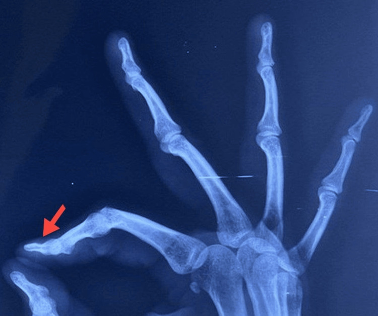 Figure 1
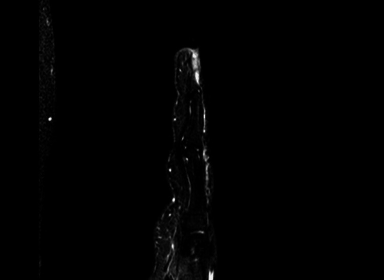 Figure 2
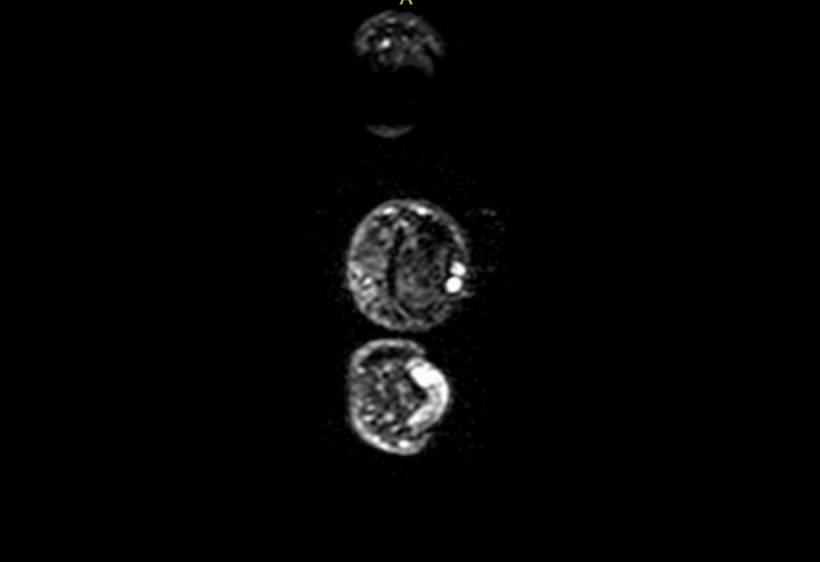 Figure 3
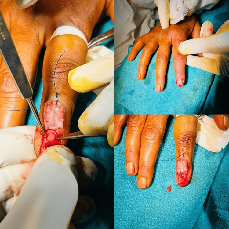 Figure 4
| Patient | Sex | Age (in years) | Location | Time to presentation (in months) | Follow-up period (in months) | Complication | Recurrence |
| 1 | F | 27 | Nail bed | 18 | 13 | Nil | No |
| 2 | F | 37 | Nail bed | 20 | 11 | Nil | No |
| 3 | F | 41 | Nail bed | 28 | 14 | Nil | No |
| 4 | F | 36 | Volar pulp | 27 | 10 | Nil | No |
| 5 | F | 38 | Nail bed | 12 | 9 | Nil | No |
| 6 | F | 34 | Nail bed | 27 | 14 | Nil | No |
| 7 | M | 29 | Nail bed | 28 | 18 | Nil | No |
| 8 | F | 42 | Nail bed | 15 | 12 | Nil | No |
| 9 | F | 39 | Volar pulp | 24 | 11 | Nil | No |
| 10 | M | 34 | Nail bed | 15 | 9 | Nil | No |
| 11 | F | 28 | Nail bed | 18 | 16 | Nil | No |
Peer Reviews
No public reviews on file for this paper yet. If you reviewed it on a platform where reviews are public (OpenReview, ICLR, NeurIPS, ICML), you can paste yours below so the community can read it here.
Videos
No videos yet. Explain this paper in a talk, walkthrough, or lecture? Add one.
Taxonomy
TopicsSoft tissue tumors and treatment · Historical Studies in Science
Introduction
Glomus tumors are relatively rare, accounting for about 1-5% of all hand tumors. Even while they can form anywhere on the body, they usually do so in the upper extremities, specifically in the subungual regions. Tenderness, cold intolerance, and acute paroxysmal pain in the fingertips are their defining characteristics [1,2]. Glomus tumors originate from the glomus body, which is a component of the dermal thermoregulation system. They were first described by Wood back in 1812 [3]. In 1924, Masson referred to glomus tumors as tumors that occur in the neuro-myo-arterial body [4]. Glomus cells, part of the body’s thermoregulation system, are responsible for managing blood flow through small arteriovenous shunts known as glomus bodies. When these cells proliferate abnormally, they form glomus tumors. These growths are usually small and well-circumscribed but can lead to severe pain and sensitivity, especially in colder environments [5]. Despite being non-cancerous, glomus tumors often present a challenging diagnostic and therapeutic scenario due to their unique characteristics.
Materials and methods
This retrospective cross-sectional study included patients diagnosed with glomus tumors of the hand at the Government Medical College and Hospital, Jammu, India, from October 2022 to August 2024. All patients presented with chief complaints of hand/digit pain, which was not relieved on taking analgesics and was tender to touch, with exacerbation in a cold environment. All patients were seen in peripheral clinics for the same, however, the case was not diagnosed and hence reported to the tertiary centre. A total of 14 patients were initially included in the study; however, two patients were lost in follow-up, and one patient’s documents/records were not recovered; hence, our final study included a total of 11 patients.
All patients were initially sent for plain X-ray radiographs (Figure 1) followed by ultrasonography. Patients detected with glomus tumor were sent for magnetic resonance imaging (MRI) (Figures 2, 3) for final definitive diagnosis. The patients were operated under ring block of the involved digit and were discharged the same day. All excised tumors were sent for histopathological examination (HPE) confirmation.
Lateral radiograph showing a scalloping effect on the bone caused by a glomus tumor (red arrow).
Sagittal MRI image showing the presence of a glomus tumor.
Axial MRI image showing a glomus tumor.
For the excision of hand glomus tumors, three surgical approaches are commonly described: trans-ungual, lateral subperiosteal, and volar. We used a trans-ungual approach for tumours of the nail bed, while the volar approach was used for volar tumours. The surgery was performed under ring block (digital anaesthesia), and a finger glove tourniquet was used (Figure 4).
En-masse removal of the tumor using the trans-ungual approach under digital block anesthesia, followed by closure.
Results
A total of 11 cases were included in the study. Of 11 patients, nine were females (81.8%) and two were males (18.2%). The mean age of presentation for both genders was 35 years. There was a history of pain for a long duration (mean: 21.1 months), with consultations for the same prior to reporting to the tertiary centre. All surgeries were done as outpatient procedures, and patients were discharged the same day with coverage of oral antibiotics and good analgesia. The samples sent for histopathological diagnosis affirmed our diagnosis. In a low-power field, the tumors were coated in fibrous capsules that were comparatively well-defined. Regularly shaped epitheloid cells were arranged in one or more layers around the dilated, thin-walled blood arteries. Epitheloid cells with a spherical or oval nucleus and essentially no mitotic signs were seen in a high-power field. Pain was relieved the very next day of surgery. There was no wound-related complication in any of the patients. Patients were followed up for a minimum of nine months. There was no case of recurrence of pain or any of the other initial presenting symptoms in any of the patients (Table 1).
Discussion
Situated in the reticular layer of the dermis, a glomus body is a unique arterio-venous anastomosis accountable for thermoregulation. The normal glomus body, consisting of glomus cells that are distorted smooth muscle cells found in the walls of the Sucquet-Hoyer canal, is the source of glomus tumors. They typically start in the skin, but they have also been known to appear in internal organs like the stomach, lung, trachea, and bone, as well as mucous membranes [6,7]. In the hand subungual region (most common), the lateral side of the digits and the palms are the most frequently affected sites.
Because glomus tumors typically arise in the extremities, particularly the digits of the upper extremities, hand or orthopaedic surgeons encounter them frequently [8]. They are also discovered in other places like the wrist, toe, hand, and knee, which is consistent with a group of 56 extra-digital glomus tumors that Schiefer et al. reported, of which 91% were detected in the extremities [9]. Glomus tumors are more common in women between the ages of 20-40, which is consistent with the findings of our study. However, males have a greater predisposition for glomus tumors at extra-digital sites [7].
Almost all patients in our study had extreme localized pain, which is consistent with their clinical presentation in the literature. There was a considerable delay in diagnosis from the time of the index complaint in most case series in the literature [7,8]. Therefore, a high index of suspicion is required in diagnosing these tumors.
Several studies have discussed radiological investigations as a supplement to clinical suspicion in forming a final diagnosis. Despite being a soft tissue tumor, glomus tumors can cause nearby bone to scallop, which can be seen on plain radiographs [10,11]. Ultrasound has been used to diagnose glomus tumor, owing to its hyper-vascular nature [10,12], and was considered the standard radiological modality in these cases. The glomus tumor, which typically manifests as a well-circumscribed lump that is hypo-intense on T1-weighted imaging and hyper-intense on T2-weighted images, can be diagnosed with an MRI scan, which is recently being commonly used for definitive diagnosis [8,11]. We, in our study, used both these modalities for diagnosing our patients.
Complete removal is thought to be curative and frequently has positive results [13]. All cases in our series had a benign glomus tumor identified by histology. While some studies claim a recurrence incidence of up to 30%, the majority of research in the literature also reports a low rate of recurrence, with extra-digital glomus tumors accounting for 85% of cases having recurrence [11,13]. A case series conducted by Sacchetti et al. in 2019 discussed a case of neoplastic distal forearm glomus tumor managed by excision along with adjuvant therapy [14]. The authors agreed that more research with longer follow-up periods will provide a more complete picture of the recurrence rate and clinical outcomes, given that the average follow-up period in this study was just 5.8 months. However, the authors across most studies in the literature have a general consensus that hand glomus tumors are benign tumors, which are treated best by complete excision with minimal/no recurrence rate [15-17].
The limiting factors in our study were a smaller number of cases, as a large number of cases or multi-institutional trials would have given a better epidemiological picture. While the minimum follow-up period in our study was nine months and the maximum follow-up was 18 months, having longer follow-up periods would have given a better and clearer picture on account of disease recurrence.
Conclusions
Hand glomus tumors, while benign, can significantly affect patients’ quality of life due to their painful and disruptive symptoms. It may be readily missed by clinicians because of its rarity and its being frequently clinically insignificant except for the presence of severe pain, which prolongs the time it takes to arrive at the correct diagnosis. Reducing discomfort and averting recurrence need early detection and effective treatment, with surgical excision offering negligible complications and minimal recurrences in expert hands.
The reference list from the paper itself. Each links out to its DOI / PubMed record.
- 1Clinical analysis of twenty cases of glomus tumor in the digits J Korean Soc Plast Reconstr Surg Lee CH Byeon JH Rhie JW 169178221995 https://scholar.google.com/scholar_lookup?journal=J+Korean+Soc+Plast+Reconstr+Surg&title=Clinical+analysis+of+twenty+cases+of+glomus+tumor+in+the+digits&author=C.H.+Lee&author=J.H.+Byeon&author=J.W.+Rhie&author=Y.J.+Kang&author=M.J.+Cho&volume=22&publication_year=1995&pages=169-178&
- 2Glomus tumors of the hand: review of the literature and report on twenty-eight cases J Bone Joint Surg Am Carroll RE Berman AT 691703541972 https://pubmed.ncbi.nlm.nih.gov/4341268/4341268 · pubmed ↗
- 3On painful subcutaneous tubercle Edinb Med Surg J Wood W 28329181812 https://pubmed.ncbi.nlm.nih.gov/30329513/PMC 574455230329513 · pubmed ↗
- 4The neuromyoarterial glomus of the tactile regions and its tumors [Article in French]Lyon Chi Masson P 257280211924 https://www.scirp.org/reference/referencespapers?referenceid=1759089
- 5Digital glomus tumors: a 29-year experience with a lateral subperiosteal approach Plast Reconstr Surg Vasisht B Watson HK Joseph E Lionelli GT 1486148911420041550993610.1097/01.prs.0000138752.36175.d 5 · doi ↗ · pubmed ↗
- 6Subungual glomus tumors: an algorithmic approach Ann Plast Surg Rohrich RJ Hochstein LM Millwee RH 300304331994798596710.1097/00000637-199409000-00011 · doi ↗ · pubmed ↗
- 7Glomus tumor Surg Gynecol Obstet Shugart RR Soule EH Johnson EW Jr 3343401171963 https://pubmed.ncbi.nlm.nih.gov/14080348/14080348 · pubmed ↗
- 8Glomus tumour Ann R Coll Surg Engl Sethu C Sethu AU 0298201610.1308/rcsann.2016.0005 PMC 523437826688416 · doi ↗ · pubmed ↗