Viability of Trichinella spiralis in traditional sour pork fermentation and its inactivation by microwave heating: Implications for zoonotic risk and food safety
Atchara Artchayasawat, Benjamabhorn Pumhirunroj, Sukhonthip Khueangchiangkhwang, Thidarut Boonmars, Parichart Boueroy, Porntip Laummaunwai, Panaratana Rattanasuwan

TL;DR
This study shows that traditional sour pork fermentation does not kill Trichinella parasites, but microwave heating can effectively eliminate them, reducing food safety risks.
Contribution
The study demonstrates that microwave heating is an effective method to inactivate Trichinella spiralis larvae in fermented pork.
Findings
Trichinella spiralis larvae remained viable during 5 days of sour pork fermentation.
Microwave heating at 400 W for 3 min or 800 W for 1 min or longer inactivated all larvae.
Boiling was confirmed as an effective control for larval inactivation.
Abstract
Cultural dietary practices involving the consumption of raw or undercooked meat, such as traditional sour fermented pork, pose significant risks for foodborne parasitic infections, particularly trichinellosis caused by Trichinella spiralis. This study aimed to evaluate the viability of T. spiralis larvae during sour pork fermentation and to assess the efficacy of microwave heating as a practical method for inactivating the larvae. Laboratory-bred hamsters were experimentally infected with T. spiralis to obtain encysted muscle larvae. Infected muscle samples were incorporated into a traditional sour pork recipe and fermented at ambient temperature (28–30°C) for 5 days. Larval viability was assessed daily using propidium iodide staining and confocal microscopy. In a separate experiment, pork slices embedded with infected muscle were subjected to microwave heating at 400 W (1–4 min) and…
Genes, proteins, chemicals, diseases, species, mutations and cell lines named across the full text — each resolved to its canonical identifier and authoritative record.
Click any figure to enlarge with its caption.
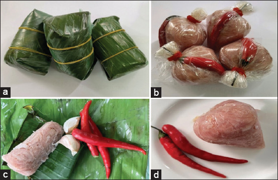 Figure 1
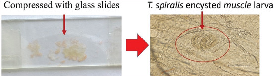 Figure 2
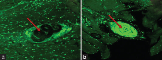 Figure 3
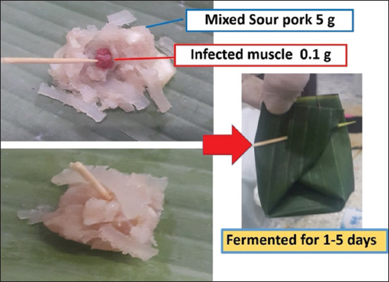 Figure 4
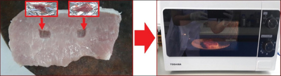 Figure 5
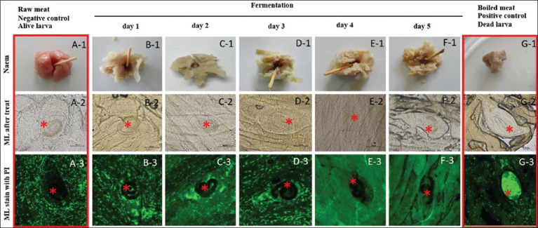 Figure 6
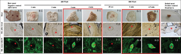 Figure 7
| Fermentation time | Groups | Total number of counts (Mean ± standard deviation) | Percentage of dead larvae | Percentage of survival rate |
|---|---|---|---|---|
| Day 1 | Sour pork | 8.33 ± 0.58 | 0 | 100 |
| Raw infected muscle | 7.67 ± 0.58 | 0 | 100 | |
| Boiled infected muscle | 9.33 ± 0.58 | 100 | 0 | |
| Day 2 | Sour pork | 8.33 ± 1.53 | 0 | 100 |
| Raw infected muscle | 8.33 ± 0.58 | 0 | 100 | |
| Boiled infected muscle | 6.67 ± 0.58 | 100 | 0 | |
| Day 3 | Sour pork | 8 ± 1 | 0 | 100 |
| Raw infected muscle | 9.67 ± 0.58 | 0 | 100 | |
| Boiled infected muscle | 9 ± 0 | 100 | 0 | |
| Day 4 | Sour pork | 8 ± 0 | 0 | 100 |
| Raw infected muscle | 8.33 ± 0.58 | 0 | 100 | |
| Boiled infected muscle | 8.67 ± 0.58 | 100 | 0 | |
| Day 5 | Sour pork | 8 ± 1 | 0 | 100 |
| Raw infected muscle | 9 ± 0 | 0 | 100 | |
| Boiled infected muscle | 8.67 ± 0.58 | 100 | 0 |
| Power of the microwave (Watt) | Time (min) | Total number of counts (Mean ± standard deviation) | Percentage of dead larvae |
|---|---|---|---|
| 400 | 1 | 8 ± 1.73 | 0 |
| 2 | 8 ± 2.65 | 6.67 | |
| 3 | 10.33 ± 3.51 | 100 | |
| 4 | 7 ± 1 | 100 | |
| 800 | 0.5 | 6.67 ± 0.58 | 0 |
| 1 | 6.67 ± 2.51 | 100 | |
| 4.5 | 7.67 ± 1.53 | 100 | |
| Positive control (boiled infected muscle) | 10 | 7.67 ± 2.52 | 100 |
| Negative control (raw infected muscle) | 6.67 ± 1.53 | 0 |
Peer Reviews
No public reviews on file for this paper yet. If you reviewed it on a platform where reviews are public (OpenReview, ICLR, NeurIPS, ICML), you can paste yours below so the community can read it here.
Videos
No videos yet. Explain this paper in a talk, walkthrough, or lecture? Add one.
Taxonomy
TopicsFood Allergy and Anaphylaxis Research · Parasitic Diseases Research and Treatment · Food Safety and Hygiene
INTRODUCTION
Trichinella, the etiological agent of trichinellosis, has been reported in human populations across 55 countries, representing 27.8% of nations globally. Infections with Trichinella spp. have also been docu-mented in domestic animals – primarily pigs – in 43 countries (21.9%) and in wildlife in 66 countries (33.3%) [1]. Human trichinellosis primarily arises from the consumption of raw or undercooked meat derived from infected domestic pigs and wild boars containing encysted larvae [2, 3]. Clinical manifestations vary with the stage of infection and typically include diarrhea, facial edema, periorbital hemorrhage, and myalgia [4–6]. The severity of disease is influenced by the infective larval dose and the host’s immune response [7]. Notably, the global incidence of outbreaks has increased in recent years, suggesting a shift in the epidemiological landscape. This rise is largely attributed to the increasing consumption of wild boar meat and the continued adherence to traditional dietary customs that involve raw pork. In Southeast Asia, traditional sour fermented pork dishes remain widely consumed and are readily available in local markets (Figure 1). While such fermented foods offer nutritional and organoleptic value, they may also act as vehicles for foodborne para-sitic infections. In response, international efforts have been initiated to improve the microbiological safety of fermented products. It is commonly assumed that the acidic conditions generated during fermentation may neutralize Trichinella larvae, similar to the reduction in viability of Opisthorchis viverrini observed in pickled fish preparations [8–10]. Nevertheless, trichinellosis continues to pose a significant public health concern, especially in areas where raw meat consumption rem-ains prevalent.
(a–d) A traditional pickled pork or sour fermented pork.
Despite longstanding awareness of Trichinella spiralis as a globally distributed zoonotic parasite, its survival in culturally significant fermented pork products remains inadequately explored. Traditional lactic acid fermentation is widely presumed to reduce the viability of various foodborne parasites due to the lowered pH and microbial competition. However, unlike extensively studied liver fluke species such as O. viverrini, there is limited empirical evidence regarding the persistence of T. spiralis larvae under real-world fermentation conditions commonly practiced in Southeast Asia. Moreover, most prior investigations have focused on Western meat preservation techniques, such as curing, freezing, and salting, leaving a critical knowledge gap regarding indigenous preparation methods that do not involve thermal treatment. In regions where raw or under-fermented pork is routinely consumed, the assumption that short-term fermentation alone ensures safety may contribute to the underestimation of zoonotic transmission risks. In addition, although thermal inactivation of T. spiralis has been well docum-ented using conventional cooking, there is a lack of standardized guidance on the use of microwave heating, a ubiquitous but often inconsistently applied method in domestic settings. These gaps hinder evidence-based public health recommendations and expose populations to preventable parasitic infections.
This study aims to (i) evaluate the viability of T. spiralis muscle larvae during traditional sour pork fermentation over a 5-day period and (ii) determine the efficacy of microwave heating at varying power levels and exposure durations as a method for larval inactivation. By simulating real-life culinary practices in Southeast Asia, the study aims to determine whether traditional fermentation can independently neutralize encysted larvae and to what extent microwave treat-ment provides a reliable alternative for enhancing food safety. The findings will contribute to a more accurate assessment of zoonotic risks associated with the consumption of fermented pork and support the development of culturally appropriate, science-based food safety guidelines.
MATERIALS AND METHODS
Ethical approval
This study was approved by the Institutional Animal Care and Use Committee of Khon Kaen University, Thailand (ACUC-KKU-51/2561).
Study period and location
This study was conducted from May 2019 to June 2023 at the Department of Parasitology, Faculty of Medicine, Khon Kaen University, Thailand.
Animal infection and maintenance
Laboratory-bred hamsters were obtained from the Animal Facility, Faculty of Medicine, Khon Kaen University, Khon Kaen, Thailand. To produce encysted muscle larvae, each hamster was orally administered 200 viable T. spiralis larvae suspended in 0.2 mL of physiological saline. The infected animals were main-tained for 45 days post-infection at the animal house of the same faculty [11–13]. Euthanasia was performed through inhalation of 1%–3% isoflurane, adhering to the Institutional Animal Ethics Guidelines.
Preparation of T. spiralis-infected muscle tissue
Following euthanasia, skeletal muscles from infected hamsters were excised, finely minced, homogenized, and weighed using a precision digital balance. A 10 mg aliquot of infected muscle was subjected to digestion using artificial gastric juice (1% hydrochloric acid and 1% pepsin) to quantify larval burden. In addition, 100 mg samples of infected tissue were compressed between glass slides and examined microscopically to enumerate encysted larvae (Figure 2). Verified infected muscle samples were allocated to experimental and control groups accordingly.
Trichinella spiralis encysted muscle larva in minced infected muscle specimens subjected to compression using glass slides and observation under a microscope.
Viability controls and propidium iodide staining
Larval viability was determined using propidium iodide staining as described by Boueroy et al. [14]. Infected muscle specimens were incubated with 0.25 mg/mL propidium iodide (Invitrogen, USA) for 30 min, followed by confocal microscopic analysis. Larvae exhibiting uptake of the stain were classified as non-viable (dead), whereas those without staining were considered viable (Figure 3).
A negative (a - alive larva) and positive (b - dead larva) control of infected muscle post propidium iodide staining. Arrow - larva.
Preparation of traditional fermented pork and larval viability assessment
Traditional sour pork was prepared using a standardized formulation: 400 g minced pork, 300 g pigskin, 25 g salt, 100 g cooked sticky rice, and 120 g garlic. Each 5 g portion of the meat mixture was supplemented with 100 mg of infected muscle, wrapped in banana leaf, and fermented under ambient conditions (temperature 28°C–30°C; relative humidity 60%–80%) for 5 days (Figure 4). Non-fermented infected muscle served as the negative control, and boiled infected muscle was used as the positive control. All groups were evaluated in triplicate. Daily assessment of larval viability was conducted through propidium iodide staining and confocal microscopy. Infected tissues were stained for 30 min with 0.25 mg/mL propidium iodide, and larvae were classified as viable or non-viable based on staining results.
A traditional fermented meat covered with banana leaf.
Microwave heating of infected muscle tissue
To evaluate the impact of microwave heating on larval viability, pork slices were cut to a uniform thickness of 0.5 inches. Each slice was modified to include two central cavities (0.25 inches deep), which were filled with 100 mg of T. spiralis-infected minced muscle. The cavities were then sealed with additional pork slices (Figure 5). Samples were microwaved at 400 W for 1, 2, 3, or 4 min, and at 800 W for 30 s, 1 min, or 4.5 min. Following treatment, 100 mg of muscle from each group was stained with 0.25 mg/mL propidium iodide to evaluate larval viability. The untreated infected muscle served as the negative control, while the boiled infected muscle functioned as the positive control. All experimental treatments and controls were performed in triplicate to ensure reproducibility.
The piece of pork with hole for containing infected muscle (red box) for microwave heating.
RESULTS
Viability of T. spiralis larvae during traditional fermented pork preparation
Microscopic examination revealed that all infected muscle samples contained approximately 6–9 encysted larvae per 100 mg of tissue, as detailed in Table 1. Negative control group consisted of raw infected muscle (Figures 6A-1 and 6A-2). The larvae did not absorb propidium iodide, indicating that they were viable (Figure 6a-3). Conversely, larvae in the positive control group (Figures 6G-1 and 6G-2) displayed clear propidium iodide uptake, confirming non-viability (Figure 6g-3). Throughout the 5-day fermentation period (Figures 6B1–6F3), larvae in all experimental groups failed to absorb the stain, demonstrating persistent viability. These results were consistent with those observed in the negative control group, suggesting that traditional fermentation conditions did not compromise larval survival.
A-1 to G-3: Representative encysted muscle larvae of Trichinella spiralis in pickled pork at various time points. The Red Box represents both the negative and positive control groups, while the Red Star indicates the T. spiralis larvae.
Larval viability following microwave heating
As shown in Table 2, the number of encysted larvae remained comparable across all microwave-treated groups. In the raw infected muscle (negative control) (Figures 7A-1 and 7A-2), no propidium iodide staining was observed, indicating larval viability (Figure 7a-3). Similarly, microwave heating at 400 W for 1 min (Figures 7B-1 and 7B-2), and 2 min (Figures 7C-1 and 7C-2), did not result in staining (Figures 7B-3 and 7C-3), indicating that the larvae remained viable under these conditions. However, exposure to 400 W for 3 min (Figures 7D-1, 7D-2 and 7E-1, 7E-2) induced clear propidium iodide staining in the larvae (Figures 7D-3 and 7E-3), signifying effective inactivation. Heating at 800 W for 30 sec did not result in any staining (Figures 7F-1, 7F-2, and 7F-3). Furthermore, extending the heating duration to 1 min (Figures 7G-1 and 7G-2) and 4.5 min (Figures 7H-1 and 7H-2) resulted in complete larval staining (Figures 7G-3 and 7H-3), thereby confirming successful inactivation. In contrast, larvae from the positive control group (boiled muscle) (Figures 7I-1 and 7I-2) also showed complete staining (Figure 7i-3), validating the staining method.
A-1 to I-3: Representative encysted muscle larvae of Trichinella spiralis after heating by microwave at various time points. The Red Box represents experimental treatment groups induced with clear propidium iodide, while the Red Star indicates the T. spiralis larvae.
DISCUSSION
Survivability of T. spiralis in fermented pork products
This study is the first to provide empirical evi-dence that traditional lactic fermentation of pork for up to 5 days is insufficient to inactivate encysted T. spiralis muscle larvae. This conclusion is supported by the absence of propidium iodide staining, which indicates that larval viability was maintained throughout the fermentation process. In contrast, microwave heating for short durations was found to effectively inactivate the larvae in pork slices of 0.5-inch thickness.
Public health risks of trichinellosis and cultural food practices
Trichinellosis, also known as trichinosis, is a parasitic infection caused by nematodes of the genus Trichinella. Human infection typically results from the consumption of undercooked or raw meat, most commonly pork, that harbors encysted larvae. T. spiralis is among the most prevalent species causing human disease through the ingestion of infected meat.
In Southeast Asia, traditional sour fermented pork – a semi-dry, lactic-acid fermented sausage – is a commonly consumed delicacy, particularly in Thailand. This dish is prepared by mixing minced raw pork with sticky rice, garlic, salt, and other seasonings. The lactic fermentation process, often employing <20% salt and using rice as the primary carbohydrate source, is also widely used for fish and other meats in the region. However, this method does not involve heat treatment, which poses a significant risk for foodborne parasitic infections. Consumption of such uncooked fermented pork has been associated with outbreaks of Taenia and Trichinella infections globally [15, 16]. This risk parallels that posed by pickled fish (Figure 1), a known trans-mission route for O. viverrini and a major contributing factor to cholangiocarcinoma in Southeast Asia [17].
Role of fermentation parameters and parasite inactivation
Despite the longstanding cultural acceptance, the consumption of raw meat remains a health hazard. Public health education is thus crucial to raise awareness of the risks associated with these traditional practices. The antimicrobial effects of fermentation depend heavily on factors such as acidity and salinity. The current findings indicate that fermentation durations of 5 days or less do not affect the viability of T. spiralis larvae. In contrast, Hill et al. [18] reported complete inactivation of T. spiralis under more stringent conditions involving a low pH (<5.2) and extended curing for ≥10 days with 1.3%–2.8% sodium chloride.
Similarly, Oh et al. [19] demonstrated the inactivation of Anisakis spp. larvae in salt-fermented squid when stored in 15% NaCl for 7 days or 20% NaCl for 6 days. However, substantial viability was still observed in lower salt concentrations: 81.7% in 5% NaCl and 26.7% in 10% NaCl after 7 days of storage. In a related study, Rodriguez-Canul et al. [20] showed that Taenia solium cysticerci were rendered inactive following salt pickling of infected pork.
Variability in fermentation efficacy across parasite species
Previous studies have shown that fermentation extending beyond 3 days can successfully eliminate O. viverrini and other intestinal flukes from cyprinid fish [9, 10]. However, the effectiveness of lactic fermentation as a decontamination method may vary depending on the parasite species and the structural resilience of their infective stages. Ducrocq et al. [21] highlighted that individuals consuming raw or undercooked meat face a 1.2–1.3-fold higher risk – and 1.7–3.0-fold higher odds – of Toxoplasma gondii infection compared to those who thoroughly cook meat, regardless of the animal species.
CONCLUSION
This study provides compelling evidence that traditional lactic fermentation of pork for up to 5 days does not inactivate encysted T. spiralis larvae. The viability of larvae throughout the fermentation process was confirmed by the absence of propidium iodide staining. In contrast, microwave heating at 400 W for at least 3 min or at 800 W for 1 min effectively inactivated larvae in pork slices of 0.5-inch thickness, as evidenced by complete larval staining.
These findings have critical practical implications for food safety in regions where traditional raw or under-fermented pork dishes are routinely consumed. Reliance on short-duration fermentation as a standalone method of parasite inactivation is insufficient and may perpetuate the risk of zoonotic transmission. Incorp-orating microwave heating as a rapid and accessible intervention could significantly reduce the risk of infe-ction in domestic settings.
A major strength of this study is its simulation of real-world culinary practices and its use of stand-ardized viability assays under both fermented and thermally treated conditions. The application of confocal microscopy, combined with propidium iodide staining, offered a reliable and sensitive approach for distinguishing between viable and non-viable larvae.
However, the study has certain limitations. First, it focused solely on a 5-day fermentation period and did not assess extended durations or variations in pH and salt concentration that may influence larval viability. Second, the microwave heating experiments were limited to specific power settings and pork thicknesses, which may not encompass all household cooking scenarios. In addition, only T. spiralis was evaluated, and the generalizability to other Trichinella species or foodborne helminths remains uncertain.
Future research should explore the interaction of fermentation parameters (e.g., pH, NaCl concentration, and fermentation duration) with larval viability and investigate the efficacy of other non-thermal preservation techniques. Expanding inactivation trials to include other Trichinella species and various fermented meat matrices will also enhance risk assessment models and inform public health recommendations.
While traditional fermented pork remains a val- ued culinary practice, it poses an underrecognized parasitic risk if not properly handled. This study highlights the urgent need for culturally sensitive food safety education and provides an evidence-based thermal guideline – microwave heating at ≥800 W for 1 min – to ensure the microbiological safety of fermented pork products.
AUTHORS’ CONTRIBUTIONS
AA, BP, SK, PB, and PL: Data curation, investigation, methodology, and formal analysis. AA and TB: Conceptualization and project administration. TB and PR: Supervision and visualization. AA, TB, and PR: Drafted, reviewed, and revised the manuscript. All authors have read and approved the final manuscript.
The reference list from the paper itself. Each links out to its DOI / PubMed record.
- 1Pozio E World distribution of Trichinella spp. Infections in animals and humans Vet. Parasitol 2007149(1-2)3211768919510.1016/j.vetpar.2007.07.002 · doi ↗ · pubmed ↗
- 2Dupouy-Camet J Raffetin A Rosca E.C Yera H Clinical picture and diagnosis of human trichinellosis In:Trichinella and trichinellosis 2021 Cambridge Academic Press 333352
- 3Khurana S Datta P Sharma B Singh C Mewara A Johnson N Pilania R.K Singh S Sehgal R Clinical and laboratory profile of trichinellosis from a non-endemic country Indian J. Med. Microbiol 20213922352393365203910.1016/j.ijmmb.2021.02.004 · doi ↗ · pubmed ↗
- 4Pavic S Andric A Sofronic-Milosavljevic L.J Gnjatovic M MitićI Vasilev S Sparic R Pavic A Trichinella britovi outbreak:Epidemiological, clinical, and biological features Med. Mal. Infect 20205065205243173224210.1016/j.medmal.2019.10.008 · doi ↗ · pubmed ↗
- 5Caron Y Bory S Pluot M Nheb M Chan S Prum S.H Lim S.B.H Sim M Sengdoeurn Y Sovann L Khieu V Vallée I Yera H Human outbreak of trichinellosis caused by Trichinella papuae nematodes, central Kampong Thom province, Cambodia Emerg. Infect. Dis 2020268175917663268702210.3201/eid 2608.191497 PMC 7392432 · doi ↗ · pubmed ↗
- 6Cash-Goldwasser S. Ortbahn, D Narayan M Fitzgerald C Maldonado K Currie J Straily A Sapp S Bishop H.S Watson B Neja M Qvarnstrom Y Berman DM Holzbauer S Outbreak of human trichinellosis - Arizona, Minnesota, and South Dakota Morb. Mortal. Wkly. Rep 2024732045645910.15585/mmwr.mm 7320 a 2PMC 1111543638781100 · doi ↗ · pubmed ↗
- 7Wang N Bai X Tang B Yang Y Wang X Zhu H Luo X Yan H Jia H Liu M Liu X Primary characterization of the immune response in pigs infected with Trichinella spiralis Vet. Res 2020511173208580810.1186/s 13567-020-0741-0PMC 7035712 · doi ↗ · pubmed ↗
- 8Aukkanimart R Boonmars T Sriraj P Sripan P Songsri J Ratanasuwan P Laummaunwai P Suwanantrai A Aunpromma S Khueangchaingkhwang S Pumhirunroj B Artchayasawat A Khuntikeo N Loilome W Namwat N Yongvanit P Boonjaraspinyo S Carcinogenic liver fluke and others contaminated in Pickled Fish of Northeastern Thailand Asian Pac. J. Cancer Prev 20171825295332834584110.22034/APJCP.2017.18.2.529PMC 5454754 · doi ↗ · pubmed ↗