Stabilization of a recalcitrant and aggressive necrobiotic xanthogranuloma with tofacitinib
Miguel Mansilla-Polo, Carlos Abril-Pérez, Daniel Martín-Torregrosa, Rafael Botella-Estrada

Abstract
Genes, proteins, chemicals, diseases, species, mutations and cell lines named across the full text — each resolved to its canonical identifier and authoritative record.
Click any figure to enlarge with its caption.
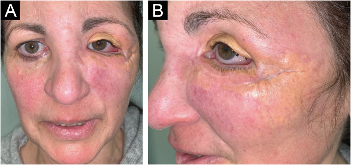 Figure 1
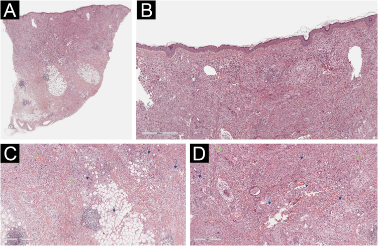 Figure 2
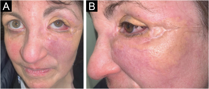 Figure 3
Peer Reviews
No public reviews on file for this paper yet. If you reviewed it on a platform where reviews are public (OpenReview, ICLR, NeurIPS, ICML), you can paste yours below so the community can read it here.
Videos
No videos yet. Explain this paper in a talk, walkthrough, or lecture? Add one.
Taxonomy
TopicsHistiocytic Disorders and Treatments · Infectious Disease Case Reports and Treatments · Autoimmune and Inflammatory Disorders
Dear Editor,
Necrobiotic Xanthogranuloma (NXG) is a form of non-Langerhans histiocytosis characterized by the development of yellow-orange infiltrated papules and plaques, with the most common location being the periorbital area. Occasionally, there is extracutaneous involvement, including ocular, respiratory, cardiac, or cerebral. Up to 80% of cases are associated with monoclonal gammopathy and other neoplasms.1 The treatment of NXG is complex and data on therapeutic options are limited, making the best approach to treatment unclear. We describe the case of a patient with refractory NXG who experienced an adequate response to tofacitinib, a non-selective inhibitor of Janus Kinases (JAK), which preferentially blocks JAK1 and JAK3.
A 54-year-old woman presented to the Department of Dermatology with refractory NXG. Specifically, the lesions had appeared at 18‒20 years of age (>30 years of evolution), and throughout her life, she had been treated with topical, intralesional, and systemic corticosteroids, methotrexate, multiple surgeries, CO_2_ laser, hydroxychloroquine, extracorporeal photochemotherapy, rituximab, Intravenous Immunoglobulins (IVIG), and cyclophosphamide. Despite the latter treatment (cyclophosphamide), combined with systemic corticosteroids, the clinical condition was progressive. There was no other cutaneous or systemic involvement, and laboratory tests, including protein electrophoresis, were normal. On examination (Fig. 1), yellowish plaques of bilateral palpebral and periocular involvement were observed, predominantly on the left side. The lesions had a shiny surface and a slightly erythematous surrounding edge. They were hard, deeply adherent, and caused some visual restriction in the left eye. The latest biopsy (Fig. 2) and all previous ones were compatible with NXG with deep involvement (muscular invasion), also confirmed by ultrasound and Magnetic Resonance Imaging (MRI). Given the refractory clinical condition, active lesions, and multiple previous treatments, it was decided by a multidisciplinary team to initiate tofacitinib therapy, initially at a dose of 5 mg/12 hours, and due to good tolerance, the dose was increased to 10 mg/12 hours after 3 months. After one year of treatment (Fig. 3), the lesions had stabilized, and even the size of some of them had reduced, especially the left upper eyelid, where the visual restriction had improved. They were also less evident on palpation, and the surrounding rim was less erythematous. The lesions remained the same at the 18-month review. Tolerability during the 1.5 years of treatment was adequate, except for an upper respiratory condition that required discontinuation of treatment for a few days at 10 months of treatment, during which time he reported worsening of the lesions. There were no other adverse effects, and follow-up laboratory tests were normal. On follow-up brain MRI at 1.5 years, the lesions remained stable. Given the adequate therapeutic response, it was decided to continue treatment.Fig. 1. Clinical presentation of the lesions before starting tofacitinib.Yellowish, shiny, and infiltrated plaques located around the eyelids and periocular region bilaterally, with more pronounced involvement in the left eye, where they caused visual restriction. Surrounding erythema around the plaques.Fig. 1. Fig. 2Histological examination of the left lower eyelid lesion (Hematoxylin & eosin; panel A 20×, panel B 80×, panel C 140×, panel D 160×).Histology showed collagenous fibrous tissue in the dermis, interspersed with increased lymphohistiocytic cellularity with abundant foamy histiocytes (blue arrows in panels C and D) and multinucleated giant cells (green arrows in panels C and D).Fig. 2. Fig. 3Clinical presentation of the lesions one year after starting tofacitinib.Improvement of the lesions, with a slightly smaller diameter, less active border, and improvement in left visual restriction. Similarly, upon palpation, the lesions felt less infiltrated.Fig. 3
The etiology of NXG, which is thought to be related to altered macrophage lipid homeostasis, has not yet been elucidated. Recently, ABCG5/G8 variants have been identified that encode heterodimers containing transporter and transmembrane domains essential for reverse cholesterol transport and sterol clearance.1 The accumulation of lipid particles can lead to the upregulation of pro-inflammatory cytokines such as Interleukin (IL) 6 and tumor necrosis factor-alpha. Animal studies have shown that JAK inhibitors can prevent IL-6 trans-signaling, which may explain the efficacy of tofacitinib on NXG in our patients.2 Similarly, the JAK pathway has been reported to play a role in the maintenance of granulomas. Thus, JAK inhibition may promote granuloma resolution, as has been described in other granulomatous diseases such as granuloma annulare3 and sarcoidosis.3, 4 On the other hand, treatment of NXG is challenging and the best approach to treatment is uncertain. Randomized clinical trials are lacking, so most recommendations are based on studies with limited evidence. Before starting treatment, it is essential to rule out monoclonal gammopathy and associated malignancy.5, 6 Generally, the first-line therapy consists of alkylating agents such as chlorambucil, cyclophosphamide, or melphalan, administered with or without systemic glucocorticoids. Other treatments used include IVIG, thalidomide, lenalidomide, interferon α-2a, plasmapheresis, dapsone, antimalarials, phototherapy, azathioprine, methotrexate, or rituximab. For localized lesions, local treatments such as surgery or CO_2_ laser therapy can be employed.5, 6, 7 Our patient had undergone most of these treatments and still had refractory lesions with progressive growth. For this reason, and based on the reported efficacy of tofacitinib in isolated cases in granulomatous diseases, including histiocytosis such as necrobiotic xanthogranuloma2 and multicentric reticulohistiocytosis,8 and non-histiocytosis such as granuloma annulare3 or sarcoidosis,3, 4 it was decided to initiate treatment with tofacitinib. Although caution must be exercised when drawing conclusions from the treatment of isolated cases, the efficacy of tofacitinib observed in our patient, a refractory case, suggests it is a therapeutic option for such NXG cases. However, it should be noted that JAK inhibitors have been associated with numerous side effects, including infections (especially herpes zoster, respiratory infections), cardiovascular events (thrombosis, infarction), hematological changes, and a possible increased risk of neoplasia.9, 10 Therefore, their use should always be justified, and safer therapeutic alternatives should be offered to the patient if available.
Financial support
None declared.
Authors’ contributions
Miguel Mansilla-Polo: Managed clinical treatment and procedures; contributed to the development of this paper, had access to the data, and played a role in writing this manuscript.
Carlos Abril-Pérez: Managed clinical treatment and procedures; contributed to the development of this paper, had access to the data, and played a role in writing this manuscript.
Daniel Martín-Torregrosa: Managed clinical treatment and procedures; contributed to the development of this paper, had access to the data, and played a role in writing this manuscript.
Rafael Botella-Estrada: Supervised the work, had access to the data, and played a role in writing this manuscript.
Conflicts of interest
None declared.
The reference list from the paper itself. Each links out to its DOI / PubMed record.
- 1Chen P.Y.Tang Z.L.Hong Y.Y.Hsieh I.J.Li Z.Y.Zhou J.Genetic aspects of necrobiotic xanthogranuloma JAMA Dermatol 1602024112511283923088210.1001/jamadermatol.2024.2921 PMC 11375517 · doi ↗ · pubmed ↗
- 2Shahriari N.Mazori D.R.Merola J.F.Intravenous immunoglobulin-refractory necrobiotic xanthogranuloma successfully treated with tofacitinib 2% cream JAAD Case Rep 32202241433663256210.1016/j.jdcr.2022.12.002PMC 9826807 · doi ↗ · pubmed ↗
- 3Damsky W.Thakral D.Mc Geary M.K.Leventhal J.Galan A.King B.Janus kinase inhibition induces disease remission in cutaneous sarcoidosis and granuloma annulare J Am Acad Dermatol 8220206126213118523010.1016/j.jaad.2019.05.098PMC 7590533 · doi ↗ · pubmed ↗
- 4Damsky W.Thakral D.Emeagwali N.Galan A.King B.Tofacitinib treatment and molecular analysis of cutaneous sarcoidosis N Engl J Med 3792018254025463058651810.1056/NEJ Moa 1805958 PMC 6351852 · doi ↗ · pubmed ↗
- 5Nelson C.A.Zhong C.S.Hashemi D.A.Ashchyan H.J.Brown-Joel Z.Noe M.H.A multicenter cross-sectional study and systematic review of necrobiotic xanthogranuloma with proposed diagnostic criteria JAMA Dermatol 15620202702793194000010.1001/jamadermatol.2019.4221 PMC 6990734 · doi ↗ · pubmed ↗
- 6Geoloaica L.G.Pătraşcu V.Ciurea R.N.Necrobiotic xanthogranuloma - case report and literature review Curr Health Sci J 47202112613110.12865/CHSJ.47.01.2134211760 PMC 8200611 · doi ↗ · pubmed ↗
- 7Hansen I.Ghandili S.Abeck F.Booken N.Schneider S.W.Therapy of necrobiotic xanthogranuloma - case series and review of the literature J Dtsch Dermatol Ges 2120231315131810.1111/ddg.1519537811744 · doi ↗ · pubmed ↗
- 8Bruscas Izu C.Hörndler Argarate C.García Latasa de Araníbar F.J.Multicentric reticulohistiocytosis: a case report treated with tofacitinib Med Clin (Barc)15620213103113217307110.1016/j.medcli.2019.12.011 · doi ↗ · pubmed ↗