Isolated Ophthalmoplegia as an Atypical Manifestation of Miller Fisher Syndrome Post-viral Conjunctivitis
Christian John S Capirig, Celine Garcia, Gerard Francis Mangubat

TL;DR
A rare case of Miller Fisher Syndrome presented with isolated eye movement issues after viral conjunctivitis, highlighting the importance of early diagnosis and treatment.
Contribution
This case report highlights an atypical presentation of Miller Fisher Syndrome with isolated ophthalmoplegia and emphasizes the role of anti-GQ1b IgG testing in diagnosis.
Findings
The patient exhibited complete bilateral ophthalmoplegia without ataxia or areflexia, an atypical MFS presentation.
Elevated anti-GQ1b IgG titers confirmed the diagnosis despite absence of the typical clinical triad.
Intravenous immunoglobulin therapy led to rapid and complete recovery of symptoms.
Abstract
Miller Fisher syndrome (MFS) is an uncommon variant of Guillain-Barré syndrome, typically identified by the presence of ophthalmoplegia, ataxia, and areflexia. While most cases follow this clinical pattern, atypical presentations lacking one or more components can complicate diagnosis. We report the case of an 81-year-old male with well-controlled hypertension who presented with acute-onset horizontal diplopia two weeks after a self-limiting episode of viral conjunctivitis. Neurological examination revealed complete bilateral ophthalmoplegia with preserved pupillary reflexes and no other focal deficits. Brain imaging and CSF analysis were unremarkable, with no evidence of albuminocytologic dissociation. A comprehensive serologic and autoimmune workup was negative, except for markedly elevated anti-GQ1b IgG titers (>1:12,800). The patient was diagnosed with an atypical form of MFS and…
Genes, proteins, chemicals, diseases, species, mutations and cell lines named across the full text — each resolved to its canonical identifier and authoritative record.
Click any figure to enlarge with its caption.
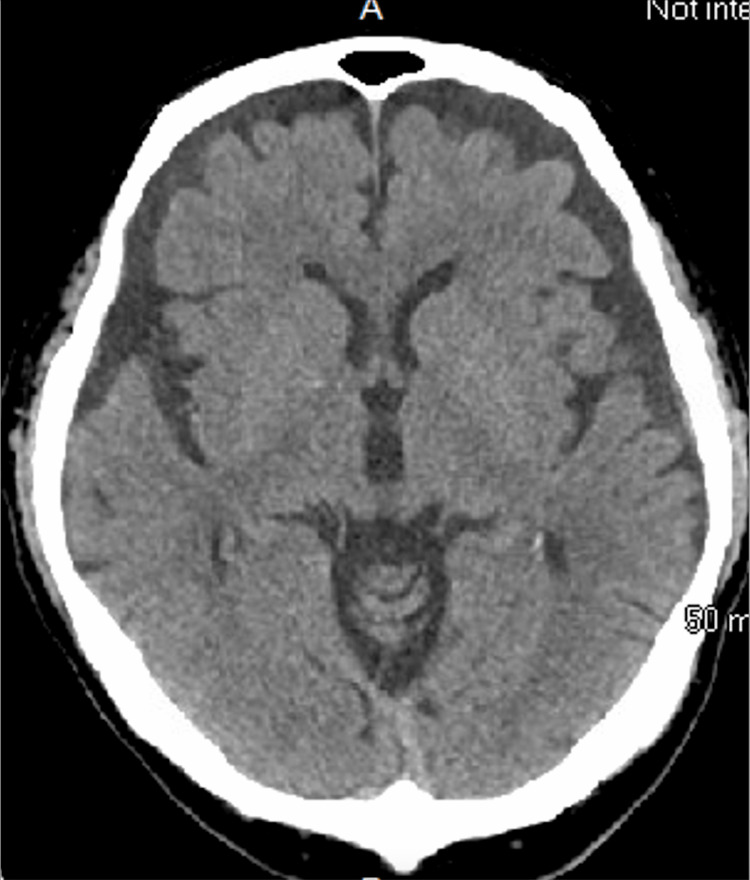 Figure 1
 Figure 2
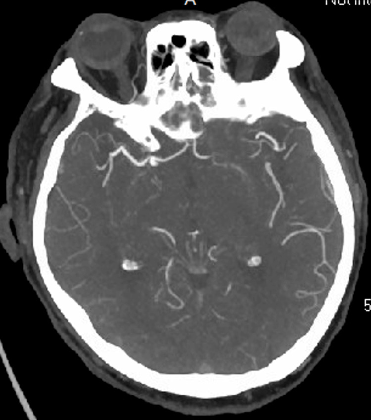 Figure 3
| Test | Result | Reference range | Units |
| WBC count | 10.31 | 3.80-10.80 | × 10³/µL |
| RBC count | 5.09 | 4.00-6.20 | × 10⁶/µL |
| Hemoglobin | 14.1 | 13.7-17.5 | g/dL |
| Hematocrit | 44 | 40.1-51.0 | % |
| MCV | 86.4 | 79.4-98.4 | fL |
| MCH | 27.7 | 26.0-34.0 | pg |
| MCHC | 32 | 32.0-36.0 | g/dL |
| RDW | 13.9 | 11.6-14.4 | % |
| Platelet count | 254 | 151-424 | × 10³/µL |
| Absolute neutrophils | 6.51 ↑ | 1.56-6.20 | × 10³/µL |
| Absolute monocytes | 0.95 ↑ | 0.24-0.82 | × 10³/µL |
| Test | Result | Reference range | Units |
| Glucose | 137 ↑ | 70-99 | mg/dL |
| BUN | 20 | 6-23 | mg/dL |
| Creatinine | 1 | 0.6-1.4 | mg/dL |
| eGFR (CKD-EPI 2021) | 76 | ≥90 | mL/min/1.73 m² |
| Sodium | 138 | 133-145 | mEq/L |
| Potassium | 4.7 | 3.3-5.1 | mEq/L |
| Chloride | 102 | 95-108 | mEq/L |
| CO₂ (bicarbonate) | 23 | 21-30 | mEq/L |
| Anion Gap | 18 | 14-20 | mEq/L |
| Calcium | 9.6 | 8.3-10.5 | mg/dL |
| AST (SGOT) | 21 | 0-40 | IU/L |
| ALT (SGPT) | 29 | 0-41 | IU/L |
| Alkaline phosphatase | 73 | 35-129 | IU/L |
| Total bilirubin | 0.6 | 0-1.2 | mg/dL |
| Total protein | 8.3 | 6.4-8.3 | g/dL |
| Albumin | 4.3 | 3.5-5.2 | g/dL |
| Test | Result | Reference range | Units |
| CSF color | Colorless | Colorless | - |
| CSF appearance | Clear | Clear | - |
| CSF supernatant | Colorless/clear | Colorless | - |
| CSF RBC | 143 | 0 | cells/µL |
| CSF WBC | 8 | 0-5 | cells/µL |
| CSF protein | 24 | 15-45 | mg/dL |
| CSF glucose | 78 | 40-70 | mg/dL |
| CSF Gram stain | No organisms seen | No cells | - |
| CSF culture | No growth after five days | No growth | - |
| Test | Result | Reference range | Units |
| CRP | 11.7 ↑ | 0.0-10.0 | mg/L |
| ESR | 54 ↑ | 0-20 | mm/hr |
| CK, total | 113 | 39-308 | IU/L |
| Vitamin B1 | 117 | 78-185 | nmol/L |
| Vitamin B1 (repeat) | 163 | 78-185 | nmol/L |
| RPR | Nonreactive | Nonreactive | - |
| Cryoglobulins | Negative | Negative | - |
| ANA | Negative | Negative | - |
| ANCA | Negative | Negative | - |
| Anti-dsDNA | <1 | <1 | - |
| Anti-SSA/Ro | <0.2 | ≤0.9 Negative | - |
| Anti-SSB/La | <0.2 | ≤0.9 Negative | - |
| C3 complement | 135 | 90-180 | mg/dL |
| C4 complement | 34 | 10-40 | mg/dL |
| ACE | 5 ↓ | 9-67 | - |
| MOG FACS | Negative | Negative | - |
| NMO/AQP4 FACS | Negative | Negative | - |
| Striated Muscle Ab | Negative | Negative | - |
| Anti-glial nuclear Ab | Negative | Negative | - |
| Amphiphysin Ab | Negative | Negative | - |
| Anti-neuronal nuclear Ab type 1 | Negative | Negative | - |
| Anti-neuronal nuclear Ab type 2 | Negative | Negative | - |
| Anti-neuronal nuclear Ab type 3 | Negative | Negative | - |
| P/Q calcium channel Ab | 0 | - | nmol/L |
| CRMP-5 IgG | Negative | Negative | - |
| Purkinje cell Ab type 1 | Negative | Negative | - |
| Purkinje cell Ab type 2 | Negative | Negative | - |
| Purkinje cell Ab type Tr | Negative | Negative | - |
| Neuronal VGKC Ab | 0 | - | nmol/L |
| Hepatitis B surface antigen | Negative | Negative | - |
| Hepatitis B core Ab (IgG/IgM) | Negative | Negative | - |
| Hepatitis B surface Ab | Negative | Negative | - |
| Hepatitis C antibody | Negative | Negative | - |
| HIV antigen/antibody | Negative | Negative | - |
| TSH | 1.36 | 0.4-4.5 | µIU/mL |
| Free T4 | 1.1 | 0.8-1.8 | ng/dL |
| Rheumatoid factor (RA) | <10 | <14 | IU/mL |
| GQ1b Ab (IgG) | >1:12,800 ↑ | <1:100 | Titer |
| GM1 Ab (IgG) | <1:800 | <1:800 | Titer |
| GM1 Ab (IgM) | <1:800 | <1:800 | Titer |
| GD1b Ab (IgM) | <1:800 | <1:800 | Titer |
| Asialo-GM1 Ab (IgM) | <1:1600 | <1:1600 | Titer |
| GD1a Ab (IgG) | <1:100 | <1:100 | Titer |
| GD1a Ab (IgM) | <1:800 | <1:800 | Titer |
| GD1b Ab (IgG) | <1:100 | <1:100 | Titer |
| Asialo-GM1 Ab (IgG) | <1:100 | <1:100 | Titer |
| MAG Ab (IgM) Western blot | Negative | Negative | - |
| Hu Ab screen | Negative | Negative | - |
Peer Reviews
No public reviews on file for this paper yet. If you reviewed it on a platform where reviews are public (OpenReview, ICLR, NeurIPS, ICML), you can paste yours below so the community can read it here.
Videos
No videos yet. Explain this paper in a talk, walkthrough, or lecture? Add one.
Taxonomy
TopicsPeripheral Neuropathies and Disorders · Hereditary Neurological Disorders · Long-Term Effects of COVID-19
Introduction
Miller Fisher syndrome (MFS) is a rare, immune-mediated variant of Guillain-Barré syndrome (GBS), first described by Charles Miller Fisher in 1956. It is classically characterized by a triad of ophthalmoplegia, ataxia, and areflexia [1]. Although MFS accounts for only 1-5% of GBS cases worldwide, it appears to be more prevalent in East Asian populations, with an estimated annual incidence of approximately 0.09 per 100,000 individuals [2].
The diagnosis of MFS is primarily clinical, based on characteristic neurological findings, and is further supported by specific serologic markers [3,4]. A key diagnostic indicator is the presence of anti-GQ1b IgG antibodies, which are detected in more than 85-90% of cases and reflect the syndrome’s underlying autoimmune mechanism [5]. Ancillary testing often includes CSF analysis, which may reveal albuminocytologic dissociation, elevated protein levels with a normal white cell count, as well as neuroimaging to exclude structural or vascular causes [2,4]. However, these findings may be absent in the early stages or in atypical variants of the disease [4,6].
We report a rare and diagnostically challenging case of atypical MFS, presenting as isolated ophthalmoplegia without accompanying ataxia or areflexia, following an episode of acute viral conjunctivitis. Despite the absence of the full clinical triad, the diagnosis was strongly supported by markedly elevated titers of anti-GQ1b IgG antibodies [5]. Extensive evaluation, including neuroimaging and CSF analysis, revealed no other abnormalities.
This case highlights the importance of maintaining a high index of suspicion for atypical MFS presentations, especially in patients with isolated cranial nerve deficits following recent viral illness. It also underscores the diagnostic utility of anti-GQ1b antibody testing in identifying MFS even in the absence of classic clinical features.
Case presentation
An 81-year-old male with a history of well-controlled hypertension presented to his primary care clinic with a two-day history of acute-onset horizontal diplopia. The diplopia was constant, binocular, and worsened on lateral gaze. He denied associated symptoms such as headache, vision loss, eye pain, photophobia, vertigo, dysphagia, nausea, vomiting, or limb weakness. Notably, he had experienced an episode of viral conjunctivitis approximately two weeks earlier, initially involving unilateral eye redness, tearing, and irritation, which progressed to bilateral involvement. These symptoms were accompanied by mild upper respiratory tract features, including rhinorrhea, sore throat, and low-grade fever, all of which resolved spontaneously within several days. He remained symptom-free until the onset of diplopia.
Given the acute cranial nerve involvement, he was referred to the emergency department for further evaluation to rule out intracranial pathology. His medical history was otherwise unremarkable, aside from hypertension managed with amlodipine 10 mg daily. He had no history of diabetes, cardiovascular disease, autoimmune conditions, or prior neurologic events. His surgical history was notable only for an appendectomy many years earlier. He was not taking any recent antibiotics or over-the-counter medications and denied any known drug allergies. He lived independently with his spouse, had retired from a career in teaching, and denied alcohol, tobacco, or illicit drug use. There was no recent travel, no known sick contacts, and no relevant family history of neurologic or autoimmune disease.
On physical examination, he was alert, oriented, and hemodynamically stable. Neurologic examination revealed complete bilateral ophthalmoplegia affecting all extraocular movements. His pupils were equal, round, and reactive to light, with preserved accommodation and no ptosis or anisocoria. Fundoscopic examination was normal, and there was no evidence of papilledema. Cranial nerves, aside from those controlling eye movement, were intact. His motor strength, reflexes, cerebellar testing, gait, and sensory examination were all within normal limits.
A comprehensive laboratory evaluation was conducted to investigate autoimmune, infectious, metabolic, and paraneoplastic causes of isolated ophthalmoplegia (Table 1, Table 2). CBC revealed only mild relative elevations in neutrophils and monocytes. CRP was mildly elevated at 11.7 mg/L (reference: 0-10), though this was nonspecific. Testing for rapid plasma reagin, cryoglobulins, hepatitis B and C, HIV, antinuclear antibody (ANA), antineutrophil cytoplasmic antibody, neuromyelitis optica/aquaporin 4 fluorescence-activated cell sorting, and myelin oligodendrocyte glycoprotein fluorescence-activated cell sorting returned negative results. Levels of vitamin B1, B12, thyroid-stimulating hormone, angiotensin-converting enzyme, and serum complements were within normal limits. Paraneoplastic panels and heavy metal screens were also unremarkable.
Non-contrast head CT (Figure 1), CT angiography of the head and neck (Figure 2), and brain MRI (Figure 3) were unremarkable, showing no evidence of infarcts, hemorrhage, mass lesions, or demyelinating changes. Lumbar puncture yielded clear CSF with normal opening pressure and no albuminocytologic dissociation (Table 3).
Non-contrast head CT scan showing no acute abnormalities
MRI of the brain demonstrating normal findings
CT angiogram of the head revealing no evidence of vascular abnormalities
Of note, anti-GQ1b IgG antibodies were strongly positive, with titers exceeding 1:12,800 (Table 4). Based on the clinical presentation and serologic findings, a diagnosis of atypical MFS was established, characterized by isolated ophthalmoplegia in the absence of areflexia, ataxia, or albuminocytologic dissociation. The patient was initiated on IVIG therapy at a dose of 0.4 g/kg/day for five consecutive days. He tolerated the treatment well and experienced no complications.
At follow-up one week after discharge, the patient reported complete resolution of diplopia. Neurologic examination confirmed full recovery of extraocular muscle function, and he remained symptom-free with no recurrence at subsequent outpatient visits.
Discussion
GBS is an immune-mediated polyradiculopathy and is the leading cause of acute flaccid paralysis globally [5,6]. Among its variants, MFS is characterized by the triad of ophthalmoplegia, ataxia, and areflexia. The global incidence of GBS is approximately 1-2 per 100,000 annually, whereas MFS represents a much smaller proportion, estimated at 1-2 cases per million [7,8]. MFS is more common in men, with a male-to-female ratio of 2:1 and a mean age of onset of 43.6 years. Its prevalence is higher in Asia (15-25% of GBS cases) compared to Western countries (1-7%) [7,9-11].
The classic MFS triad - ophthalmoplegia, ataxia, and areflexia - is seen in about 80% of cases. Similar to GBS, MFS is typically preceded by an infection. The interval between the preceding infection and the onset of neurological symptoms typically ranges from eight to 10 days but may vary from one to 30 days [7,12]. Diplopia is often the initial symptom, followed by the gradual development of ophthalmoplegia and ataxia, with areflexia occurring later in the disease course [7,10]. Less frequent symptoms accompanying the triad may include limb dysesthesias, ptosis, and cranial nerve involvement affecting the face, bulbar muscles, or pupils [7,11,13].
The diagnosis of MFS is supported by the clinical triad, a history of antecedent infection, and the presence of anti-ganglioside antibodies, most notably anti-GQ1b IgG, which is present in over 90% of patients and is highly specific for the condition [12-14]. Beyond the classic triad, atypical presentations of MFS have been reported in the literature [15]. These include bilateral internal ophthalmoplegia, unilateral oculomotor nerve paralysis, severe bilateral headache, and partial Parinaud’s syndrome, highlighting the clinical variability of the disorder.
Our case, featuring isolated bilateral ophthalmoplegia without ataxia or areflexia, represents an atypical variant of MFS. Such cases fall within the broader spectrum of anti-GQ1b antibody syndromes and have been described in previous literature. Due to the rarity of MFS and its typically self-limiting nature, limited data exist regarding treatment, and no randomized controlled trials have been published to date [7,14,16].
In our patient, laboratory abnormalities such as an elevated ESR (54 mm/hr) and mildly increased CRP (11.7 mg/L) suggested an underlying inflammatory process. Neuroimaging (brain MRI and CT angiography) was normal, ruling out central causes like stroke or mass lesions. Although CSF analysis did not show albuminocytologic dissociation, differential diagnoses - including myasthenia gravis, brainstem stroke, Wernicke’s encephalopathy, and paraneoplastic syndromes - were excluded based on imaging, serologic results, preserved mental status, and the absence of malignancy. While the classic MFS triad was not fully present, our findings support an atypical variant of MFS with isolated ophthalmoplegia following viral conjunctivitis.
The pathophysiology of MFS involves molecular mimicry following infection, leading to autoimmune targeting of ganglioside components in peripheral nerves. Among the anti-ganglioside antibodies, anti-GQ1b plays a pivotal role. It is not only a reliable diagnostic marker but also correlates with disease severity and progression [17]. GQ1b ganglioside is found predominantly in cranial nerves (oculomotor, abducent, and trochlear) and in the dorsal root ganglia [6,18]. The binding of anti-GQ1b IgG to these sites triggers complement-mediated injury and conduction block, resulting in ophthalmoplegia, proprioceptive dysfunction (ataxia), and areflexia. The high concentration of GQ1b in the oculomotor nerve explains the frequent involvement of ocular motility in MFS [17].
Other implicated anti-ganglioside antibodies in MFS include anti-GM1, anti-GD1a, anti-GD1b, anti-GT1a, anti-LM1, and anti-GD3 [17,19]. Atypical MFS presentations may be due to diverse antibody reactivities beyond GQ1b [9]. Serum antibody profiles vary among patients; some have antibodies against both GQ1b and GM1, but not GQ1b or GT1a alone. These antibody differences may account for the varied clinical manifestations. For instance, sensory deficits and paresthesias are uncommon in patients with both GQ1b and GM1 reactivity but more frequent in those with antibodies specific to GQ1b or GT1a. Furthermore, patients lacking ophthalmoplegia or other motor symptoms may possess antibodies to GD1b and/or GD3 rather than to GQ1b or GT1a [19,20]. Thus, the clinical features of MFS are likely influenced by the specific profile of anti-ganglioside antibodies and the anatomical distribution of their target antigens.
A prospective case series from Japan found that IVIG led to earlier symptom resolution, while plasmapheresis did not significantly hasten recovery [16]. IVIG may facilitate clinical improvement by blocking anti-GQ1b binding and preventing complement activation and subsequent nerve damage. This case highlights the importance of early recognition of atypical MFS presentations and the need for a comprehensive evaluation to ensure appropriate management and prevent complications. Prompt diagnosis and confirmation with anti-GQ1b testing are essential for guiding timely and effective treatment.
Conclusions
MFS can present with atypical features, including isolated ophthalmoplegia without ataxia, areflexia, or CSF abnormalities. This case underscores that high anti-GQ1b antibody titers are a key diagnostic indicator, even when the full clinical triad is absent. Clinicians should consider this diagnosis in post-viral neurologic syndromes involving selective cranial nerve dysfunction. Early recognition and treatment with IVIG can result in rapid recovery and excellent outcomes.
The reference list from the paper itself. Each links out to its DOI / PubMed record.
- 1An unusual variant of acute idiopathic polyneuritis (syndrome of ophthalmoplegia, ataxia and areflexia)N Engl J Med Fisher M 576525519561333479710.1056/NEJM 195607122550201 · doi ↗ · pubmed ↗
- 2Ganglioside mimicry and peripheral nerve disease Muscle Nerve Yuki N 6917113520071737370110.1002/mus.20762 · doi ↗ · pubmed ↗
- 3Recent developments in Miller Fisher syndrome and related disorders Curr Opin Neurol Overell JR Willison HJ 5625661820051615544110.1097/01.wco.0000173284.25581.2f · doi ↗ · pubmed ↗
- 4Serum anti-GQ 1b Ig G antibody is associated with ophthalmoplegia in Miller Fisher syndrome and Guillain-Barré syndrome: clinical and immunohistochemical studies Neurology Chiba A Kusunoki S Obata H Machinami R Kanazawa I 19111917431993841394710.1212/wnl.43.10.1911 · doi ↗ · pubmed ↗
- 5Guillain-Barré syndrome mimics Brain Behav Tham SL Prasad K Umapathi T 08201810.1002/brb 3.960PMC 594372929761013 · doi ↗ · pubmed ↗
- 6Miller Fisher's syndrome Semin Neurol Teener JW 5125163220122367765910.1055/s-0033-1334470 · doi ↗ · pubmed ↗
- 7Guillain-Barre syndrome in southern Chinese children: 32 year experience in Hong Kong Pediatr Int Ma YM Liu TK Wong V 13195220101970264910.1111/j.1442-200X.2009.02951.x · doi ↗ · pubmed ↗
- 8Miller-Fisher syndrome at King Chulalongkorn Memorial Hospital J Med Assoc Thai Lolekha P Phanthumchinda K 471477922009 https://pubmed.ncbi.nlm.nih.gov/19374296/19374296 · pubmed ↗