Correction: The correlation of fibrinogen‑like protein‑1 expression with the progression and prognosis of hepatocellular carcinoma
Nanni Hua, Anxian Chen, Chen Yang, Hui Dong, Xianglei He, Guoqing Ru, Xiangmin Tong, Feifei Zhou, Shibing Wang

Abstract
Genes, proteins, chemicals, diseases, species, mutations and cell lines named across the full text — each resolved to its canonical identifier and authoritative record.
Click any figure to enlarge with its caption.
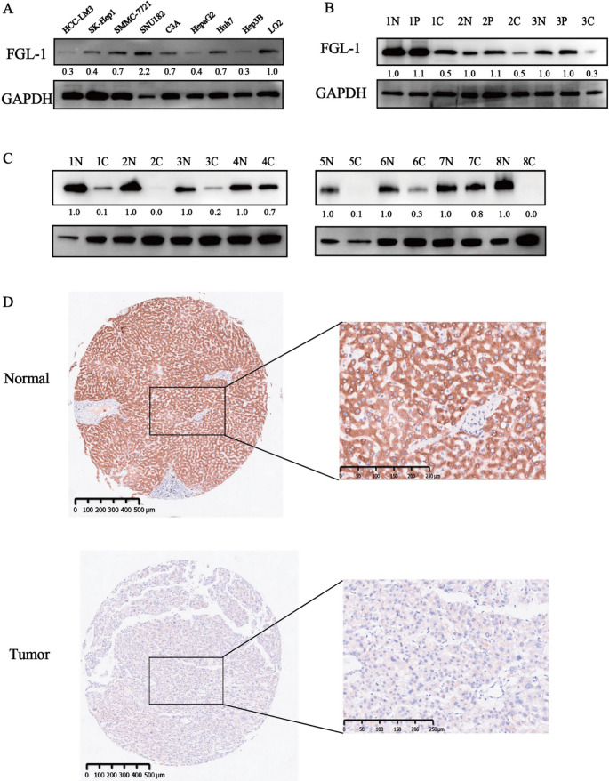 Figure 1
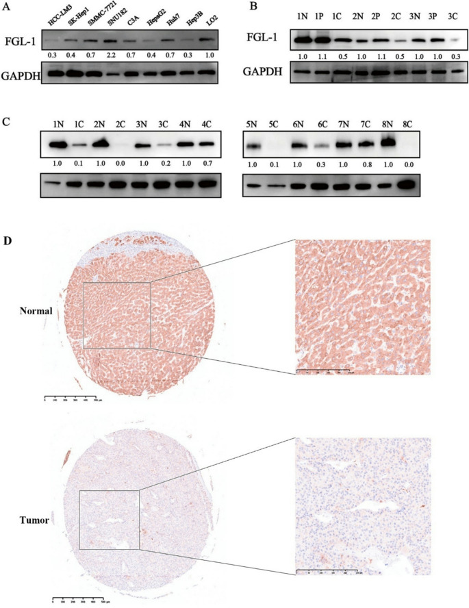 Figure 2
Peer Reviews
No public reviews on file for this paper yet. If you reviewed it on a platform where reviews are public (OpenReview, ICLR, NeurIPS, ICML), you can paste yours below so the community can read it here.
Videos
No videos yet. Explain this paper in a talk, walkthrough, or lecture? Add one.
Taxonomy
TopicsBlood properties and coagulation · Cancer, Lipids, and Metabolism · Lipid metabolism and disorders
Correction to: Molecular Biology Reports (2022) 49:7911–7919
10.1007/s11033-022-07624-6
In this article, Fig. 2D appeared incorrectly and has now been corrected in the original publication. For completeness and transparency, the correct and old incorrect versions are displayed below.
The original article has been corrected.
Incorrect version:
Fig. 2FGL1 expression showed obvious downregulation in several human HCC cell lines and HCC tissues. A Determination of FGL1 expression in several human HCC cell lines and the normal liver cell line via western blot analysis. B Determination of FGL1 expression with 3 pairs of HCC tissues and peri-tumor tissues and paired normal liver tissues via western blot analysis. C Determination of FGL1 expression with 8 pairs of HCC tissues and paired normal liver tissues via western blot analysis. D IHC staining for tumor tissues and adjacent normal liver tissues from HCC patients in the TMA. N normal liver tissue, P peri-tumor tissue, C cancer tissue; the numbers before N, P and C represent the group number
Correct version:
Fig. 2FGL1 expression showed obvious downregulation in several human HCC cell lines and HCC tissues. A Determination of FGL1 expression in several human HCC cell lines and the normal liver cell line via western blot analysis. B Determination of FGL1 expression with 3 pairs of HCC tissues and peri-tumor tissues and paired normal liver tissues via western blot analysis. C Determination of FGL1 expression with 8 pairs of HCC tissues and paired normal liver tissues via western blot analysis. D IHC staining for tumor tissues and adjacent normal liver tissues from HCC patients in the TMA. N normal liver tissue, P peri-tumor tissue, C cancer tissue; the numbers before N, P and C represent the group number