Neuromyelitis Optica Diagnosis in Two Elderly Patients with Systematic Lupus Erythematosus: A Case Series
Kyriaki Astara, Maria Lypiridou, Konstantinos Kalafatakis, Georgios Nikolaou, Georgios Stouraitis

TL;DR
This case series reports two elderly women with both systemic lupus erythematosus and neuromyelitis optica, highlighting the importance of early diagnosis and treatment to prevent disability.
Contribution
The paper presents two rare cases of NMO co-occurring with SLE in elderly patients, emphasizing diagnostic and treatment considerations.
Findings
NMO can coexist with SLE, even in elderly patients.
Myelitis in patients with connective tissue diseases should prompt suspicion of NMO.
Timely treatment is crucial to prevent cumulative disability in NMO.
Abstract
Background and Clinical Significance: Neuromyelitis optica (NMO) is a chronic demyelinating inflammatory disease of the central nervous system (CNS), mediated by autoantibodies against aquaporin-4 (AQ4) receptors. In the spectrum of NMO, other autoimmune diseases also coexist, though their association with systemic lupus erythematosus (SLE) is rare. Case Presentation: We present two cases of patients in their 70s who were diagnosed with NMO in the context of SLE. The first case concerns a 78-year-old woman with drug-induced SLE and thoracic myelitis who developed T4-level incomplete paraplegia over three weeks. The second case involves a 71-year-old woman with a history of SLE and myasthenia gravis, presenting with cervical myelitis with progressive worsening of walking and C4-level paraparesis over two months. In both cases, elevated serum anti-AQ4 titers were detected, establishing…
Genes, proteins, chemicals, diseases, species, mutations and cell lines named across the full text — each resolved to its canonical identifier and authoritative record.
Click any figure to enlarge with its caption.
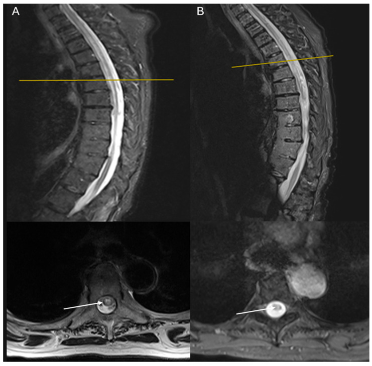 Figure 1
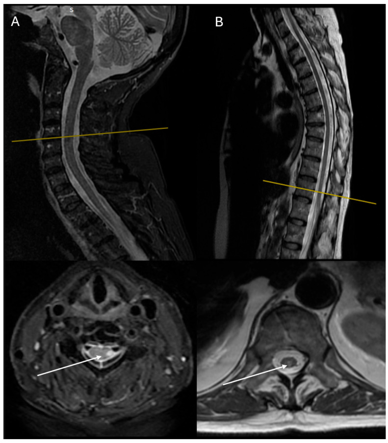 Figure 2
Peer Reviews
No public reviews on file for this paper yet. If you reviewed it on a platform where reviews are public (OpenReview, ICLR, NeurIPS, ICML), you can paste yours below so the community can read it here.
Videos
No videos yet. Explain this paper in a talk, walkthrough, or lecture? Add one.
Taxonomy
TopicsMultiple Sclerosis Research Studies · Peripheral Neuropathies and Disorders · Systemic Lupus Erythematosus Research
1. Introduction and Clinical Significance
Neuromyelitis optica (NMO) is a central nervous system (CNS) autoimmune disorder characterized by optic neuritis and longitudinally extensive transverse myelitis, typically involving more than three contiguous spinal cord segments. The disease is associated with pathogenic IgG antibodies targeting aquaporin-4 (AQ4) water channels on astrocyte podocytes [1]. Systematic lupus erythematosus (SLE), a multisystem autoimmune disorder, may involve the CNS, occasionally presenting with myelitis. However, myelitis in SLE affects fewer vertebral segments, has a monophasic course, and tends to present earlier in life than NMO [2].
We report two rare and diagnostically complex presentations of neuromyelitis optica spectrum disorder (NMOSD) in two elderly female patients with SLE, illustrating the rare coexistence of these conditions and highlighting diagnostic complexities involved.
2. Case Presentation
Case 1: A 78-year-old Caucasian female with a medical history of type 2 diabetes mellitus presented with a sudden-onset three-week history of progressive gait. Neurological examination revealed preserved higher cortical functions and cranial nerves II–XII but demonstrated quadriparesis, with proximal lower limb weakness (2/5), distal lower limb weakness (4/5), and upper limb weakness (3/5), along with hyperreflexia in the lower limbs and bilateral Babinski sign. Tone was normal, but muscle atrophy was present in both the trapezoids and vastus muscles of the left foot. Abdominal reflexes were absent. A level of sensory perception change at T6 was indicated, and urinary retention was reported. Her modified Rankin Scale (mRS) score at admission was 5, while before the onset of symptoms, it was 0. Initial laboratory and imaging (CT brain and lumbar spine) were unremarkable. Laboratory findings were significant for normocytic anemia. The patient was admitted to the Neurology Department for further investigation.
She underwent an electrodiagnostic investigation, which showed mild left carpal tunnel syndrome and non-specific low muscle contraction of the lower extremities. CT scans of the cervical and thoracic spine were also performed, with unremarkable results. Magnetic resonance imaging (MRI) scans of the brain and cervical spine showed no significant results, while the thoracic spine demonstrated a well-defined, multi-segment lesion extending from T3 to T8, having low-intensity signal on T1-weighted imaging with no gadolinium enhancement and high-intensity signal on T2-weighted imaging (Figure 1). Lumbar puncture (LP) showed no abnormal results (Table 1). Serum anti-AQ4 antibodies were positive, using an enzyme-linked immunosorbent assay (ELISA) (320 U/mL). Extended autoimmune workup showed elevated anti-nuclear antibodies (ANAs) (157.00 AU/mL) and ds-DNA antibody (97.90 IU/mL), raising suspicion for lupus spectrum disease. Normocytic anemia was attributed to autoimmune hemolysis, as indicated by elevated LDH and reticulocytes, along with a positive direct Coombs test. Given the patient’s history of prior antibiotic exposure, drug-induced lupus was initially considered; however, the presence of high-titer ANA, ds-DNA positivity, and the absence of anti-histone antibodies, along with the involvement of the CNS, pointed to SLE. The diagnosis was supported by the EULAR 2019 Classification Criteria for SLE, which require positive ANA as an entry criterion, followed by the involvement of the hematologic domain, due to autoimmune hemolysis [3]. Cerebrospinal fluid (CSF) PCR was positive for HHV-6 infection; the patient received intravenous acyclovir accordingly. Cancer biomarkers and thyrotropin were normal. Hence, a diagnosis of NMO with SLE was documented.
The patient was initially treated with intravenous infusions of 1gr methylprednisolone, which was discontinued due to a pancreatic reaction, followed by two cycles of 1gr of cyclophosphamide. Then, she was scheduled for semiannual sessions of intravenous infusion of 1gr RTX.
One year later, the patient remained functionally dependent (mRS 5), with stable MRI findings and decreased anti-AQ4 titers (100 U/mL) (Table 1).
Case 2: A 71-year-old Caucasian female with a medical history of arterial hypertension, prior thyroidectomy (on levothyroxine) and a history of SLE (with musculoskeletal and hematologic involvement of joint pain and morning stiffness, and autoimmune hemolytic anemia-treated with hydroxychloroquine and methylprednisolone) presented with a 2-month history of sudden-onset progressive gait disturbance, neuropathic pain in the lower extremities, and urinary incontinence. Neurological examination revealed preserved cognitive function and cranial nerves II–XII, with paraparesis more pronounced on the left (4/5 proximally, 3/5 distally with drop foot) than on the right (4/5) and normal muscle strength of upper extremities. Reflexes were brisk in the left lower limb, while bilateral Hoffman and Babinski signs were present. Tone was normal in all extremities. Sensory findings included reduced pinprick sensation below C3 and impaired proprioception in the right lower limb. Her mRS at presentation was 4, while before the onset of symptoms, it was 0. The laboratory evaluation of biofluids on admission was unremarkable. She was admitted to the Neurology Department for further investigation.
Outpatient MRI scans of the cervical and lumbar spine demonstrated a well-defined, multi-segment lesion extending from C3 to C7, having low-intensity signal on T1-weighted imaging with no gadolinium enhancement and high-intensity signal on T2-weighted imaging (Figure 2). She underwent additional MRI scans of the brain and thoracic spine (no notable findings), while LP showed no abnormal findings (Table 1). Serum anti-AQ4 receptor antibodies were markedly elevated, using ELISA (3200 U/mL), while extended serological workup was only indicative of SLE. Hence, a diagnosis of NMO with SLE was documented.
The patient was treated with intravenous infusions of 1gr methylprednisolone for 5 days, followed by two cycles of 1gr cyclophosphamide while continuing with tapering oral steroids. Then, she was scheduled for semiannual sessions of intravenous infusion of 1gr RTX.
One year later, the patient showed significant clinical improvement (mRS 1), with minimal residual left lower limb weakness (4/5). Follow-up cervical and thoracic spine MRI displayed reduced lesion extent with a weaker increase in T2 signal intensity and no gadolinium enhancement (C4–C7), though a new demyelinating lesion extending to T9–T12 was detected. Anti-AQ4 receptor antibody titer declined to 100 U/mL (Table 1).
3. Discussion
In this paper, we present two cases of SLE and NMO, highlighting the heterogeneity in disease onset, progression, and response to treatment. The first case was initially suspected to be drug-induced lupus, due to prior antibiotic use [4]. Nevertheless, persistent neurological symptoms months after discontinuation of the offending agents, relevant coexistent clinical domains, and the serological profile suggested an alternative diagnosis [5]. The second case, with a prior diagnosis of SLE, was maintained on hydroxychloroquine and low-dose methylprednisolone. The first case presented with a rather abrupt and severe NMO onset, with limited therapeutic response, whereas the second exhibited gradual progression and excellent response to the same standard immunosuppressive therapy. One could attribute the gradual progression and favorable response of the second case to the immunosuppressive effect maintained methylprednisolone. Several factors have been associated with poor prognosis, including age >50 years, female sex, African ethnicity, late pregnancy/postpartum period, high AQ-4 antibody titers, longitudinally extensive transverse myelitis, and spinal cord atrophy [6,7].
A recent systematic review of 46 cases with NMOSD in SLE demonstrated female predominance and NMO onset after lupus, as in our cases, but earlier age of onset and at least one relapse after treatment [8]. To our knowledge, these are the first reported cases of SLE with late-onset NMO and such a different clinical course.
Immunosuppressive therapy is fundamental for NMO management. In particular, RTX, a CD20-targeting monoclonal antibody, depletes CD20+ B cells, suppressing antibody-mediated immunity and reducing AQ4 antibodies, which have been implicated in the pathogenesis of NMO [9]. In fact, AQ4 antibodies demonstrate 94% specificity and 76% sensitivity for NMOSD [10]. RTX is a highly efficient option in reducing relapse rates and long-term disability, while it is well tolerated by patients, despite the lack of large randomized controlled trials [11,12].
A subset of patients, such as our first case, have shown a limited response to RTX. In a study by Dai et al. [13], NMO cases with suboptimal response to RTX were reviewed, proposing possible mechanisms like suboptimal depletion, the production of neutralizing antibodies of several B cell subsets [14], the involvement of cell-mediated immunity by various T cell subsets [15], and complement activation [16]. In such cases, tocilizumab (anti-interleukin-6 receptor) and eculizumab (complement inhibitor) have displayed promising results [16,17]. However, to our knowledge, refractory NMO in the context of SLE has not been previously reported.
4. Conclusions
Although rare, the coexistence of SLE with NMO should be considered in patients with connective tissue disorder presenting with myelitis, especially in women over 65 years of age, even several years after the diagnosis of SLE. Prompt recognition is crucial, as time-to-treatment is critical and may result in irreversible neurological damage. While RTX therapy is generally effective, clinical vigilance is essential for cases with suboptimal response.
The reference list from the paper itself. Each links out to its DOI / PubMed record.
- 1Wingerchuk D.M. Lucchinetti C.F. Neuromyelitis Optica Spectrum Disorder N. Engl. J. Med.202238763163910.1056/NEJ Mra 190465536070711 · doi ↗ · pubmed ↗
- 2Hryb J.P. Chiganer E. Contentti E.C. Di Pace J.L. Lessa C. Perassolo M.B. Myelitis in Systemic Lupus Erythematosus: Clinical Features, Immunological Profile and Magnetic Resonance Imaging of Five Cases Spinal Cord. Ser. Cases 201621600510.1038/scsandc.2016.528053749 PMC 5129439 · doi ↗ · pubmed ↗
- 3Aringer M. Costenbader K.H. Daikh D.I. Brinks R. Mosca M. Ramsey-Goldman R. Smolen J.S. Wofsy D. Boumpas D. Kamen D.L. 2019 EULAR/ACR Classification Criteria for Systemic Lupus Erythematosus Arthritis Rheumatol.2019711400141210.1002/art.4093031385462 PMC 6827566 · doi ↗ · pubmed ↗
- 4He Y. Sawalha A.H. Drug-Induced Lupus Erythematosus: An Update on Drugs and Mechanisms Curr. Opin. Rheumatol.20183049049710.1097/BOR.000000000000052229870500 PMC 7299070 · doi ↗ · pubmed ↗
- 5Vaglio A. Grayson P.C. Fenaroli P. Gianfreda D. Boccaletti V. Ghiggeri G.M. Moroni G. Drug-Induced Lupus: Traditional and New Concepts Autoimmun. Rev.20181791291810.1016/j.autrev.2018.03.01630005854 · doi ↗ · pubmed ↗
- 6Palace J. Lin D.-Y. Zeng D. Majed M. Elsone L. Hamid S. Messina S. Misu T. Sagen J. Whittam D. Outcome Prediction Models in AQP 4-Ig G Positive Neuromyelitis Optica Spectrum Disorders Brain 20191421310132310.1093/brain/awz 05430938427 PMC 6487334 · doi ↗ · pubmed ↗
- 7Ma X. Kermode A.G. Hu X. Qiu W. Risk of Relapse in Patients with Neuromyelitis Optica Spectrum Disorder: Recognition and Preventive Strategy Mult. Scler. Relat. Disord.20204610252210.1016/j.msard.2020.10252233007726 · doi ↗ · pubmed ↗
- 8Kopp C.R. Prasad C.B. Naidu S. Sharma V. Misra D.P. Agarwal V. Sharma A. Overlap Syndrome of Anti-Aquaporin-4 Positive Neuromyelitis Optica Spectrum Disorder and Systemic Lupus Erythematosus: A Systematic Review of Individual Patient Data Lupus 2023321164117210.1177/0961203323119118037487596 · doi ↗ · pubmed ↗