Cerebrotendinous Xanthomatosis With a Heterozygous Frameshift Mutation Involving CYP27A1(C.526del)
Ajitesh Roy, Sounak Kumar Roy, Soumyadip Das

Abstract
Genes, proteins, chemicals, diseases, species, mutations and cell lines named across the full text — each resolved to its canonical identifier and authoritative record.
Click any figure to enlarge with its caption.
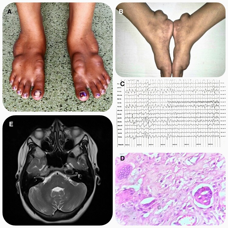 Figure 1
Peer Reviews
No public reviews on file for this paper yet. If you reviewed it on a platform where reviews are public (OpenReview, ICLR, NeurIPS, ICML), you can paste yours below so the community can read it here.
Videos
No videos yet. Explain this paper in a talk, walkthrough, or lecture? Add one.
Taxonomy
TopicsUrticaria and Related Conditions · Cholesterol and Lipid Metabolism
Image Legend
A 31-year-old female presented with painless, progressive focal swelling in both feet over the past 4 years. There was no history suggestive of any cardiac or neurologic abnormalities. On examination, multiple lumps were observed in each foot, including over the Achilles tendon, which were soft and nontender (Fig. 1A and 1B). Neurologic and cardiac examinations revealed no abnormalities. Investigations showed subtle dyslipidemia [cholesterol: 207 mg/dL (5.36 mmol/L) (normal reference range: less than 200 mg/dL; 5.17 mmol/L); triglycerides: 351 mg/dL (3.96 mmol/L) (normal reference range: less than 150 mg/dL; 1.7 mmol/L); low-density lipoprotein: 103 mg/dL (2.67 mmol/L) (normal reference range:100 mg/dL; 2.6 mmol/L)]. Electroencephalogram revealed generalized sharp wave discharges (Fig. 1C). Histopathological examination revealed foamy histiocytes and Touton giant cells (Fig. 1D). Magnetic resonance imaging demonstrated T2 hyperintensities in the bilateral dentate nuclei (Fig. 1E). Exome sequencing showed a pathogenic heterozygous frameshift mutation involving c.526del in exon 3 of the CYP27A1 gene, confirming the diagnosis of cerebrotendinous xanthomatosis. This mutation has been previously reported in a few cases, all of which had associated clinical neurological abnormalities [1, 2]. In our case, however, there were no neurological symptoms. Therefore, a patient with tendon xanthomas should not be automatically diagnosed with familial hypercholesterolemia. Conversely, the absence of clinical neurological abnormalities should not exclude the diagnosis of cerebrotendinous xanthomatosis.
(A) Multiple tendon xanthomas. (B) Tendon xanthomas involving Achilles tendon. (C) Electroencephalogram showing generalized sharp wave discharges in both hemisphere. (D) Tendon xanthoma histopathological examination showing foamy histiocytes and Touton giant cells. (E) T2 weighted magnetic resonance imaging showing hyperintensities in the bilateral dentate nuclei.
The reference list from the paper itself. Each links out to its DOI / PubMed record.
- 1Katragadda P, Holla VV, Kamble N, Saini J, Yadav R, Pal PK. Clinical and imaging profile of patients with cerebrotendinous xanthomatosis—a video case series from India. Tremor Other Hyperk. 2024;14(1):10.10.5334/tohm.851PMC 1092927738476584 · doi ↗ · pubmed ↗
- 2Dutta AK, Danda S, Muthusamy K, et al Cerebrotendinous xanthomatosis: possibility of founder mutation in CYP 27A 1 gene (c.526del G) in Eastern Indian and Surinamese population. Mol Genet Metab Rep. 2015;3:33‐35.26937392 10.1016/j.ymgmr.2015.03.002PMC 4750635 · doi ↗ · pubmed ↗