Successful removal of B3 bile duct stones from B2 bile duct endoscopic ultrasound-guided hepaticogastrostomy route using wire-loop and bridge technique
Hiroaki Tsuji, Ryota Sagami, Takao Sato, Hidefumi Nishikiori, Yasuhisa Hiroshima, Kazuhiro Mizukami, Kazunari Murakami

Abstract
Genes, proteins, chemicals, diseases, species, mutations and cell lines named across the full text — each resolved to its canonical identifier and authoritative record.
Click any figure to enlarge with its caption.
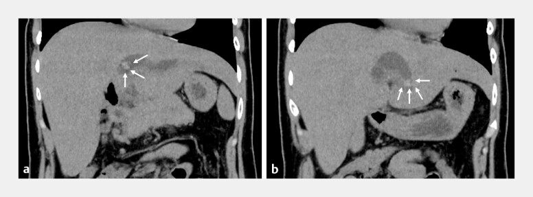 Fig. 1
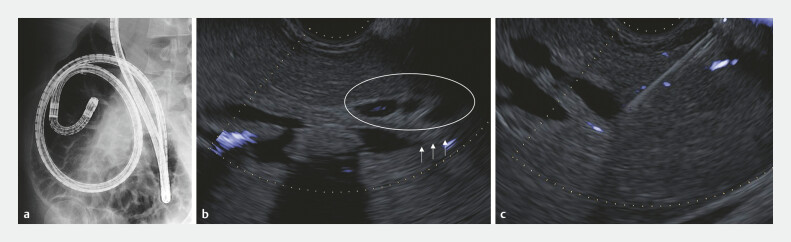 Fig. 2
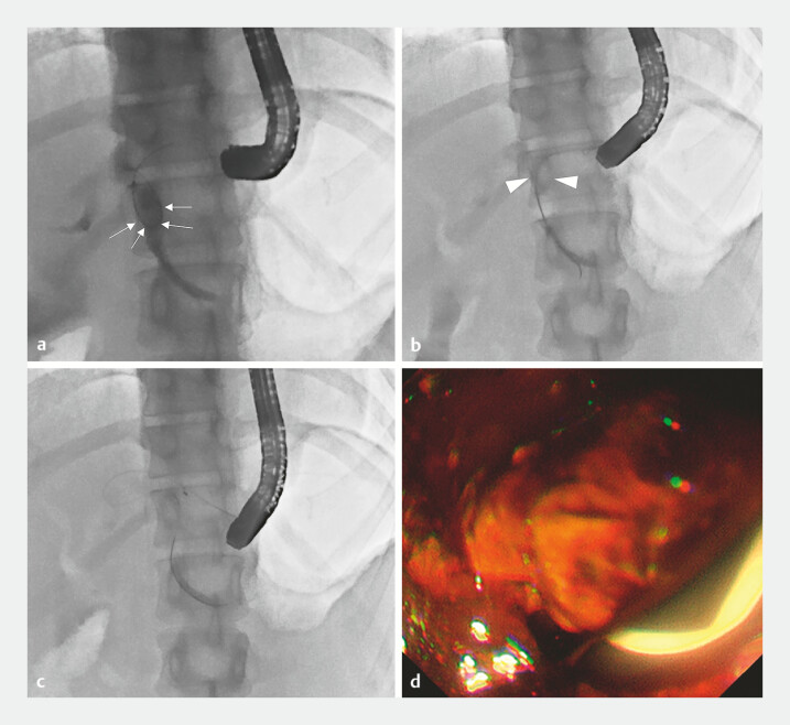 Fig. 3
Peer Reviews
No public reviews on file for this paper yet. If you reviewed it on a platform where reviews are public (OpenReview, ICLR, NeurIPS, ICML), you can paste yours below so the community can read it here.
Videos
No videos yet. Explain this paper in a talk, walkthrough, or lecture? Add one.
Taxonomy
TopicsPediatric Hepatobiliary Diseases and Treatments · Gallbladder and Bile Duct Disorders · Cholangiocarcinoma and Gallbladder Cancer Studies
Endoscopic ultrasound-guided hepaticogastrostomy (EUS-HGS) is an effective drainage technique for difficult-to-access cases by usual endoscopic retrograde cholangiopancreatography (ERCP) 1 . Technical tips and reviews have been shown 2 3 , however, difficult situations exist.
A 36-year-old woman underwent extrahepatic bile duct resection and hepaticojejunostomy for congenital biliary duct dilatation at 3 years old. Percutaneous transhepatic biliary drainage (PTBD) and balloon enteroscope-assisted ERCP (BE-ERCP) were performed several times for repeated cholangitis. However, cholangitis caused by intrahepatic bile duct stones (IBDS) recurred. Computed tomography revealed IBDS in the B3 and B2 bile ducts ( Fig. 1 ). Repeated BE-ERCP was technically challenging/invasive because of the severe adhesion ( Fig. 2 a ).
Imaging of computed tomography. a The left intrahepatic bile duct dilation and intrahepatic bile duct stones in the B2 branch. b The intrahepatic bile duct stone was also found in the B3 branch.
Imaging of BE-ERCP and EUS. a Severe adhesion would be expected from past BE-ERCP imaging. b No punctured route from the B3 bile duct because of no bile duct downstream dilation (white arrows) and many intervening vessels (white circle). c EUS-HGS was conducted from the B2 bile duct. Abbreviations: BE-ERCP, balloon enteroscope-assisted ERCP; EUS, endoscopic ultrasound; EUS-HGS, endoscopic ultrasound-guided hepaticogastrostomy.
First, EUS-HGS was conducted using a dedicated plastic stent (Through & Pass Type-IT; Gadelius Medical) from the B2 bile duct (5 mm) because there is no puncturing route from B3 with no bile duct downstream dilation and many intervening vessels ( Fig. 2 b, c ). Two weeks after the initial drainage, a dumbbell-shaped covered metal stent (M-Intraductal; Medicos Hirata) was deployed to dilate ESCR (EUS-guided created route). One week later, B3 IBDS removal was planned from the ESCR; however, the insertion of the basket catheter into the B3 was difficult because of the sharp angle between B2 and B3 bile ducts.
A 0.025-inch soft guide wire (TRU wire; Medicos Hirata) passed from the B2 EUS-HGS route to B3 using the wire-loop and bridge technique ( Video 1 ).
Making a loop of a guidewire and swinging the EUS scope could be useful for passing the wire between the sharp angles of B2 and B3 bile ducts and stone removal.Video 1
After the balloon catheter (Extraction, Xemex, Zeon Medical) was inserted, B3 IBDSs could be moved to the B2 bile duct without dislocating the wire ( Fig. 3 b ). Finally, all IBDSs were removed from the ESCR ( Fig. 3 c, d ).
The X-ray fluorescence and endoscopic image of endoscopic stone removal. a X-ray image of wire manipulation using wire-loop and bridge technique (white arrows). b X-ray image of B3 stone movement to B2 bile duct by a balloon catheter (white arrow heads). c X-ray image of stone removal by the basket catheter. d Endoscopic image of the removal of stones from the ESCR. Abbreviation: ESCR, EUS-guided created route.
This guidewire manipulation technique with a balloon catheter may help remove IBDSs from the other EUS-HGS route for whom the EUS-puncturing route is restricted.
Endoscopy_UCTN_Code_TTT_1AS_2AH
The reference list from the paper itself. Each links out to its DOI / PubMed record.
- 1Paik WH Park DH Outcomes and limitations: EUS-guided hepaticogastrostomy Endosc Ultrasound 20198 S 44S 4910.4103/eus.eus_51_1931897379 PMC 6896431 · doi ↗ · pubmed ↗
- 2Ogura T Higuchi K Endoscopic Ultrasound-Guided Hepaticogastrostomy: Technical Review and Tips to Prevent Adverse Events Gut Liver 20211519620510.5009/gnl 2009632694240 PMC 7960972 · doi ↗ · pubmed ↗
- 3Kadkhodayan K Irani SEUS-guided hepaticogastrostomy: practical tips and tricks Video GIE 2024941742410.1016/j.vgie.2024.05.01539429912 PMC 11489514 · doi ↗ · pubmed ↗