When the brain fades before the eye: encephalopathy as a rare presentation of direct carotid-cavernous fistula
Leonardo Furtado Freitas, Eduardo J. Labat, Robert T. Wicks, Charif Sidani, Kevin J. Abrams

Abstract
Genes, proteins, chemicals, diseases, species, mutations and cell lines named across the full text — each resolved to its canonical identifier and authoritative record.
Click any figure to enlarge with its caption.
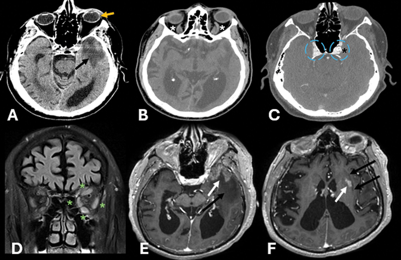 Figure 1
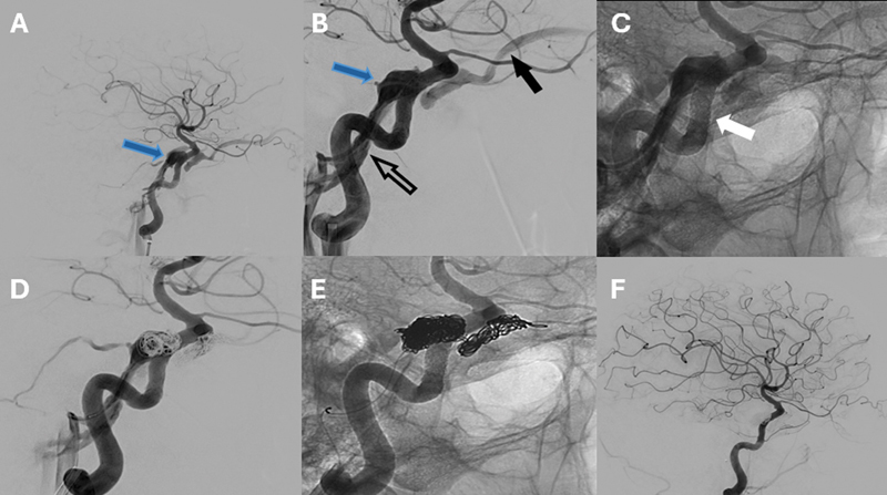 Figure 2
Peer Reviews
No public reviews on file for this paper yet. If you reviewed it on a platform where reviews are public (OpenReview, ICLR, NeurIPS, ICML), you can paste yours below so the community can read it here.
Videos
No videos yet. Explain this paper in a talk, walkthrough, or lecture? Add one.
Taxonomy
TopicsVascular Malformations Diagnosis and Treatment · Intracranial Aneurysms: Treatment and Complications · Intracerebral and Subarachnoid Hemorrhage Research
We herein report a rare case of a 76-year-old male patient with progressive cognitive decline, acute encephalopathy, and visual symptoms 6 months after cavernous internal carotid artery (ICA) stenting for intracranial stenosis. The initial workup was negative. Neuroimaging ( Figure 1 ) showed left temporal and orbital edema with cavernous sinuses abnormalities. The ophthalmologic exam revealed palsy of the left cranial nerve VI and elevated intraocular pressure. Cerebral angiography ( Figure 2 ) confirmed a direct left-sided, type-A carotid-cavernous fistula (CCF) with bilateral venous drainage. Endovascular embolization resulted in clinical improvement. This case highlights an uncommon presentation of direct CCF with predominant encephalopathy 1 2 and emphasizes the importance of considering vascular causes in atypical cognitive decline following ICA stenting.
Axial non-contrast computed tomography (CT) scan ( A, B ) and CT angiography (CTA) ( C ). Coronal orbit magnetic resonance imaging (MRI) scan on fluid-attenuated inversion recovery (FLAIR)-weighted images with fat suppression ( D ) and brain MRI scan on post-contrast axial T1 gradient echo-weighted images ( E,F ). Left temporal hypoattenuation with proptosis (black and orange arrows), dilation of the superior ophthalmic veins (white asterisks), and a stent in the cavernous segment of the left internal carotid artery (black asterisk) with cavernous sinuses engorgement (dashed blue circles). MRI scan showing prominent left temporal vasogenic edema extending to the subinsular region and putamen (black arrows), edema in the left extraocular muscles (green asterisks). There is also thick cortical enhancement in the left temporal pole and nodular enhancement in the ipsilateral putamen (white arrows).
Cerebral angiogram. Left internal carotid artery, lateral projection ( A ), revealing direct carotid cavernous fistula with contrast opacification of the cavernous sinus (blue arrow). Left internal carotid artery injection, magnified lateral projection ( B ) – the solid arrow denotes dilated superior ophthalmic vein. The hollow and blue arrows show dilated inferior petrosal sinus and cavernous sinus, respectively. Left Internal carotid artery injection, unsubtracted lateral projection ( C ), which enables the visualization of internal carotid artery cavernous segment stent (white arrow). Left internal carotid artery injection, lateral projection ( D ). Intraprocedural coiling of carotid cavernous fistula via the transvenous approach through the inferior petrosal sinus. Left internal carotid artery injection, unsubtracted lateral projection ( E ). Posttransvenous coil embolization of carotid cavernous fistula. Left internal carotid artery injection, ateral projections ( F ), revealing complete coil embolization of the carotid cavernous fistula.
The reference list from the paper itself. Each links out to its DOI / PubMed record.
- 1Oie A K Herrmann A A Rosenbloom M H Left dural carotid cavernous fistula mimicking an inflammatory process: A case report Radiol Case Rep 202419062558256010.1016/j.radcr.2024.03.02138596179 PMC 11001617 · doi ↗ · pubmed ↗
- 2Klevtsova E Nguyen-Min C Lalani T Carlan S J Madruga M Posttraumatic carotid cavernous fistula that presented as seizure and focal neurological deficits with symptom resolution after therapeutic coil embolization J Emerg Med 2015480218619010.1016/j.jemermed.2014.09.04325453851 · doi ↗ · pubmed ↗