2D-cranial T1-black-blood MRI in suspected giant cell arteritis—measurement of vessel wall thickness does not give a diagnostic advantage compared to visual scoring alone
Pascal Seitz, Susana Bucher, Lukas Bütikofer, Britta Maurer, Harald Marcel Bonel, Fabian Lötscher, Luca Seitz

TL;DR
This study compares two MRI scoring methods for diagnosing giant cell arteritis and finds that visual scoring alone is as effective and faster than adding wall thickness measurements.
Contribution
The study demonstrates that visual scoring alone is sufficient for diagnosing giant cell arteritis using 2D-T1-black-blood MRI, without the need for additional quantitative measurements.
Findings
Visual scoring (T1-BB-VISUAL) had higher sensitivity and similar specificity compared to the composite method (T1-BB-COMP).
The overall agreement between the two methods was very good (91.6%) with higher agreement in larger arterial segments.
Visual scoring was significantly faster to perform than the composite method.
Abstract
To compare two established scoring schemes for the 2D-T1-weighted “black-blood” MRI sequence (T1-BB) for superficial cranial arteries (SCA) in the diagnosis of giant cell arteritis (GCA). Ten arterial segments were evaluated in T1-BB images with two different methods: a visual semiquantitative scheme (T1-BB-VISUAL) and a composite scheme that included both the semiquantitative assessment and a quantitative wall thickness measurement (T1-BB-COMP). The expert clinical diagnosis after ≥6 months of follow-up was the diagnostic reference standard. Diagnostic accuracy and agreement on the segment and patient levels were evaluated for the two different rating schemes. Retrospectively, 151 consecutive patients with clinically suspected GCA were included. The study cohort consisted of 82 patients with and 69 without GCA. For the T1-BB-COMP and the T1-BB-VISUAL, the sensitivity was 81.7% vs.…
Genes, proteins, chemicals, diseases, species, mutations and cell lines named across the full text — each resolved to its canonical identifier and authoritative record.
Click any figure to enlarge with its caption.
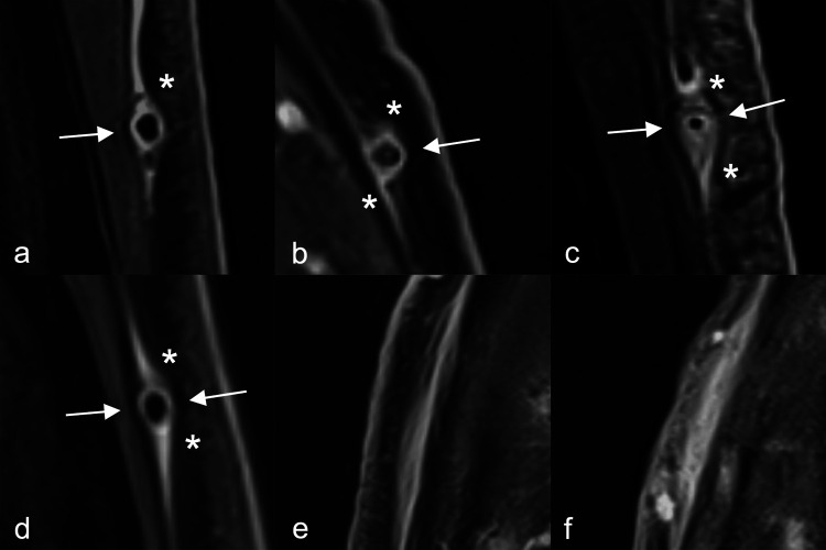 Figure 1
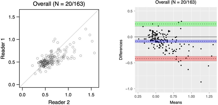 Figure 2
Peer Reviews
No public reviews on file for this paper yet. If you reviewed it on a platform where reviews are public (OpenReview, ICLR, NeurIPS, ICML), you can paste yours below so the community can read it here.
Videos
No videos yet. Explain this paper in a talk, walkthrough, or lecture? Add one.
Taxonomy
TopicsVasculitis and related conditions · Cerebrovascular and Carotid Artery Diseases · Coronary Artery Anomalies