Star of the Diabetic Macular Show: Stellate Nonhereditary Idiopathic Foveomacular Retinoschisis (SNIFR)
Cory A. Christensen, Neha Gupta, Mark P. Breazzano

Abstract
Genes, proteins, chemicals, diseases, species, mutations and cell lines named across the full text — each resolved to its canonical identifier and authoritative record.
Click any figure to enlarge with its caption.
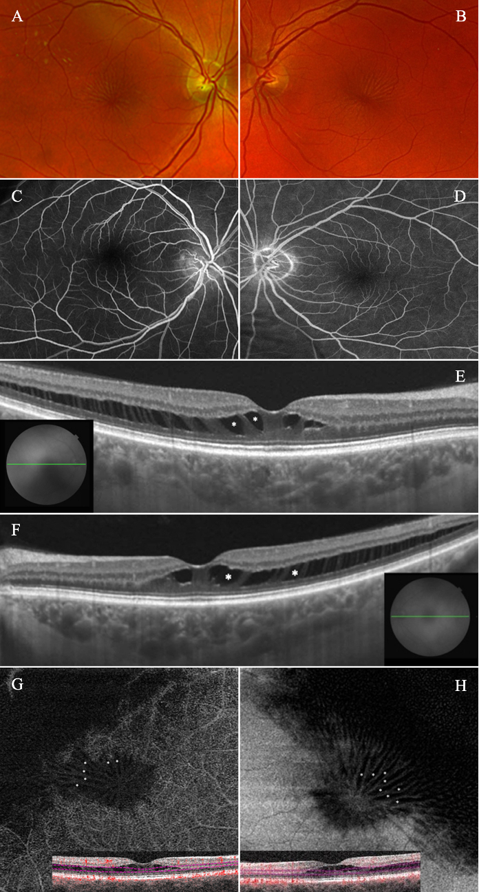 Figure 1
Peer Reviews
No public reviews on file for this paper yet. If you reviewed it on a platform where reviews are public (OpenReview, ICLR, NeurIPS, ICML), you can paste yours below so the community can read it here.
Videos
No videos yet. Explain this paper in a talk, walkthrough, or lecture? Add one.
Taxonomy
TopicsOcular Diseases and Behçet’s Syndrome · Retinal and Optic Conditions · Retinal Diseases and Treatments
PRESENTATION
A 66-year-old man with recently diagnosed type 2 diabetes mellitus presented with concern for macular edema. He reported no visual complaints and had no family history of ocular disease. Examination revealed bilateral 20/20 Snellen acuity, with blunting of the foveal reflexes and macular star-like appearances [Figures 1A & 1B]. Intravenous fluorescein angiography in late phase revealed no evidence of staining or leakage [Figures 1C & 1D]. Swept-source optical coherence tomography (OCT) showed perifoveal hyporeflective cavities OU [Figures 1E & 1F]. Spectral-domain OCT angiography (OCTA) revealed classic stellate patterns of radially orientated macular spoking and lack of flow signal within the pseudocystic spaces, indicative of avascular outer plexiform layer [Figures 1G & 1H].
(A & B) Fundus photography of the right and left eyes, respectively, with evidence of retinal folds radiating from the foveae. (C & D) Intravenous fluorescein angiography in late phases of the right and left eyes, respectively, with no evidence of staining, leakage, or microaneurysms, despite the history of diabetes mellitus. (E & F) Swept-source optical coherence tomography B-scan through the foveae of the right and left eyes, respectively, revealing hyporeflective cavities (asterisks) involving the outer plexiform layers with extension into the temporal maculae. (G & H) Spectral-domain optical coherence tomography angiography of the right and left eyes, respectively, displaying an absence of flow signal through pseudocystic spaces in stellate configurations (asterisks) upon en-face structural imaging with segmentation through the outer plexiform layers (pink lines, B-scans).
DISCUSSION
Patients with a history of diabetes and OCT findings of perifoveal pseudocystic spaces can easily bias the clinician toward diabetic macular edema. In this case, a negative family history, lack of diabetic retinopathy, presence of macular pseudo-folds, and characteristic OCT and structural OCTA findings were consistent with stellate nonhereditary idiopathic foveomacular retinoschisis (SNIFR).
OCT alone helps exclude vitreomacular interface abnormalities for tractional causes of foveoschisis.^[1]^ However, OCTA adds utility in distinguishing SNIFR from X-linked retinoschisis, which classically demonstrates vascularity from the flow signal within the outer plexiform layer.^[2]^ Taxane- and niacin-induced retinoschisis should be excluded with history-taking, and as in this case, similarly lack leakage on intravenous fluorescein angiography.
Ethical Considerations
Institutional Review Board approval and associated contingencies were waived per guidelines for this case report as it does not qualify as human subjects research.
Financial Support and Sponsorship
None.
Conflicts of Interest
MPB has served as an advisor for Iveric Bio/Astellas and as a consultant for ONL Therapeutics.
The reference list from the paper itself. Each links out to its DOI / PubMed record.
- 1Breazzano MP Chang S Split down the middle of the fovea JAMA Ophthalmol 2021139103710383423639410.1001/jamaophthalmol.2020.6869 · doi ↗ · pubmed ↗
- 2Fragiotta S Leong BCS Kaden TR Bass SJ Sherman J Yannuzzi LA etal A proposed mechanism influencing structural patterns in X-linked retinoschisis and stellate nonhereditary idiopathic foveomacular retinoschisis Eye 2019337247283051897510.1038/s 41433-018-0296-8PMC 6707275 · doi ↗ · pubmed ↗