Endoscopic ultrasound-guided fine-needle aspiration for the diagnosis of laryngeal mucosa-associated lymphoid tissue lymphoma
Wenwen Xiao, Yemei Wu, Jiaqi Dai, Haitao Zhao, Mingyao Xu, Hongbo Wang

Abstract
Genes, proteins, chemicals, diseases, species, mutations and cell lines named across the full text — each resolved to its canonical identifier and authoritative record.
Click any figure to enlarge with its caption.
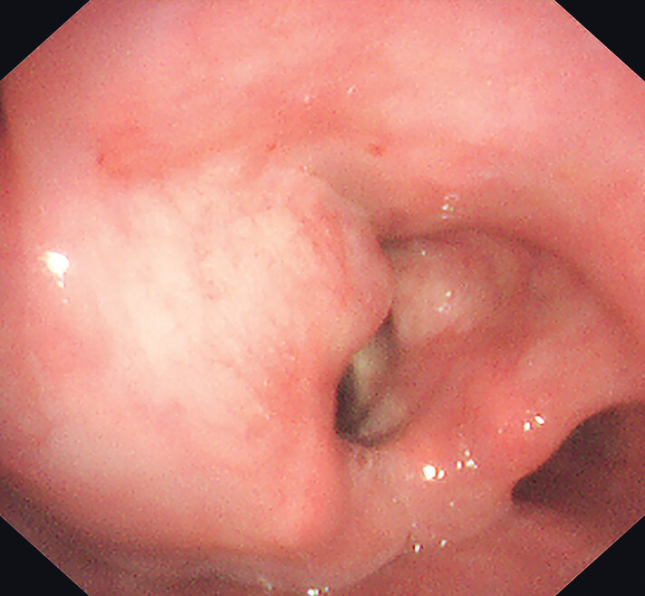 Fig. 1
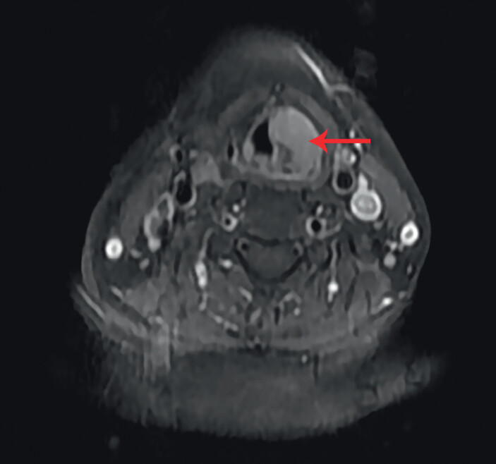 Fig. 2
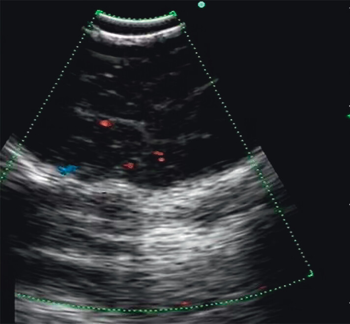 Fig. 3
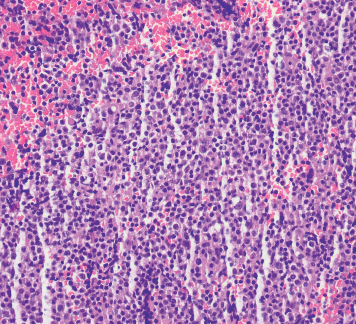 Fig. 4
Peer Reviews
No public reviews on file for this paper yet. If you reviewed it on a platform where reviews are public (OpenReview, ICLR, NeurIPS, ICML), you can paste yours below so the community can read it here.
Videos
No videos yet. Explain this paper in a talk, walkthrough, or lecture? Add one.
Taxonomy
TopicsLymphoma Diagnosis and Treatment · Head and Neck Cancer Studies · Medical Imaging and Pathology Studies
This case demonstrates how endoscopic ultrasound-guided fine-needle aspiration (EUS-FNA) can safely and effectively overcome the limitations of traditional biopsy in submucosal laryngeal lesions.
A 61-year-old woman underwent surgical treatment for a left orbital nodule 4 years ago. Postoperative pathology confirmed extranodal marginal zone lymphoma of the mucosa-associated lymphoid tissue (MALT lymphoma). She subsequently received four cycles of cyclophosphamide, doxorubicin, vincristine, and prednisolone (CHOP) chemotherapy and local radiotherapy. One month ago, she underwent laryngoscopy for investigation of throat discomfort. The examination revealed swelling in the left aryepiglottic fold, left vestibular wall, and left laryngeal compartment, with smooth surface mucosa ( Fig. 1 ). Magnetic resonance imaging of the throat showed an abnormal signal in the left piriform fossa, involving the left aryepiglottic fold ( Fig. 2 ).
Laryngoscopy showed swelling of the left aryepiglottic fold, left vestibular wall, and left laryngeal compartment, with smooth surface mucosa.
Magnetic resonance imaging revealed an abnormal signal (arrow) in the left piriform fossa involving the left aryepiglottic fold.
Given the smooth mucosal surface of the tumor and the narrow supraglottic space, direct biopsy under laryngoscopy risked a lower diagnostic yield and airway obstruction. Therefore, we opted for EUS-FNA to confirm the diagnosis. EUS revealed a hypoechoic area in the aryepiglottic fold, with clear boundaries and low internal vascularity ( Fig. 3 ). Under real-time Doppler ultrasound guidance (BF-UC260FW; Olympus, Tokyo, Japan), a 21-gauge needle (NA-201SX-4021; Olympus) was used to puncture the lesion ( Video 1 ). The procedure caused minimal bleeding, and histopathology confirmed MALT lymphoma ( Fig. 4 ). Finally, the patient received repeat chemotherapy and local radiotherapy.
Endoscopic ultrasound revealed a hypoechoic area in the left aryepiglottic fold, with well-defined boundaries and low internal vascularity.
Hematoxylin and eosin staining confirmed mucosa-associated lymphoid tissue lymphoma.
Endoscopic ultrasound revealed a hypoechoic area in the left aryepiglottic fold, with clear margins and low internal blood flow. Under real-time Doppler ultrasound guidance, a 21-gauge needle was used to puncture the lesion.Video 1
MALT lymphoma involving the larynx is relatively rare, and such lesions are typically located partially or entirely beneath the mucosa 1 . Conventional endoscopic biopsy is limited to the mucosal layer, and inadequate sampling depth often renders definitive diagnosis challenging. The application of EUS-FNA to submucosal lesions in the oropharynx is safe 2 ; therefore, it is also suitable for use in the larynx. We present this case to offer new perspectives on the diagnosis and management of submucosal laryngeal lesions. EUS-FNA provides a relatively safe and effective approach for obtaining tissue samples from these lesions.
Endoscopy_UCTN_Code_CCL_1AB_2AB
The reference list from the paper itself. Each links out to its DOI / PubMed record.
- 1Siddiqui NA Branstetter BF 4th Hamilton BE Imaging characteristics of primary laryngeal lymphoma AJNR Am J Neuroradiol 2010311261126510.3174/ajnr.A 208520360337 PMC 7965484 · doi ↗ · pubmed ↗
- 2Zhang Z Luo Y Shi MEUS-FNA to diagnose a submucosal oropharyngeal carcinoma Endosc Ultrasound 20241327327510.1097/eus.000000000000006839318750 PMC 11419498 · doi ↗ · pubmed ↗