Meloxicam mitigated methylglyoxal-induced glycative stress in rats
Talha bin Fayyaz, Ghulam Abbas, Hammad Ahmed, Najeeb Khatian, Shumaila Usman, Uzair Nisar, Noor Ul Ain, Yamna Khurshid, Syed Abid Ali

TL;DR
Meloxicam, a common NSAID, was found to reduce damage caused by glycative stress in rats, suggesting it could be repurposed as a treatment for related conditions.
Contribution
The study demonstrates meloxicam's potential as a repurposed drug to combat glycative stress in a rat model.
Findings
Meloxicam significantly protected against cognitive, liver, and kidney impairments caused by methylglyoxal.
Meloxicam normalized carboxymethyllysine levels and reduced gene expression linked to glycative stress.
No significant histopathological changes were observed in meloxicam-treated rats.
Abstract
Glycation is one of the primary underlying processes attributed to senescence and related diseases. No medicine currently targets this harmful manifestation. Drug repurposing is an efficient and cost-effective way of developing drugs. The present study evaluated meloxicam, a clinically used NSAID, for its ability to offer protection against glycative stress. Methylglyoxal (MGO; 17.25 mg/kg) was administered for two weeks to create a rat model of glycative stress. Aminoguanidine (AG; 50 mg/kg) and Meloxicam (MEL; 0.15, 0.3, and 0.6 mg/kg) were used as standard and test agents, respectively. Afterward, the cognitive (Morris Water Maze), liver (LFT), and kidney (Creatinine) functioning were evaluated. The expression of genes of interest (TNF-α, RAGE, BACE, Glyoxalase, and VEGF) were estimated (qPCR) in the liver, brain, and kidney along with histopathology (H&E staining).…
Genes, proteins, chemicals, diseases, species, mutations and cell lines named across the full text — each resolved to its canonical identifier and authoritative record.
Click any figure to enlarge with its caption.
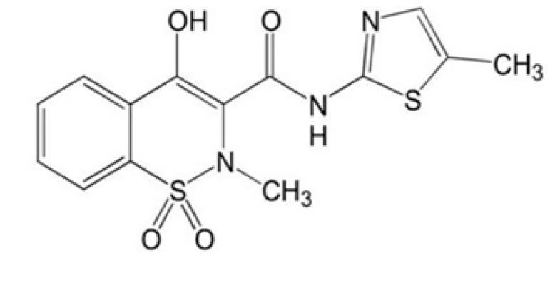 Figure 1
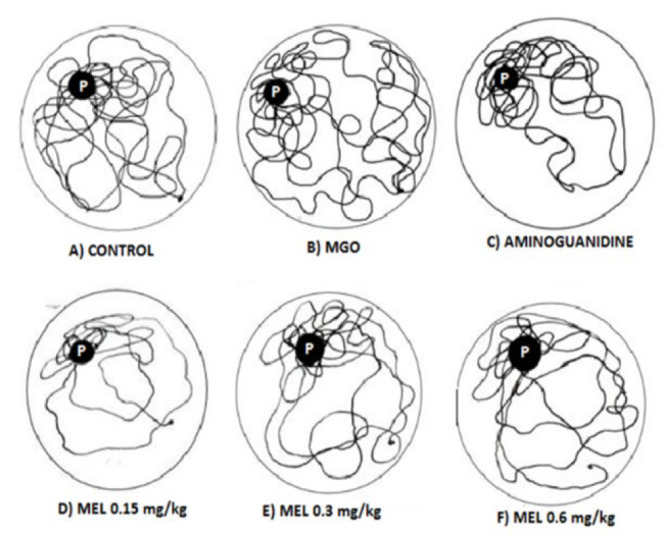 Figure 2
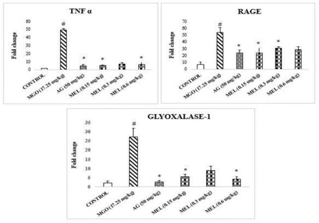 Figure 3
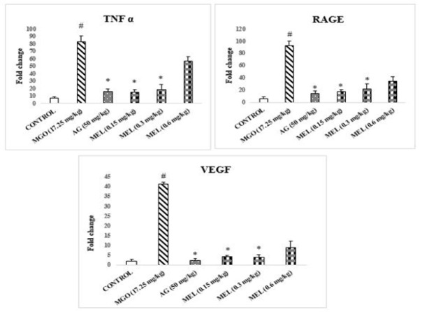 Figure 4
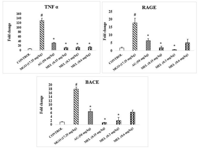 Figure 5
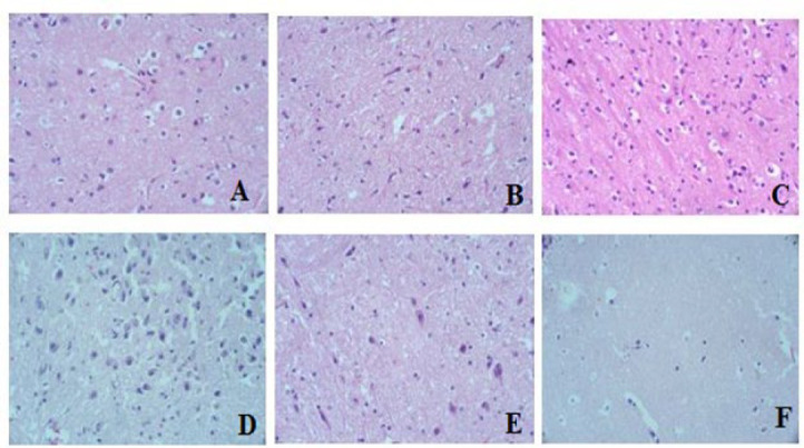 Figure 6
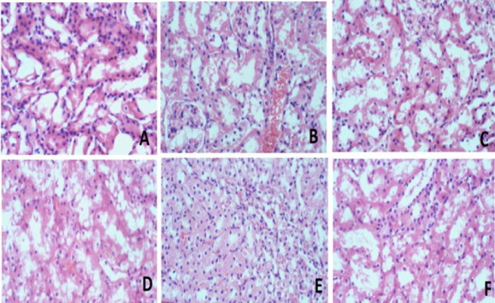 Figure 7
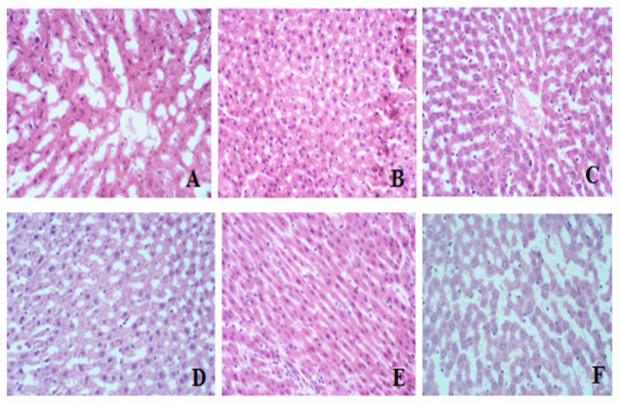 Figure 8
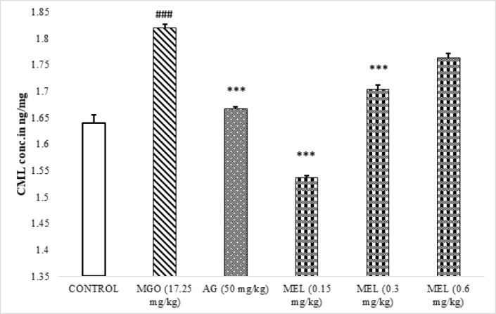 Figure 9
| GENE | PRIMER SEQUENCE | |
|---|---|---|
| BACE | F | AGGGCTACTATGTGGAGATG |
| R | CATACACAGACTTTCGGAGG | |
| RAGE | F | AGTCCGAGTCTACCAGATTC |
| R | TCTCCTCCTTCACAACTGTC | |
| GLYOXALASE-1 | F | CAAGATCCTGATGGCTACTG |
| R | CAGAATGGCTTGAACTGGAG | |
| VEGF | F | GGAGTACCCCGATGAGATA |
| R | TCATCTCTCCTATGTGCTGG | |
| TNF-α | F | CACGCTCTTCTGTCTACTG |
| R | CTGCTTGGTGGTTTGCTA | |
| GAPDH | F | GGATGGAATTGTGAGGGAGA |
| R | GTGGACCTCATGGCCTACAT | |
| Groups | Time to reach target quadrant (sec) | Time spent in target quadrant (sec) | Crossings through platform position (Count) |
|---|---|---|---|
| Control | 9 ± 2 | 43 ± 3 | 8 ± 1 |
| Model group | 20 ± 1 # | 25 ± 2 ## | 5 ± 0.3 # |
| AG (50 mg/kg) | 3 ± 1 *** | 72 ± 6 *** | 17 ± 2 *** |
| MEL (0.15 mg/kg) | 4 ± 1 *** | 56 ± 1 *** | 10 ± 0.3 ** |
| MEL (0.3 mg/kg) | 3 ± 1 *** | 37 ± 4 ** | 9 ± 1 * |
| MEL (0.6 mg/kg) | 4 ± 0.3 *** | 34 ± 3 * | 10 ± 1 * |
| Treatment groups | Renal function | Hepatic function | ||||
|---|---|---|---|---|---|---|
| Creatinine | Bilirubin | Direct bilirubin | ALP | SGPT | SGOT | |
| Control | 0.7 ± 0.02 | 0.6 ± 0.06 | 0.14 ± 0.02 | 177 ± 2 | 23 ± 2 | 19 ± 2 |
| MGO | 0.9 ± 0.06 # | 0.7 ± 0.07 | 0.16 ± 0.01 | 218 ± 5 # | 30 ± 1 # | 24 ± 1 # |
| AG (50 mg/kg) | 0.6 ± 0.01* | 0.7 ± 0.03 | 0.15 ± 0.02 | 190 ± 5* | 23 ± 1* | 18 ± 1* |
| MEL (0.15 mg/kg) | 0.6 ± 0.01** | 0.6 ± 0.01 | 0.13 ± 0.01 | 162 ± 3** | 19 ± 1** | 14 ± 1** |
| MEL (0.3 mg/kg) | 0.6 ± 0.04* | 0.6 ± 0.04 | 0.14 ± 0.00 | 169 ± 4 ** | 20 ± 2** | 17 ± 1* |
| MEL (0.6 mg/kg) | 0.6 ± 0.05* | 0.6 ± 0.05 | 0.14 ± 0.01 | 197 ± 3* | 22 ± 3* | 17 ± 2* |
Peer Reviews
No public reviews on file for this paper yet. If you reviewed it on a platform where reviews are public (OpenReview, ICLR, NeurIPS, ICML), you can paste yours below so the community can read it here.
Videos
No videos yet. Explain this paper in a talk, walkthrough, or lecture? Add one.
Taxonomy
TopicsAdvanced Glycation End Products research · Biochemical and biochemical processes · Natural Antidiabetic Agents Studies
Introduction
Aging, also known as senescence, is the physiological process of getting old concomitant with the emergence of various ailments (1). One of the possible underlying explanations is the glycation theory, which states that age co-relates with the burden of Advanced glycation end products, i.e., AGEs (2). They contribute to the progressive loss of body functions with time (3). AGEs are formed upon non-enzymatic interaction between carbohydrates (particularly the carbonyl group) and protein (amino acid, particularly the N-terminal of lysine and arginine’s side chain). This post-translational phenomenon is called the Maillard reaction. Glycation happens in each cell of our body; however, glycation becomes heightened in numerous pathological conditions, especially during hyperglycemia (4). They are reported to underlie cognitive decline (5), diabetes-related issues (6), kidney (7) and liver (8) abnormalities. Furthermore, AGEs interact with their receptors, known as RAGE, and cause stress, termed glycative stress (9). Methylglyoxal (MGO), alternatively known as 2-oxopropanal or pyruvaldehyde, possesses two carbonyl groups. Its formation is attributed to glycolysis, lipid metabolism, and amino acid metabolism (10). The accumulation of dicarbonyl compounds leads to heightened modification of DNA and proteins, potentially resulting in tissue and cell dysfunction and aging (11). MGO can also alter proteins, particularly lysine and arginine residues, producing AGEs (12).
Numerous efforts have been made to develop the inhibitors of glycation. The first AGE inhibitor that received some success was aminoguanidine, a scavenger of the di-carbonyl intermediates formed during glycation reaction (13). Unfortunately, it could also not pave the way to bedside for various reasons, particularly unpleasant outcomes. Under these conditions, the notion of repurposing enables a viable and cheap way to introduce novel compounds for the process of drug development (14). Initial screening of lead compounds for drug development initiatives relies heavily on the concept of structure-activity relationship (15). Regarding this, sulfur-containing compounds have been reported to possess anti-glycation properties, potentially exerting their effects through various mechanisms such as antioxidant activity, trapping reactive carbonyl species, and modulating pathways involved in AGE formation (16). Meloxicam is an enolic acid that belongs to the class of “oxicam .”MEL contains a thiazole ring, a five-membered aromatic heterocyclic compound with nitrogen and sulfur atoms, and a cyclic tertiary sulfonamide moiety (17, 18). Based on the aforementioned structural similarity, the current work aimed to detect and analyze MEL (Figure 1), a clinically used NSAID (19), for its potential utility as an anti-glycation drug.
Materials and Methods
Animals
Wistar rats (150–200 g) were purchased from the Animal Resource Facility of the International Centre for Chemical and Biological Sciences, University of Karachi. They were housed in standard conditions, i.e., 12-hour cycles of darkness and light, and a constant temperature of 25±1 °C. The rats were housed in recommended cages, given free access to pelleted rat chow, and cleaned tap water ad libitum. Approval for all experiments was granted by the Institution’s Animal Ethics Committee (Approval No: 2021–007/MM). Additionally, the principles outlined in the “Guide for the Care and Use of Laboratory Animals” published by the National Research Council (NRC) were followed during the entire study.
Chemicals
Formalin, methylglyoxal, and sodium chloride were purchased from Sigma-Aldrich (Germany). Meloxicam was a gift from Hilton Pharma Ltd (Pakistan).
Grouping of animals
Wistar rats were randomized into six groups of 3 rats:
Group 1: (Vehicle control): Normal saline 0.9% (5 ml/kg, IP) for 14 days.
Group 2: (Model Group): Methylglyoxal (MGO, 17.25 mg/kg, IP) for 14 days (20).
Group 3: (Positive Control): Aminoguanidine (AG, 50 mg/kg, IP) was administered 30 min before MGO administration for 14 days.
Group 4, 5, and 6: (Treatment Groups): Meloxicam (MEL; 0.15 mg/kg, 0.3 mg/kg, or 0.6 mg/kg, IP) was administered 30 min before MGO injection for 14 days.
It is of note that the present work is part of a bigger project whereby multiple drugs were tested for their ability to be repurposed against glycation. A part of this work was published earlier which contains the data on vehicle and positive control (20), which is also used in this manuscript.
After 14 days, the following tests were performed:
Assessment of cognitive performance
The Morris Water Maze (MWM) experiment was used to evaluate spatial memory development (21). The MWM pool (black color), with a diameter of 180 cm and a height of 50 cm, was partitioned into four equal sections (quadrants). A disc-shaped platform (black color) was positioned within one of these sections for escape purposes. This platform’s location remained fixed throughout the experiment. The pool was filled with water and kept at 25 ± 2 °C. Distinct cues were strategically positioned along the pool’s walls to aid the animals in locating the platform. Initially, the platform rested 1 cm above the water’s surface during the familiarization session on day one. Four trials, each lasting 120 sec, were conducted to train the animals in this session. During these sessions, rats were placed in one of the pool’s quadrants facing the wall. If an animal found the platform within the allotted time, it could only remain on the elevated surface for 5 sec; alternatively, it was gradually led to the platform and permitted to stay about 30 sec. In subsequent training (acquisition) sessions conducted over the next three days, the platform was gradually submerged below the water’s surface, following the same procedure as day one, with five trials for each animal. On the fifth day, the platform was removed from the pool to assess spatial memory retention to perform a probe trial. The animals had 120 sec to locate the platform. A video camera mounted above the MWM recorded the experiment. Various parameters were analyzed to evaluate spatial memory retention, including latency to reach the target quadrant, duration spent in the target quadrant, and the number of crossings across the platform’s prior position.
Assessment of renal and hepatic functions
After collecting blood via cardiac puncture, it was permitted to coagulate at room temperature for 15 min. Later, the serum was obtained by centrifugation at 4250×g (or about 3500 RPM) for 20 min. For renal assessment, creatinine was measured, while for hepatic assessment, SGPT, SGOT, ALP, direct, and total bilirubin levels were measured via a clinical chemistry analyzer (AU5800, Beckman Coulter, Germany).
Expression Study
The gene expression was assessed using RT-qPCR (quantitative Polymerase Chain Reaction). The genes of interest from the liver were RAGE (receptor for advanced glycation end products), TNF-α (tumor necrosis factor-alpha), and Glyoxalase-1. From the kidney, genes of interest were RAGE, TNF-α, and VEGF (vascular endothelial growth factors), while TNF-α, RAGE, and BACE (beta-secretase) were from the brain. Glyceraldehyde-3-phosphate dehydrogenase (GAPDH) was used as a reference housekeeping gene. Total RNA from the liver, kidney, and brain was extracted with Trizol reagent and then converted to cDNA (cDNA Synthesis Kit, Thermo Scientific). The reaction mixture (25 µl) underwent thermal cycling in three steps, i.e., denaturation for 10 min at 95 °C, followed by a second denaturation for 15 sec at 95 °C, annealing for 30 sec at 60 °C, and a final extension for 30 sec at 72 °C. These denaturation, annealing, and extension steps were repeated for 40 cycles. The Thermo Scientific Maxima SYBR Green/ROX qPCR master mix (2X) kit was utilized with the respective primers (Table 1). Gene expression variation between control and treated groups was determined using the delta-delta cycle threshold (Ct) method (22).
Histology
The liver, brain, and kidney were extracted, cut, and preserved in 10% formalin. Following dehydration in higher ethanol concentrations, the organs were cleaned in xylene and embedded in paraffin. Thin 4 µm slices were prepared and stained with H&E (Haematoxylin & Eosin). A bright field microscope was used to analyze structural and morphological alterations in identified cells from each tissue (Olympus U-MDOB3, Japan).
CML Quantification
The serum obtained was also used to estimate CML using an ELISA kit (CEB977Ge 96 Tests, Cloud Clone Corp, China) following the manufacturer protocol. The test specimens were placed on a plate frame, and 50 μl was applied to each well. A membrane was placed over the plate and then maintained for 30 min. After the incubation, wells were washed thoroughly 3-4 times using a multichannel pipette, ensuring no well contamination, and then allowed to dry. Later, horseradish peroxidase (HRP, 50 μl) conjugate was applied to every well (except the blanks) and incubated for 30 min. Afterward, wells were rewashed, and chromogen solutions A and B (50 μl each) were added. The membrane was firmly sealed, and the plate was incubated for an additional 15 min. Finally, a stop solution was introduced, changing the color from blue to yellow. Absorbance readings were taken immediately at 450 nm via a spectrophotometer (Biotek Synergy HTX Multi-mode reader, Japan).
Statistical analysis
The data is provided as mean ± SEM (n=3 per group). For comparing groups statistically, one-way ANOVA was utilized, accompanied by post-hoc analysis (Least Significant Difference, LSD). The statistical analysis was performed using IBM SPSS software (version 21.0, USA). P<0.05 serves as the minimal level of significance.
Results
Evaluation of cognitive function
MGO-treated rats demonstrated a significant enhancement in escape latency time and time to reach the target quadrant, whereas a significant reduction in the time spent in the target quadrant and number of crossings through platform position was observed in comparison with control rats (Table 2). A vice-versa effect was noted in AG and MEL treatment rats compared to the MGO group. Among the various tested doses (0.15 mg/kg, 0.3 mg/kg, and 0.6 mg/kg), the lowest showed the most prominent effect.
The navigation patterns revealed the movement of control rats in the platform quadrant, while rats in the model group displayed erratic movement in the MWM pool (Figure 2). In contrast, the treatment groups (Aminoguanidine and meloxicam-treated rats) exhibited a pattern in their navigation plots more similar to that of the control.
Assessment of renal and hepatic function
The concentrations of creatinine, ALP, SGPT, and SGOT in the Model group (MGO) exhibited a significant elevation compared to the control group (Table 3). In contrast, both AG and MEL (0.15 mg/kg, 0.3 mg/kg, and 0.6 mg/kg) demonstrated a notable decrease in the amounts of markers in contrast to MGO.
Quantitative polymerase chain reaction (qPCR)
The qPCR of various samples are as follows:
** Liver **
The RT-qPCR analysis exhibited a significant increase in the fold change of RAGE, TNF-α, and Glyoxalase-I genes in methylglyoxal-administered livers compared with control samples (Figure 3). On the contrary, significant reductions in the expression of these genes were found in AG and MEL-treated rats.
** Kidney **
TNF-α, RAGE, and VEGF gene expression exhibit a significant increase compared to control rats (Figure 4). However, expression of these genes appears to be decreased in AG and MEL-administered rats compared to MGO rats.
** Brain **
The concentrations of RAGE, TNF-α, and BACE genes were significantly elevated in the brains of MGO-injected rats compared to the control group (Figure 5). However, their expression showed a significant decline in AG and MEL-treated brains compared to the MGO group.
Histopathological study
The histological examination of brain, kidney, and liver tissue revealed no discernible pathological alterations in any of the treatment groups compared to the control (Figures 6, 7, and 8, respectively).
CML Quantification
The results of CML quantification unveiled significantly (P<0.005) elevated CML levels in the serum of rats in the model group in comparison with the control (Figure 9). The AG and MEL-treated serums demonstrated a significant reduction in the levels of CML in comparison with MGO-treated rats.
Discussion
Glycation is one of the pathogenic pathways implicated in aging and related morbidities. Unfortunately, no medicine exists to fight this harmful anomaly. Repurposing is a quick and cost-effective way to introduce candidate compounds into drug research process. Having this in view, the current investigation was aimed to explore the influence of meloxicam, a clinically used NSAID, on glycative stress.
MGO, a di-carbonyl compound, has been reported to modify DNA and proteins, resulting in tissue and cell dysfunction, diseases, and aging (23). It can alter proteins, particularly their lysine and arginine residues, producing AGEs (12). Hence, it induced accelerated aging in rats via enhancing glycative stress (24). Morris water maze experiment is an effective tool for evaluating cognitive abilities and spatial memorization in rodents (25). Our findings revealed that MGO-treated rats showed severe deficits in visual learning and memory, as shown by higher latency in getting to the target quadrant, less time stayed in the target quadrant, and lowered frequency of crossings in contrast to the control group (Table 2). A similar effect of MGO on cognition was reported earlier, which authenticates our experimental setup (26). Both AG and MEL, especially their lower tested doses (0.15 and 0.3 mg/kg), have mitigated this MGO-induced cognitive decline. The literature revealed that MEL might display concentration-independent behavior. At lower concentrations, it was found to be more effective in certain studies than in higher doses (27-29). The most probable explanation is that excessive drug molecules may compete for binding or self-associate at higher doses, potentially reducing their availability to interact with reactive carbonyl species efficiently, thus attenuating the anti-glycation activity. However, further study is required to delineate this unusual outcome. Furthermore, our data involving navigation maps also exhibited erratic movement in MGO-treated rats, as observed earlier (30). However, like control rats, the treatment groups maintained more focused navigation in the platform quadrant (Figure 2). This indicates enhanced memory retention, suggesting its utility as a therapeutic intervention for cognitive dysfunction associated with glycative stress.
The renal and hepatic function was also altered in MGO-treated rats as the serum levels of creatinine, ALP, SGOT, and SGPT were higher in comparison with control (Table 3). A similar effect of MGO in serum biomarkers of renal and hepatic function was reported earlier following the administration of MGO, thereby validating our experimental setup (31). It is of note that the AG and MEL, especially lower tested doses, were found to protect the renal and hepatic function against the harmful effects of MGO.
In our experimental design, along with the biochemical, the expression of certain pathologically important genes from the liver, kidney, and brain were also quantified to explore the molecular changes in these tissues. Our data reveals a considerable elevation in the expression of all genes of interest, i.e., RAGE, TNF α, and Glyoxalase-1 in MGO-exposed hepatic tissues compared to control (Figure 3). This indicates a heightened glycation stress in the liver. A similar effect of MGO in hepatic tissues was reported earlier (32, 33). The search of the literature revealed that RAGE activation triggers phosphorylation of ERK1/2, PI3-K/AKT, JAK2, and RhoGTPases, culminating in NF-κB stimulation and pro-inflammatory cytokine production, such as TNF-α (32, 34). Conversely, the concomitant treatment with AG and MEL has significantly attenuated the aforementioned up-regulation of genes.
The VEGF expression was also assessed in the case of the kidney, along with TNF-α and RAGE. A literature search revealed that MGO triggers the synthesis of VEGF in endothelial and mesothelial cells, consequently fostering angiogenesis (35). Our investigation found considerable TNF-α, RAGE, and VEGF elevation in MGO-administered rats’ kidneys comparable to control rats (Figure 4). In conformity with AG, Meloxicam (MEL) administration decreased TNF-α, RAGE, and VEGF levels in the kidneys, comparable to the MGO-administered group, highlighting its potential as a therapeutic agent against MGO-induced renal injury.
In the case of MGO-treated brains, substantial up-regulation of RAGE, TNF α, and BACE in contrast to the control group was noted (Figure 5). In conformity, BACE activity was earlier associated with MGO-induced neurodegenerative processes (36). Conversely, simultaneous treatment with AG and MEL resulted in significant reductions in the fold change of RAGE, TNF α, and BACE expression in comparison with the MGO group, thereby underscoring their efficacy in ameliorating MGO-induced neuroinflammation and β-amyloid production.
Our histopathological evaluation did not reveal any structural changes in the model group’s brain, kidney, and liver compared to the control (Figures 6, 7, and 8). The lack of detectable pathological changes suggests that the duration of exposure may have been insufficient to produce significant histological alterations (37). Histological transformations typically require more time to manifest (38). However, the absence of overt histological abnormalities does not necessarily imply the absence of underlying physiological or biochemical alterations.
Carboxymethyllysine (CML) is one of the major AGEs that was reported to co-relate with the aging process and associated morbidities (39, 40). As a result, its concentration in serum was determined. Our data shows the significantly raised CML in the serum of MGO-treated rats (Figure 9). This advocates the role of CML in mediating the cognitive, hepatic, and renal impairment observed in the present study. In contrast, both AG and MEL administrations have significantly hampered the MGO-induced increase in CML levels. This supports the notion that the protective effect shown by AG and MEL in the present study was most likely because of their ability to inhibit glycation.
Chemical structure of meloxicam
Navigation Mapping in the Morris Water Maze
Expression of genes in liver tissue of rats
Expression of genes in Kidney tissue of rats
Expression of genes in Brain tissue of rats
Histology of Brain tissue of rats
Histology of Kidney tissue of rats
Histology of Liver tissue of rats
Effects of meloxicam on CML levels in the serum of rats
Conclusion
Our study demonstrates that meloxicam, a clinically used NSAID, mitigated rats’ MGO-induced cognitive, renal, and hepatic impairments. Hence, it is a potential lead molecule for repurposing as an anti-glycation agent.
The reference list from the paper itself. Each links out to its DOI / PubMed record.
- 1Li Z Zhang Z Ren Y Wang Y Fang J Yue H Aging and age-related diseases: From mechanisms to therapeutic strategies Biogerontology 2021221651873350263410.1007/s 10522-021-09910-5PMC 7838467 · doi ↗ · pubmed ↗
- 2Anguizola J Matsuda R Barnaby OS Hoy K Wa C De Bolt E Glycation of human serum albumin Clin Chim Acta 201342564762389185410.1016/j.cca.2013.07.013PMC 3795802 · doi ↗ · pubmed ↗
- 3Chaudhuri J Bains Y Guha S Kahn A Hall D Bose N The role of advanced glycation end products in aging and metabolic diseases: Bridging association and causality Cell Metab 2018283373523018448410.1016/j.cmet.2018.08.014PMC 6355252 · doi ↗ · pubmed ↗
- 4Biedrzycki G Wolszczak-Biedrzycka B Dorf J Michalak DŻendzian-Piotrowska M Zalewska A Antioxidant and anti-glycation potential of H 2 receptor antagonists—in vitro studies and a systematic literature review Pharmaceuticals 202316127312923776508110.3390/ph 16091273 PMC 10535796 · doi ↗ · pubmed ↗
- 5Lotan R Ganmore I Shelly S Zacharia M Uribarri J Beisswenger P Long term dietary restriction of advanced glycation end-products (AG Es) in older adults with type 2 diabetes is feasible and efficacious-results from a pilot RCT Nutrients 202012314331563307621710.3390/nu 12103143 PMC 7602440 · doi ↗ · pubmed ↗
- 6Rungratanawanich W Qu Y Wang X Essa MM Song B-J Advanced glycation end products (AG Es) and other adducts in aging-related diseases and alcohol-mediated tissue injury Exp Mol Med 2021531681883356875210.1038/s 12276-021-00561-7PMC 8080618 · doi ↗ · pubmed ↗
- 7Damrath JG Creecy A Wallace JM Moe SM The impact of advanced glycation end products on bone properties in chronic kidney disease Curr Opin Nephrol 20213041141710.1097/MNH.0000000000000713 PMC 815470633928911 · doi ↗ · pubmed ↗
- 8Pereira RM Botezelli J Dda Cruz Rodrigues KC Mekary RA Cintra DE Pauli JR Fructose consumption in the development of obesity and the effects of different protocols of physical exercise on the hepatic metabolism Nutrients 201794054262842593910.3390/nu 9040405 PMC 5409744 · doi ↗ · pubmed ↗