Ultrasonographic aspects of sural nerve anatomy
Márcio Luís Duarte, Ocacir de Souza Reis Soares, Jean-Louis Brasseur

TL;DR
This paper describes the anatomy of the sural nerve using ultrasound, helping improve surgical outcomes and nerve studies.
Contribution
The paper provides a detailed ultrasonographic description of the sural nerve's anatomy.
Findings
Ultrasonography is a practical method to visualize the sural nerve's anatomy.
Precise pre-surgery assessment of the sural nerve improves postoperative success.
Ultrasonography aids in conducting nerve conduction studies and biopsies.
Abstract
The anatomy of the sural nerve is highly variable, and the nerve can present injuries of various etiologies, including iatrogenic injury during surgery. Precise knowledge of the course and morphology of the sural nerve is valuable, and the ability to assess the nerve properly before surgery increases the postoperative success rate, as well as facilitating the execution of nerve conduction studies and biopsies. The purpose of this article is to describe and illustrate the anatomy of the sural nerve, as seen on ultrasonography, which is a practical and economical imaging method.
Genes, proteins, chemicals, diseases, species, mutations and cell lines named across the full text — each resolved to its canonical identifier and authoritative record.
Click any figure to enlarge with its caption.
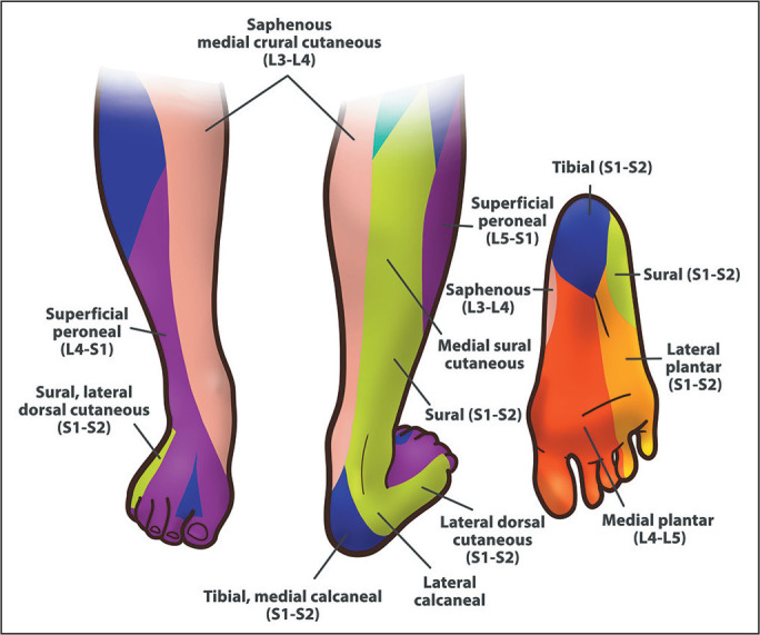 Figure 1
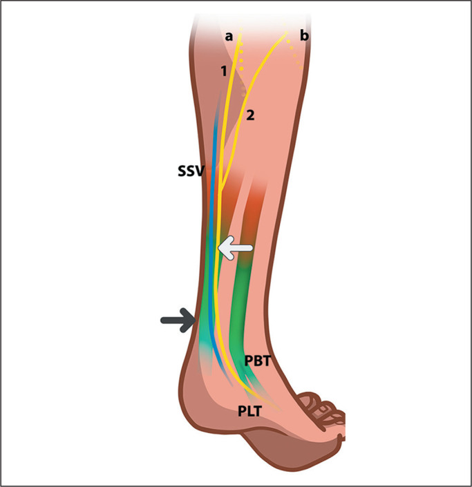 Figure 2
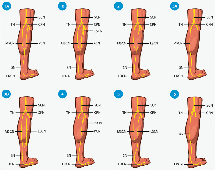 Figure 3
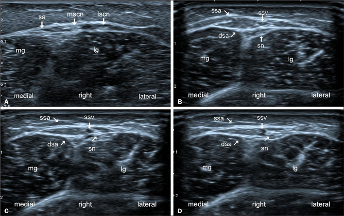 Figure 4
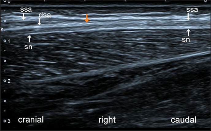 Figure 5
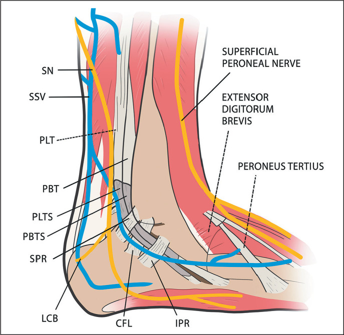 Figure 6
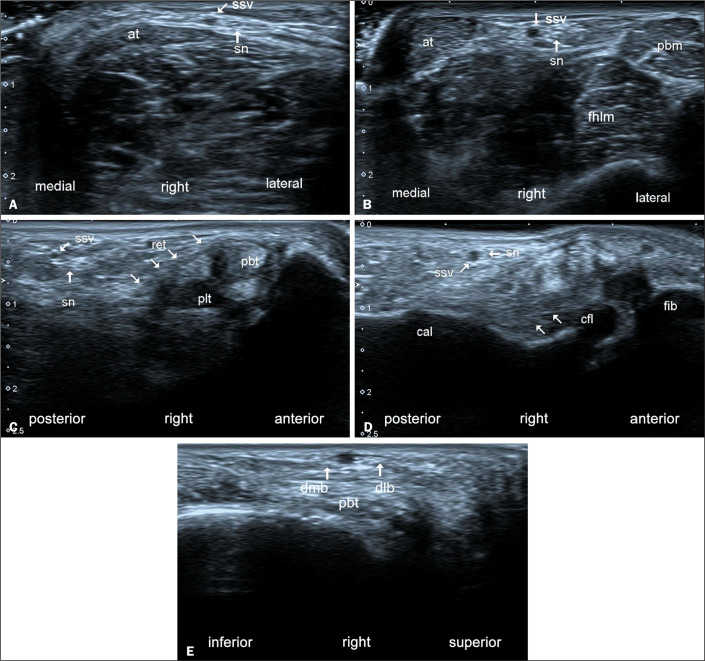 Figure 7
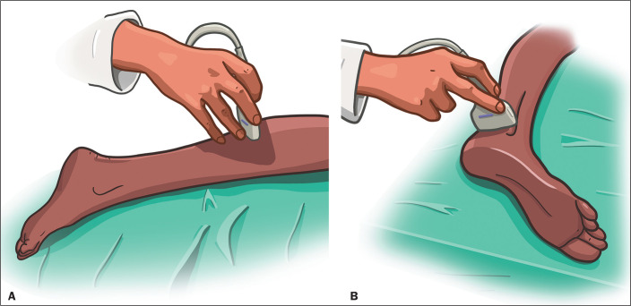 Figure 8
Peer Reviews
No public reviews on file for this paper yet. If you reviewed it on a platform where reviews are public (OpenReview, ICLR, NeurIPS, ICML), you can paste yours below so the community can read it here.
Videos
No videos yet. Explain this paper in a talk, walkthrough, or lecture? Add one.
Taxonomy
TopicsPeripheral Nerve Disorders · Nerve Injury and Rehabilitation · Tendon Structure and Treatment