Wide-angle fluid reservoir thickness changes during short-term scleral lens wear
Feifu Wang, Stephen J. Vincent, Pauline Cho, Yi Shen, Zihao Sheng, Meixiao Shen, Jun Jiang

TL;DR
This study used OCT imaging to show how fluid thickness under scleral lenses changes over time across different corneal regions and conditions.
Contribution
A new method for analyzing fluid reservoir thickness using wide-angle OCT and customized software during short-term scleral lens wear.
Findings
Fluid reservoir thickness decreased exponentially over four hours with the fastest reduction in the first two hours.
The thinnest fluid reservoir was in the superior mid-periphery for myopia and post-keratoplasty groups, and central for keratoconus.
The fluid reservoir was thickest inferiorly, showing the most asymmetry along the vertical meridian.
Abstract
To analyze the fluid reservoir thickness over the whole cornea during scleral lens settling using wide-angle optical coherence tomography (OCT) images and customized computer software. A total of 75 participants were recruited – 29 (myopes) with regular corneas and 46 with irregular corneas (35 with keratoconus, and 11 post-keratoplasty). All participants were fitted with customized scleral lenses and anterior segment OCT (Tomey Casia 2) images were taken 0, 30, 60, 120, and 240 min after lens application at the dispensing visit. Customized software was used to automatically segment the anterior cornea and the posterior surface of the scleral lens and determine the fluid reservoir thickness at 17 corneal regions across a 12 mm diameter. Fluid reservoir thickness decreased over time (P < 0.001) following an exponential decay, with no differences observed over time between the three…
Genes, proteins, chemicals, diseases, species, mutations and cell lines named across the full text — each resolved to its canonical identifier and authoritative record.
Click any figure to enlarge with its caption.
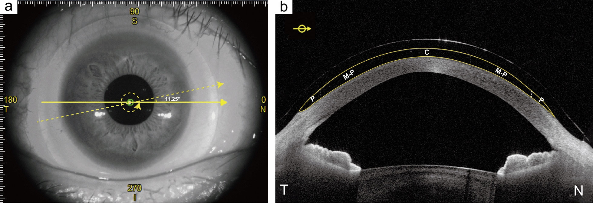 Figure 1
 Figure 2
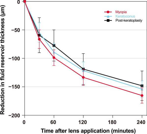 Figure 3
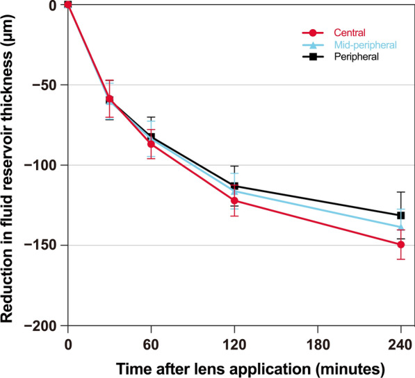 Figure 4
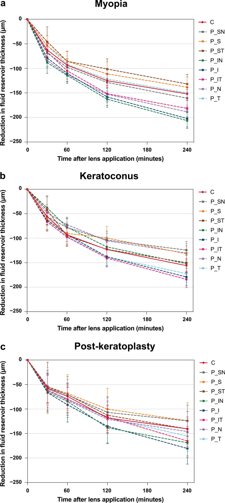 Figure 5
Peer Reviews
No public reviews on file for this paper yet. If you reviewed it on a platform where reviews are public (OpenReview, ICLR, NeurIPS, ICML), you can paste yours below so the community can read it here.
Videos
No videos yet. Explain this paper in a talk, walkthrough, or lecture? Add one.
Taxonomy
TopicsCorneal surgery and disorders · Intraocular Surgery and Lenses · Ophthalmology and Visual Impairment Studies