Foot Morphology Profile in Indonesian Recreational Runners
Astuti Pitarini, Andi Kurniawan, Mitchel Mitchel, Karina S Gani, Jeremy M Sudjaka, Erica Kholinne

TL;DR
This study describes foot shape characteristics in Indonesian recreational runners, finding differences in foot length between males and females but similar arch structures.
Contribution
Provides first detailed 3D foot morphology data for Indonesian recreational runners, including arch index and heel angle distributions.
Findings
Male runners had significantly longer feet than females (mean 256 mm vs 229 mm).
Most participants had low arch index values, with similar arch structures between genders.
Heel angles showed mild variations with only one case of severe angle among all participants.
Abstract
Background Foot morphology is influenced by factors such as physical activity, footwear habits, gender, and body mass index (BMI) and plays an important role in fields like orthopedics and sports science. However, data on the foot morphology of Indonesian recreational runners remain limited. This study aimed to describe the foot morphology of Indonesian recreational runners using a 3D foot scanner. Methods A cross-sectional study including 50 recreational runners was included. Data were collected using a self-developed questionnaire and foot scanning using a 3D foot scanner UPOD-S during static standing to assess a few parameters, including BMI, arch index (AI), heel angle, foot length, toe type, shoe size, shoe brand, vitamin D supplementation intake, history, and recent foot and leg pain, which were conducted via a physical examination by a foot and ankle surgeon. Result This…
Genes, proteins, chemicals, diseases, species, mutations and cell lines named across the full text — each resolved to its canonical identifier and authoritative record.
Click any figure to enlarge with its caption.
 Figure 1
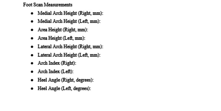 Figure 2
| Variable | Male | Female |
| Total patients | 32 | 18 |
| Age (years) | 33.1±10.6 | 35.0±9.9 |
| Body weight (kg) | 70.6±11.0 | 60.1±11.0 |
| Height (cm) | 171.7±6.0 | 157.8±4.7 |
| BMI (kg/m2) | 25.0±7.3 | 24.1±4.1 |
| Shoe size | 42.4±1.7 | 38.1±1.5 |
| Regular core training (n) | 10 | 8 |
| Regular aerobic exercise (n) | 8 | 6 |
| Sex | Left Foot | Right Foot | ||||||
| Neutral | Mild | Moderate | Severe | Neutral | Mild | Moderate | Severe | |
| Male | 6 | 18 | 6 | 2 | 7 | 17 | 8 | 0 |
| Female | 1 | 7 | 9 | 1 | 1 | 10 | 7 | 0 |
| Sex | Left Foot | Right Foot | ||||||||
| High | Normal | Low | High | Normal | Low | |||||
| + | ++ | +++ | + | ++ | +++ | |||||
| Male | 0 | 17 | 1 | 8 | 6 | 1 | 15 | 6 | 6 | 4 |
| Female | 1 | 9 | 2 | 3 | 3 | 1 | 7 | 2 | 4 | 4 |
| Variable | Arch Index | |
| Without pain | With pain | |
| Left foot | ||
| Male | 0.28±0.04 | 0.26±0.04 |
| Female | 0.27±0.02 | 0.27±0.06 |
| Right foot | ||
| Male | 0.28±0.03 | 0.25±0.04 |
| Female | 0.27±0.03 | 0.28±0.06 |
| Location | n (%) |
| Forefoot | |
| Metatarsalgia | 3 (6%) |
| Hallux valgus | 1 (2%) |
| Sesamoiditis | 1 (2%) |
| Midfoot | |
| Midfoot strain | 1 (2%) |
| Lisfranc injury | 1 (2%) |
| Tibialis posterior tendinitis | 3 (6%) |
| Plantar fibromatosis | 1 (2%) |
| Hindfoot | |
| Plantar fasciitis | 4 (8%) |
| Achilles tendinitis and/or tendinopathy | 2 (4%) |
| Baxter nerve lesion | 1 (2%) |
| Ankle | |
| Achilles tendinitis and/or tendinopathy | 2 (4%) |
| Knee | |
| Patellofemoral instability | 1 (2%) |
Peer Reviews
No public reviews on file for this paper yet. If you reviewed it on a platform where reviews are public (OpenReview, ICLR, NeurIPS, ICML), you can paste yours below so the community can read it here.
Videos
No videos yet. Explain this paper in a talk, walkthrough, or lecture? Add one.
Taxonomy
TopicsLower Extremity Biomechanics and Pathologies · Sports Performance and Training · Sports Dynamics and Biomechanics
Introduction
Running is a popular sport and recreational activity worldwide, including in Indonesia. Almost 60% of adults stay active by running, with the number of people participating in organized races increasing by 50% over the past decade [1]. Running is one of the most accessible physical activities, offering significant benefits for improving fitness and preventing obesity, cardiovascular diseases, and other chronic conditions [2]. Despite its surge in popularity, running-related injuries (RRI) and tendinopathy are common, particularly overuse injuries [3]. The injury incidence among short-distance runners (<15 km) ranges from 14.3% to 44.7%, whereas for long-distance runners (half-marathons or marathons), it ranges from 16.7% to 79.3% [4]. Many risk factors contribute to the RRI, including extrinsic factors (such as age, sex, limb mechanics, and genetics) and intrinsic factors (activity level, training factors, loading characteristics, footwear, and medication intake). However, previous research revealed that RRI was the strongest risk factor for injury in long-distance runners. Additionally, significant risk factors for short-distance runners included previous non-RRI (musculoskeletal complaints), higher body mass index (BMI), older age, male sex, lack of running experience, and running less than two hours per week [1].
Human foot morphology is a key focus in physical anatomical studies across various biomedical fields, such as orthopedics, orthotic development, and sports science. Factors such as environmental conditions, daily activities (including the frequency of sports participation and shoe-wearing habits), as well as gender, BMI, and age, have all been shown to significantly influence foot shape and structure in adults [5]. A widely used method for analyzing foot morphology is through 2D footprints, as they are easy to obtain. However, this approach lacks vertical shape information, which limits its accuracy [6]. Foot types such as flat feet or high arches are typically classified based on visual inspection, and numerous footprint-based indices have been developed, primarily targeting the medial longitudinal arch. Despite their practicality, 2D measurements fail to capture the foot's complex 3D structure. Research has shown that attempts to infer 3D morphology from 2D data often result in significant inaccuracies [7].
Materials and methods
This cross-sectional study included recreational runners participating in a running event. All participants underwent interviews and physical examinations to screen for foot or gait abnormalities. The inclusion criteria consisted of running at least 5 km per week for three consecutive months, either indoors or outdoors, regardless of whether participants were competing in the event. Consent was obtained from each participant. This study employed a simple random sampling approach to select participants and schedule measurements, ensuring unbiased data collection. In this study, we aimed to describe the foot morphology of Indonesian recreational runners using a 3D foot scanner. The parameters evaluated included arch index (AI), heel angle, foot length, and toe type. Additional information, such as BMI, shoe size and brand, vitamin D supplementation intake, and the presence of foot or leg pain, was collected through a combination of physical examination by a foot and ankle surgeon and a self-reported questionnaire. The Research Ethics Committee of Fakultas Kedokteran Universitas Trisakti approved the study (registration number 006/KER/FK/02/2025), and all participants were fully informed of the study's procedures and provided written consent.
Foot morphology
In this study, we measured the AI, heel angle, foot length, and toe type separately for the left and right feet to evaluate foot morphology. The heel angle consists of neutral (0), mild (<4), moderate (4-8), and severe (>8). For the AI, values below 0.21 were classified as high, 0.21-0.26 as normal, and above 0.26 as low. The low category is further subdivided into three levels: low (+) < 0.28; low (++) < 0.30; and low (+++) > 0.30 [8]. The toe type will be classified as either Roman, Greek, or Egyptian. The heel angle was defined as the angle formed between the medial and lateral borders of the posterior heel region. Foot length was defined as the linear distance from the most posterior aspect of the calcaneus to the tip of the longest toe.
Foot morphology data were collected under static standing conditions using a 3D foot scanner UPOD-S (Wuhan City, China) with an accuracy of ±1 mm. The participants needed to follow the steps: (1) Roll up their trousers above the knee and remove their shoes and socks; (2) stand with both feet in a natural posture, eyes and head straight facing front, and place one barefoot (right or left) on the UPOD-S foot scanner within the measuring range; (3) distribute body weight evenly between both feet and maintain balance for three to five seconds until the foot morphology scan is fully recorded; and (4) switch to the other foot and repeat the process.
Pain assessment and self-report survey
A foot and ankle surgeon conducted the foot pain assessments and self-reported surveys of recreational runners following foot morphology measurement. The survey included medical history, physical examination, and personal data (including sex, age, height, weight, email address, and phone number). For foot and leg pain assessment, knee pain was categorized as leg pain, while foot and ankle pain were classified as foot pain. The participants were asked to describe the quality of pain (e.g., burning, stabbing, dull ache), as well as the aggravating and relieving factors, including physical activities (e.g., walking, prolonged standing), footwear, orthotic use, and rest. Clinical examination assessed for associated foot conditions, such as hallux valgus (bunion), calluses, hammer toes, and skin changes.
Additionally, foot pain was divided into forefoot, midfoot, and hindfoot based on anatomical location. The analysis excluded muscle cramps, dermatological conditions, digital calluses, and nighttime paresthesia. Physical examinations were conducted to evaluate the recreational runner's foot and leg pain based on the following steps: (1) The surgeon reviewed the self-reported survey and pain complaints, then guided participants to stand barefoot with their trouser legs rolled up above the knees. (2) The lower extremities were examined through palpation and pressure testing of the feet, ankles, patellae, knees, hips, tibias, fibulas, and femurs, cross-checking the contralateral side for consistency. (3) To precisely localize pain, the surgeon assessed (i) soft tissues: plantar fascia, Achilles tendon, gastrocnemius, tibialis anterior and posterior, biceps femoris, quadriceps, medial/lateral ankle ligaments, anterior cruciate ligament, medial/lateral collateral ligaments, abductor hallucis, abductor digiti minimi, and lower back; (2) bone structures: navicular, cuboid, phalanges, metatarsals, and calcaneus; and (3) joints and key anatomical sites: ankles, patella, knees, hips, and tibia.
Results
This study involved 50 recreational runners (32 males and 18 females). The participants' ages ranged from 22 to 45 years, with mean body weights of 70.6±11.0 kg for males and 60.1±11.0 kg for females. Shoe sizes varied from 36 to 44 (European sizing). Detailed information is provided in Table 1. The average weekly running distance among participants was 13.8±13.1 km, reflecting considerable variation in training levels.
Participants wore various running shoe brands, including Hoka, Adidas, Asics, Nike, On Running, Diadora, and New Balance. Seven participants reported regular consumption of vitamin D supplements. Seven left feet and eight right feet exhibited a neutral heel angle, 25 left feet and 27 right feet showed a mild heel angle, and three left feet displayed a severe heel angle (Table 2).
Table 2: Heel angleMild <4; moderate 4-8; severe >8
Most participants had normal and low AI in both feet, as detailed in Table 3. The mean AI of the male left foot in those without pain was 0.28±0.04, while in those with pain, it was 0.26±0.04 (mean AI = 0.27±0.04). On the right foot, the AI in males without pain was 0.28±0.03, compared to 0.25±0.04 in those with pain (mean AI = 0.27±0.04). For females, the left foot AI was 0.27±0.02 in individuals without pain and 0.27±0.06 in those with pain (mean AI = 0.27±0.04). Similarly, on the right foot, the AI in females without pain was 0.27±0.03, while in those with pain, it was 0.28±0.06 (mean AI = 0.27±0.04). The details can be seen in Table 4.
Table 3: Type of arch indexHigh <0.21; normal 0.21-0.26; low >0.26Low (+) <0.28; low (++) ≤0.30; low (+++) >0.30
The toe types were identified among the participants: Roman, Egyptian, and Greek. In the male group, the mean foot lengths were 257.3±10.3 mm for the right foot and 257.5±11.2 mm for the left foot. Similarly, in the female group, the mean foot lengths were 231.3±8.5 mm for the right foot and 232.5±8.7 mm for the left foot. Foot and leg pain was reported by 21 participants, with the most common pathology being plantar fasciitis, which was found in four participants (8%). Detailed pathology is presented in Table 5.
Discussion
Running is one of the most widely practiced physical activities worldwide. However, although the health benefits of running for physically active individuals have been demonstrated, recreational runners are often affected by RRI at a rate of approximately 79% [4]. Various intrinsic and extrinsic factors, including foot morphology, have been implicated in the development of RRI [1]. However, there is limited data on the specific foot characteristics of runners across different populations. Based on our knowledge, this is the first study that describes the foot morphology in Indonesian recreational runners.
In our recent study, we observed that the AI values of feet with pain and those without pain were nearly identical. This suggests that foot and leg pain do not consistently correlate with variations in foot morphology, particularly when assessed using the AI. Furthermore, arch height variations may not be a key factor in foot pain development. This finding contrasts with previous research linking abnormal AI to foot pain. A possible explanation is that foot pain is multifactorial, involving neuromuscular function, inflammation, and individual pain perception [9]. Additionally, compensation mechanisms such as altered gait patterns or muscle adaptations may help mitigate plantar pressure abnormalities, thereby reducing their direct impact on pain. Esculier et al. reported that gait modifications in runners with patellofemoral pain, such as increasing the step rate by 10% (to 180 steps per minute), decreasing the step rate by 10%, striking with the forefoot or heel, and running softer, are associated with reduced movement-related pain. Additionally, gait modifications may induce analgesic effects that are not solely related to mechanical loading but also involve central pain mechanisms [10].
Previous research has highlighted the utility of the AI derived from footprints as a predictor of foot arch height and a tool for classifying footprint morphology [11]. According to the literature by Cavanagh and Rodgers [8], the AI value for normal arches typically falls within the range of 0.21-0.26. A retrospective study conducted by Chow et al. found the mean AI of both feet in recreational rugby players to be within the normal range. The elite group's arch type fell into the category of low arch, based on 57 male college students (mean age, 20 years). This recent study reported that the elite rugby group more commonly experienced lower limb pain, particularly pain in the metatarsophalangeal joints and the cuboid bone [12]. Our study found that most of the participants were classified as having low-arched feet or flat feet. This finding is consistent with a study by Chow, which reported that Indigenous Taiwanese have lower arched feet compared to non-Indigenous Taiwanese [13].
The data collected in this study show that males generally had longer feet than females on both sides. These gender-based differences in foot morphology align with findings from previous research, which consistently report that men tend to have longer feet than women. Zhao et al. noted that gender has a greater influence on foot dimensions, such as length, width, height, and girth, than BMI or age [14]. Similarly, Castro et al., using calipers and footprint analysis in older Brazilian adults, found that men had significantly higher instep height, forefoot width, and rearfoot width than women [15]. A study utilizing a 3D foot scanner in 291 older adults also confirmed that men exhibited significantly larger values across all major foot dimensions, including length, width, height, and girth, compared to women [16]. Wearing et al. observed that individuals with higher BMIs tend to have a lower plantar arch height. They attributed this finding to fat under the foot, making it difficult to measure arch height [17]. A similar study reported by Domjanic et al. found that a high BMI was associated with wide and flat feet [18]. Kim et al. reported that gender differences in lower limb biomechanics were observed following a 5 km barefoot run. In female runners, there was an increase in loading under the lateral forefoot and lateral midfoot, whereas male runners experienced a decrease [19]. Anatomical variations may influence these differences, as females exhibit greater hip internal rotation, while males show external rotation. Additionally, female runners demonstrated significantly higher hip adduction, hip internal rotation, and knee abduction angles than their male counterparts [20].
In this study, we found that most participants have normal or low-arched feet, which are commonly referred to as flat feet. A study conducted by Powell et al. indicated that individuals with high- or low-arched or flat feet experience a higher incidence of lower extremity injuries than those with normal arches. Furthermore, high-arched feet are associated with an increased risk of bony injuries on the lateral side of the lower extremity. In contrast, low-arched feet are more prone to injuries on the medial side [21]. Low- and normal-arch feet have better shock absorption than high-arch feet. Flat feet result in an uneven distribution of weight across the foot. Over time, this altered weight loading can lead to thickening of the plantar fascia due to increased mechanical stress. Consequently, individuals may rely on excessive knee and hip joint flexion to compensate for limited dorsiflexion during walking, which can ultimately result in joint pain and discomfort [22]. Feger et al. found that subjects with low-arched feet are more prone to an ankle sprain and ankle instability due to weak muscle strength in dorsiflexion and eversion [23]. In marathon and half-marathon runners, toe injuries such as bruised toenails, nail thickening, and subungual hematomas are common. These injuries result from the continuous friction between the toe and the shoe's toe box and can significantly affect a runner's training and performance [24]. Menz [25] also found that the wrong-fitted shoe size was associated with toe corns, hallux valgus deformity, and foot pain.
Marathon running presents a significant challenge for amateur runners. The gait characteristics of marathon runners are crucial, as the feet endure substantial force during running. Research indicates that the prolonged physical exertion associated with marathons can lead to fatigue in the soft tissues supporting the plantar arch, which may, in turn, alter pressure distribution across specific regions of the foot [26]. A study by Toresdahl et al. found that overuse injuries are the primary factors contributing to marathon noncompletion, and strength training did not reduce the incidence of overuse injuries, minor injuries, pain during the race, or the need for medical attention in the marathon medical tent [27]. Foot shape is dynamic and influenced by factors such as obesity and prolonged physical activity, which can result in deformities and variations in morphology. These changes may cause shoes to fit poorly, leading to dermatological issues such as blisters and corns. Additionally, improper footwear can lead to abnormal compensatory movements, thereby increasing the risk of musculoskeletal injuries.
A healthy runner's lifestyle is grounded in balanced nutrition, effective training routines, adequate recovery, and mental well-being. Proper nutrition is essential, emphasizing a diet rich in carbohydrates, proteins, and fats, along with sufficient hydration, to support optimal performance and recovery. Moreover, supplements like vitamin D can play a crucial role in preventing skeletal muscle injuries in athletes after exercise [28]. A similar study reported that lower serum vitamin D levels are associated with increased muscle weakness, fatigue, and a higher incidence of injuries [29]. Furthermore, dietary vitamin D supplementation has been shown to benefit immune system function by reducing exercise-induced proinflammatory cytokines in elite athletes [28]. However, in this study, we did not measure the serum 25(OH)D concentrations due to logistical limitations and the focus of our study.
The primary limitation of our study is the small sample size, which restricts the generalizability of our findings to the broader Indonesian population. This limitation may have reduced the statistical power of our analysis, potentially contributing to the lack of a significant association between the AI and foot and leg pain. Second, although the study focused on foot morphology in recreational runners, we did not provide detailed descriptions of the characteristics related to their running, including duration, intensity, frequency, and warm-up and cool-down protocols. This lack of specificity may hinder reproducibility and limit the ability of clinicians or researchers to implement or compare similar interventions in future studies.
Conclusions
Male Indonesian recreational runners generally have longer foot lengths compared to females. Interestingly, the AI values in feet with and without pain were remarkably similar between men and women, with most of the genders exhibiting a low arch. This suggests that foot pain in runners with low arches or flat feet is not exclusively attributable to arch structure. As such, clinicians should explore other possible underlying foot conditions during assessment. These findings may suggest a general trend toward mild biomechanical variations in foot morphology among Indonesian recreational runners. Although these differences may seem subclinical, they could hold clinical relevance for orthopedic and sports medicine specialists when designing training programs for runners, as well as for long-term musculoskeletal health monitoring and preventive care.
The reference list from the paper itself. Each links out to its DOI / PubMed record.
- 1Risk factors for overuse injuries in short- and long-distance running: a systematic review J Sport Health Sci van Poppel D van der Worp M Slabbekoorn A 14281020213253527110.1016/j.jshs.2020.06.006PMC 7856562 · doi ↗ · pubmed ↗
- 2Biomechanical variables and perception of comfort in running shoes with different cushioning technologies J Sci Med Sport Dinato RC Ribeiro AP Butugan MK Pereira IL Onodera AN Sacco IC 93971820152444475410.1016/j.jsams.2013.12.003 · doi ↗ · pubmed ↗
- 3Effects of running-induced fatigue on plantar pressure distribution in novice runners with different foot types Gait Posture Anbarian M Esmaeili H 52564820162747770810.1016/j.gaitpost.2016.04.029 · doi ↗ · pubmed ↗
- 4Injuries in runners; a systematic review on risk factors and sex differences P Lo S One van der Worp MP ten Haaf DS van Cingel R de Wijer A Nijhuis-van der Sanden MW Staal JB 010201510.1371/journal.pone.0114937 PMC 433821325706955 · doi ↗ · pubmed ↗
- 5Three-dimensional quantitative analysis of healthy foot shape: a proof of concept study J Foot Ankle Res StankovićK Booth BG Danckaers F 81120182954116210.1186/s 13047-018-0251-8PMC 5845135 · doi ↗ · pubmed ↗
- 6Foot type classification: a critical review of current methods Gait Posture Razeghi M Batt ME 2822911520021198350310.1016/s 0966-6362(01)00151-5 · doi ↗ · pubmed ↗
- 73D foot shape generation from 2D information Ergonomics Luximon A Goonetilleke RS Zhang M 6256414820051608749810.1080/0014013050070970 · doi ↗ · pubmed ↗
- 8The arch index: a useful measure from footprints J Biomech Cavanagh PR Rodgers MM 547551201987361112910.1016/0021-9290(87)90255-7 · doi ↗ · pubmed ↗