Symmetrically Erupted Upper Fourth Molars and an Impacted Fifth Molar: A Case Report
Jian Shi, Ye Bi, Zhi Wu

TL;DR
A rare case of symmetrically erupted upper fourth molars and an impacted fifth molar is reported, with treatment based on patient symptoms and preferences.
Contribution
A unique clinical case of bilaterally erupted maxillary fourth molars and an impacted fifth molar is documented.
Findings
Symmetrically erupted maxillary fourth molars were identified in a 38-year-old male.
An impacted fifth molar was found in the right upper jaw through radiographic imaging.
Extraction of affected molars was performed, with follow-up care recommended for remaining teeth.
Abstract
Introduction: Supernumerary teeth are often asymptomatic and impacted within the jawbone, typically identified during routine radiographic examinations. While cases of impacted fourth molars have been reported, bilaterally erupted fourth molars are exceedingly rare. This case report presents a unique instance of symmetrically erupted maxillary fourth molars, along with an impacted fifth molar in the right upper jaw. Case Presentation: A 38-year-old male presented with food impaction and discomfort in the area of the left maxillary third molar. Clinical examination revealed bilaterally erupted fourth molars, and a panoramic radiograph incidentally uncovered an impacted fifth molar in the right maxilla. Cone beam computed tomography confirmed these findings. The left maxillary fourth molar was diagnosed with periapical periodontitis. Both the left maxillary third and fourth molars were…
Genes, proteins, chemicals, diseases, species, mutations and cell lines named across the full text — each resolved to its canonical identifier and authoritative record.
Click any figure to enlarge with its caption.
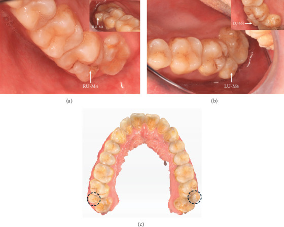 Figure 1
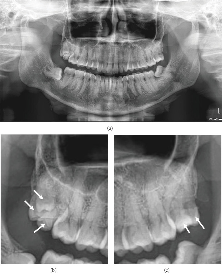 Figure 2
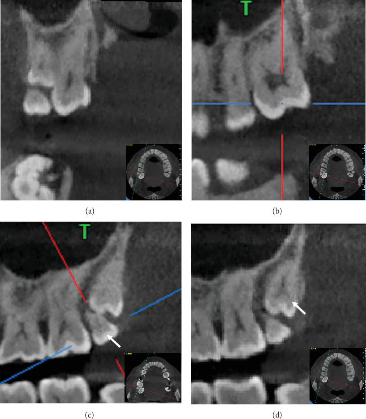 Figure 3
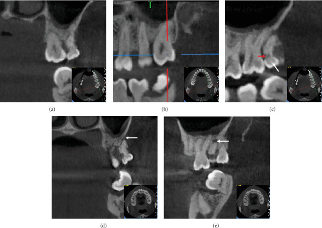 Figure 4
 Figure 5
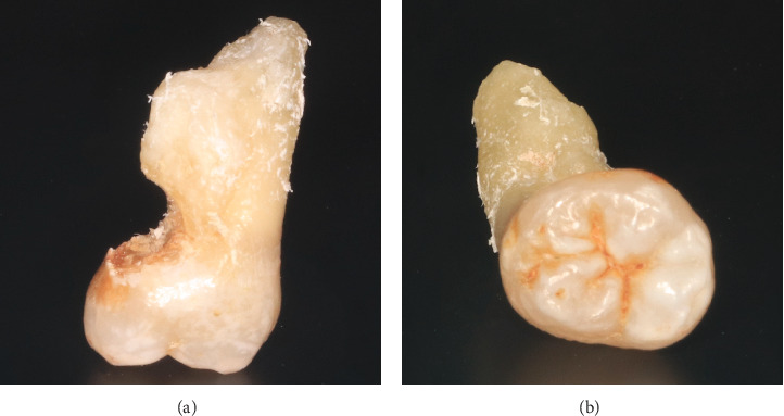 Figure 6
Peer Reviews
No public reviews on file for this paper yet. If you reviewed it on a platform where reviews are public (OpenReview, ICLR, NeurIPS, ICML), you can paste yours below so the community can read it here.
Videos
No videos yet. Explain this paper in a talk, walkthrough, or lecture? Add one.
Taxonomy
Topicsdental development and anomalies · Oral and Maxillofacial Pathology · Bone and Dental Protein Studies
1. Introduction
Supernumerary teeth (ST) are developmental anomalies characterized by an excess number of teeth in either the primary or permanent dentition. These additional teeth can occur unilaterally or bilaterally, as a single tooth or multiple teeth, and may be present in one or both jaws. ST are classified based on their location: mesiodens (situated between the two maxillary central incisors), paramolars (located between the second and third maxillary molars), and distomolars (positioned distal to the third molar) [1, 2].
The prevalence of ST ranges from 0.3% to 3.8% in the general population, with a slightly lower prevalence of 1.5% to 3.5% in individuals with permanent dentition. ST are more common in males, with a male-to-female ratio of approximately 2:1 [2, 3].
ST may erupt normally or remain impacted within the jawbone, often remaining asymptomatic. They are often discovered incidentally during routine imaging, such as panoramic radiography, usually prompted by dental complications like swelling, dental caries, occlusal interference, and pericoronitis or by other odontological complaints from patients at the dental clinic.
Based on searching on electronic databases of PubMed and Chinese databases (CNKI, Wanfang, and VIP databases), no similar case was found.
2. Case Presentation
A 38-year-old male presented to the Department of Stomatology with a chief complaint of food impaction and occasional discomfort in the region of the left maxillary third molar, persisting for several months. The patient reported no significant personal or family medical history and was generally in good health.
2.1. Clinical Examination
Vital signs included a blood pressure of 122/78 mmHg and a pulse rate of 70 beats per minute. Intraoral examination unexpectedly revealed symmetrically positioned bilateral maxillary fourth molars in the buccal embrasures between the second and third molars (Figure 1). The third molars appeared fully formed with no apparent decay, though the patient reported discomfort during percussion. Laboratory tests revealed that the patient's complete blood count and coagulation indices were within normal reference ranges.
2.2. Imaging Studies
Panoramic radiography showed that the left upper third and fourth molars overlapped in the two-dimensional image (Figure 2). Notably, three additional tooth-like structures were observed in the region of the right upper third molar. The patient was unaware of these ST and had no knowledge of similar conditions in his family.
To clarify the morphology, three-dimensional positioning, root structure, and relationships with adjacent anatomical structures, a cone beam computed tomography (CBCT) scan was performed (Figures 3 and 4). The CBCT confirmed the presence of an impacted fifth molar in the right upper maxilla (RU-M5). Coronal views showed the impacted fifth molar positioned buccal to the third molar, while sagittal views revealed it was distal to the fourth molar. The crowns of both the fourth and fifth molars exhibited molariform characteristics.
2.3. Final Diagnosis
Impacted teeth, ST, and periapical periodontitis of the left upper fourth molar (LU-M4) were diagnosed based on the clinical and radiographic examinations.
2.4. Treatment and Results
Considering the patient's preferences and symptomatology, along with a detailed analysis of the CBCT scan and 3D reconstruction (Figure 5), it was decided to extract the left maxillary third and fourth molars. The procedure was performed under local anesthesia, with the patient in aseptic conditions. Anesthetic was infiltrated into the left upper posterior maxilla, and the extractions were completed without perioperative complications. Examination of the extracted left maxillary fourth molar revealed decay on the proximal and lingual surfaces (Figure 6). The extraction socket was curetted and irrigated with saline, and platelet-rich fibrin (PRF) prepared from the patient's blood was packed into the socket to promote healing. The surgical site was closed with 3-0 sutures, and the patient was prescribed chlorhexidine mouth rinse (0.12%) twice daily for 5 days, along with an analgesic (diclofenac sustained-release capsules, 50 mg). Sutures were removed 1 week postoperatively. The patient chose not to proceed with the preventive extraction of the right upper wisdom tooth and the supernumerary molars, as there were no symptoms present. Regular follow-up appointments were recommended. The patient reported minimal postsurgical pain, and at the 1-week follow-up, the wound exhibited normal healing.
3. Discussion
The etiology and pathogenesis of ST remain unclear. Proposed theories include genetic predisposition, atavism, hyperactivity of the dental lamina, and environmental factors [4, 5]. There is evidence suggesting a correlation between ST and certain hereditary conditions, such as Ehlers–Danlos syndrome, Gardner syndrome, and cleidocranial dysplasia [6]. Notably, the patient in this case did not present with any of these conditions. Variants in the FER1L6 and PDGFRB genes have been linked to nonsyndromic ST [7, 8]. Recent studies have also identified a connection between heterozygous variants in FREM2 and the presence of mesiodens and ST [9]. These findings highlight the importance of retaining blood samples for further genetic testing and research in future cases of ST, particularly paramolars.
ST can be classified by shape into conical, tuberculate, molariform, and supplemental types [10, 11]. The conical supernumerary tooth, often peg-shaped, is the most common in permanent dentition and typically appears as a mesiodens, which may cause rotation or displacement of the permanent incisor but rarely delays eruption. Tuberculate STs, characterized by one or more cusps or tubercles and delayed root formation, are often found on the palatal side of the central incisor and are associated with delayed incisor eruption. Supplemental STs are duplications of teeth at the end of a tooth series, with the permanent maxillary lateral incisor being the most common, followed by supplemental premolars and molars [12]. The bilateral maxillary fourth molars (paramolars) and the impacted fifth molar in this case are molariform in shape and smaller than typical molars, though STs are often rudimentary in shape.
A review of the literature revealed only three original case reports [13–15] in English regarding fifth molars, based on searches of the PubMed database complemented by additional manual efforts. Searches conducted within Chinese databases (CNKI, Wanfang, and VIP databases) yielded no cases of supernumerary fifth molars. The rarity of fifth molars is evident. The details of the reported cases are summarized in Table 1. All reported patients were female, aged 20 to 33 years. Among the five impacted ST, one was impacted in soft tissue. Two cases [13, 15] exhibited symptoms unrelated to ST, while one case [14] reported pressure in the maxillary third molar region bilaterally. Two cases underwent preventive removal [13, 14], and one was recommended for periodic follow-up [15]. Due to the limited number of published cases, no definitive conclusions can be drawn about the prevalence of fifth molars by sex.
ST are typically impacted and asymptomatic, often discovered incidentally during panoramic or CBCT imaging, and may remain impacted throughout an individual's life. However, complications can arise from impacted STs, including tooth decay, root resorption of adjacent teeth, periapical periodontitis, and dentigerous cysts. Prophylactic extraction of ST is recommended to prevent such complications, especially in symptomatic cases. CBCT is an essential tool for accurate preoperative assessment and surgical planning. For asymptomatic patients who are hesitant to undergo surgery or are at higher surgical risk, periodic follow-up and monitoring should be considered.
4. Conclusion
ST are frequently impacted, and the occurrence of a fifth molar is exceptionally rare. This case report details a unique instance of symmetrically erupted fourth molars on both sides of the maxilla, along with a hidden fifth molar on the right. Treatment should be guided by the patient's symptoms and preferences. Additionally, reporting of similar cases is encouraged to enhance the understanding of supernumerary molars.
The reference list from the paper itself. Each links out to its DOI / PubMed record.
- 1Syriac G. Joseph E. Rupesh S. Philip J. Cherian S. A. Mathew J. Prevalence, Characteristics, and Complications of Supernumerary Teeth in Nonsyndromic Pediatric Population of South India: A Clinical and Radiographic Study Journal of Pharmacy & Bioallied Sciences 20179 Supplement 1S 231S 23610.4103/jpbs.JPBS_154_172-s 2.0-8503656944129284970 PMC 5731020 · doi ↗ · pubmed ↗
- 2Mukhopadhyay S. Mesiodens: A Clinical and Radiographic Study in Children Journal of Indian Society of Pedodontics and Preventive Dentistry 2011291343810.4103/0970-4388.799282-s 2.0-7995586524921521916 · doi ↗ · pubmed ↗
- 3Mahabob M. N. Anbuselvan G. J. Kumar B. S. Raja S. Kothari S. Prevalence Rate of Supernumerary Teeth Among Non-Syndromic South Indian Population: An Analysis Journal of Pharmacy and Bioallied Sciences 20124 Supplement 2S 373S 3752306629310.4103/0975-7406.100279 PMC 3467922 · doi ↗ · pubmed ↗
- 4Rahnama M. Szyszkowska A. Pulawska M. Szczerba-Gwozdz J. A Rare Case of Retained Fourth Molar Teeth in Maxilla and Mandible. Case Report Current Issues in Pharmacy and Medical Sciences 201427211812010.2478/cipms-2014-00282-s 2.0-84939785524 · doi ↗
- 5Primosch R. E. Anterior Supernumerary Teeth - Assessment and Surgical Intervention in Children Pediatr Dent. 1981322042156945564 · pubmed ↗
- 6Cassetta M. Altieri F. Giansanti M. Di-Giorgio R. Calasso S. Morphological and Topographical Characteristics of Posterior Supernumerary Molar Teeth: An Epidemiological Study on 25,186 Subjects Medicina Oral, Patologia Oral y Cirugia Bucal 2014196 e 545e 54910.4317/medoral.197752-s 2.0-8490839452825129242 PMC 4259368 · doi ↗ · pubmed ↗
- 7Hua W. Gan Z. Wu Y. Zhao L. Identification of a Novel Missense Mutation in Non-Syndromic Familial Multiple Supernumerary Teeth Archives of Oral Biology 202214310554210.1016/j.archoralbio.2022.10554236108431 · doi ↗ · pubmed ↗
- 8Bae D. H. Lee J. H. Song J. S. Jung H. S. Choi H. J. Kim J. H. Genetic Analysis of Non-Syndromic Familial Multiple Supernumerary Premolars Acta Odontol Scand. 201775535035410.1080/00016357.2017.13125152-s 2.0-8501725595328393601 · doi ↗ · pubmed ↗