Compressed sensing-based image reconstruction for discrete tomography with sparse view and limited angle geometries
Haytham A. Ali, Essam A. Rashed, Hiroyuki Kudo

TL;DR
This paper introduces a new image reconstruction method for CT scans that works well even with limited data and noise.
Contribution
A novel framework combining compressed sensing and parametric level sets for discrete tomography is proposed.
Findings
The proposed method preserves boundary sharpness and discrete intensity levels in undersampled and noisy conditions.
Quantitative metrics show superior performance over conventional methods in sparse-view and limited-angle scenarios.
The approach demonstrates robustness and accuracy on both synthetic and real CT datasets.
Abstract
This paper addresses the image reconstruction problem in discrete tomography, particularly under challenging imaging conditions such as sparse-view and limited-angle geometries commonly encountered in computed tomography (CT). These conditions often result in low-quality reconstructions due to insufficient projection data and incomplete angular coverage. To overcome these limitations, we propose a novel reconstruction framework that integrates compressed sensing (CS) with a parametric level set (PLS) method tailored for discrete images. The proposed approach leverages prior knowledge of discrete gray-level values and employs a parametric level set function to represent boundaries in both binary and multi-gray-level images. Unlike previous methods, our PLS is constructed using a dictionary of basis functions composed of single-scale or multiscale Gaussian functions. Reconstruction is…
Genes, proteins, chemicals, diseases, species, mutations and cell lines named across the full text — each resolved to its canonical identifier and authoritative record.
Click any figure to enlarge with its caption.
 Figure 1
 Figure 2
 Figure 3
 Figure 4
 Figure 5
 Figure 6
 Figure 7
 Figure 8
 Figure 9
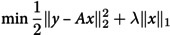 Figure 10
 Figure 11
 Figure 12
 Figure 13
 Figure 14
 Figure 15
 Figure 16
 Figure 17
 Figure 18
 Figure 19
 Figure 20
 Figure 21
 Figure 22
 Figure 23
 Figure 24
 Figure 25
 Figure 26
 Figure 27
 Figure 28
 Figure 29
 Figure 30
 Figure 31
 Figure 32
 Figure 33
 Figure 34
 Figure 35
 Figure 36
 Figure 37
 Figure 38
 Figure 39
 Figure 40
 Figure 41
 Figure 42
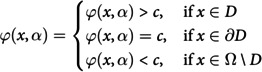 Figure 43
 Figure 44
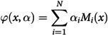 Figure 45
 Figure 46
 Figure 47
 Figure 48
 Figure 49
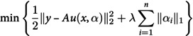 Figure 50
Peer Reviews
No public reviews on file for this paper yet. If you reviewed it on a platform where reviews are public (OpenReview, ICLR, NeurIPS, ICML), you can paste yours below so the community can read it here.
Videos
No videos yet. Explain this paper in a talk, walkthrough, or lecture? Add one.
Taxonomy
TopicsMedical Imaging Techniques and Applications · Digital Image Processing Techniques · Advanced X-ray and CT Imaging