Artemisinin alleviates arsenic-induced myocardial injury in rats by modulating oxidative stress and inflammatory responses: Artemisinin alleviates arsenic-induced myocardial injury
Wenjuan Qin, Yifei Zhou, Chuncui Chen, Xueting Guo, Ruimeng Tian, Ruoxi Chen, Wenrong Shi, Lei Huang, Caiyun Zhang, Shanshan Dong, Guilin Lu

Abstract
Genes, proteins, chemicals, diseases, species, mutations and cell lines named across the full text — each resolved to its canonical identifier and authoritative record.
Click any figure to enlarge with its caption.
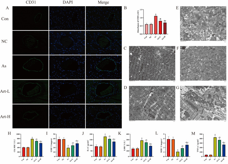 Figure 1
- —the grants from the National Natural Science Foundation of China
- —the Guiding Science and Technology Project of Xinjiang Production and Construction Corps
- —Self-funded by Shihezi University
- —the Youth Fund of the First Affiliated Hospital of Shihezi University
Peer Reviews
No public reviews on file for this paper yet. If you reviewed it on a platform where reviews are public (OpenReview, ICLR, NeurIPS, ICML), you can paste yours below so the community can read it here.
Videos
No videos yet. Explain this paper in a talk, walkthrough, or lecture? Add one.
Taxonomy
TopicsArsenic contamination and mitigation · Heavy Metal Exposure and Toxicity · Retinoids in leukemia and cellular processes
Arsenic is widely present in nature, and its compounds are extensively used in industrial, agricultural, and medical fields [1]. Arsenic trioxide (As _2_O 3) is specifically used as a therapeutic agent for acute promyelocytic leukemia because it induces cancer cell differentiation and apoptosis, significantly reduces the cancer cell count and has unique medical value [2]. However, owing to its high toxicity and carcinogenicity, long-term use can induce cardiovascular diseases such as arrhythmia and myocardial contractile dysfunction [3]. However, research on the treatment of arsenic-induced cardiotoxicity remains relatively scarce. Notably, artemisinin has anti-inflammatory and antioxidative effects on various heart diseases, effectively inhibiting reactive oxygen species (ROS) production, preventing myocardial damage and apoptosis caused by arsenic poisoning, and improving cardiac contractile and diastolic functions, thus enhancing cardiac function [4]. This study aims to discuss the impact of artemisinin on the myocardium of arsenic-poisoned rats.
Forty 12-week-old male SD rats [provided by SPF (Beijing) Biotechnology Co. Ltd, Beijing, China; SCXK-2024-0010] were randomly divided into five groups (8 rats per group): the control group (Con group), arsenic poisoning group (As group), drug control group (NC group, 30 mg/kg), low-dose artemisinin group (Art-L group, 30 mg/kg), and high-dose artemisinin group (Art-H group, 60 mg/kg). As _2_O 3 was intraperitoneally injected at 5 mg/kg/day for 10 days in the arsenic poisoning group, low-dose group, and high-dose group, whereas the control group and drug control group received equal volumes of physiological saline. Artemisinin was subsequently injected at the corresponding doses for three weeks. After the rat model was established, myocardial contrast images were obtained via the EPIQ7C ultrasound system (Philips, Eindhoven, Netherlands) and loaded into the QLab 12.0 workstation to obtain the time to peak (TTP), peak intensity (PI), wash-in slope (WIS), and area under the curve (AUC). Myocardial perfusion blood flow was represented by WIS×PI, with values measured three times and averaged. Blood samples were collected from the inner canthus vein, and myocardial injury marker and inflammatory factor levels were determined via kits from Nanjing Jiancheng Bioengineering Institute (Nanjing, China). Rats were then sacrificed to obtain myocardial tissue samples, and myocardial peroxidase levels were measured according to the instructions of superoxide dismutase (SOD) and reduced glutathione (GSH) kits. Myocardial tissue samples were fixed, embedded, sectioned, stained, and observed under an electron microscope to collect relevant images. CD31 expression in myocardial cells was measured via immunofluorescence technology. All animal experiments were conducted according to the “Guidelines and Operational Procedures for Experimental Animals” approved by the Experimental Animal Ethics Committee of the First Affiliated Hospital of Shihezi University School of Medicine.
Myocardial contrast echocardiography (MCE) is a non-invasive method for assessing myocardial blood perfusion and is an effective tool for monitoring myocardial microcirculation [5]. Compared with the control and drug control groups, the As group presented a significantly lower AUC and WIS × PI, indicating decreased myocardial blood perfusion ability. With artemisinin intervention, the AUC and WIS × PI values increased to a certain extent, more notably at higher concentrations. This is due to myocardial cell damage and cardiac dysfunction induced by arsenic poisoning, coupled with increased myocardial oxygen consumption from tachycardia and increased cardiac output caused by inflammation. Artemisinin antagonizes this reaction, effectively alleviating myocardial ischemia ( Table 1).
** Table 1 ** MCE parameters of each group of rats
Con
NC
As
Art-L
Art-H
F
P
WIS (dB/s)
28.32 ± 4.28
27.92 ± 3.34
5.55 ± 1.10*
10.55 ± 3.11* ^,+^
15.67 ± 3.01* ^,+,#^
84.93
< 0.05
PI (dB)
128.67 ± 3.73
127.84 ± 5.83
19.14 ± 4.67*
46.47 ± 5.83* ^,+^
79.81 ± 3.79* ^,+,#^
802.15
< 0.05
WIS×PI (dB ^2^/s)
3477.89 ± 215.52
3506.05 ± 203.72
136.95 ± 81.47*
517.97 ± 209.46* ^,+^
1259.54 ± 233.91* ^,+,#^
539.68
< 0.05
TTP (s)
13.05 ± 3.10
13.44 ± 3.03
14.37 ± 3.36
13.08 ± 2.18
13.65 ± 3.85
0.24
0.92
AUC
2746.62 ± 179.03
2724.73 ± 196.43
427.83 ± 148.36*
844.48 ± 91.74* ^,+^
1301.52 ± 189.95* ^,+,#^
336.38
< 0.05 * P < 0.05 compared with the Con and NC groups; ^+^ P < 0.05 compared with the As group; ^#^ P < 0.05 compared with the Art-L group. PI: peak intensity; WIS: wash-in slope; AUC: area under the curve; TTP: time to peak.
CD31 is a specific vascular endothelial marker that is positively correlated with myocardial angiogenesis and is often used to assess microvascular density [6]. In this study, the control and drug control groups presented lower CD31 (green) expression in myocardial cells. The As group exhibited extensive myocardial cell damage, oxidative reactions, and inflammation, promoting microvascular formation and resulting in significant fluorescence reactions. After intervention with different concentrations of artemisinin, the fluorescence reactions weakened to varying degrees with increasing concentrations, indicating the protective effect of artemisinin on myocardial cell damage induced by arsenic ( Figure 1A,B).
Figure 1 The effects of artemisinin on contrast-enhanced ultrasound, myocardial tissue structure, and biochemical markers in arsenic-poisoned rats(A) The effect of artemisinin on the immunofluorescence staining of myocardial tissue in arsenic-poisoned rats CD31 (green) and DAPI (blue). Magnification, ×400. (B) CD31 immunofluorescence absorbance values in each group of rats. (C–G) Electron microscopy results of myocardial tissue in rats treated with artemisinin. (C) Con group. (D) NC group. (E) As group. (F) Art-L group. (G) Art-H group. (H–M) Blood serum samples were collected from each group to measure the following parameters: (H) CK-MB, creatine kinase-MB; (I) LDH, lactate dehydrogenase; (J) GSH, glutathione; (K) SOD, superoxide dismutase; (L) IL-6, interleukin-6; and (M) TNF-α, tumor necrosis factor-α. a P < 0.05 compared to the Con and NC groups; b P < 0.05 compared to the As group; c P < 0.05 compared to the Art-L group.
Additionally, electron microscopy results were analyzed to verify the experimental conclusions [7]. The control and drug control groups presented clear myocardial myofibrils, neatly arranged myocardial cells with clear contours and full shapes, and no noticeable necrotic cells. The As group presented disordered myocardial myofibrils with many fractures, separated myofilament bundles, tortuous Z-line structures, no H bands, myocardial cells of varying sizes and shapes, some shrunken and dissolved nuclei, swollen and enlarged mitochondria, irregular shapes, increased autophagic bodies, and various degrees of endoplasmic reticulum shrinkage. In the low-dose and high-dose groups, there was some improvement in the myofibrils, myocardial cells, and organelles, which became more pronounced with increasing artemisinin dosage ( Figure 1C–G).
Previous studies indicated that arsenic exposure leads to cardiac lipid peroxidation, generating large amounts of inflammatory factors, mediating oxidative stress, reducing myocardial contractility, and affecting the extracellular matrix, leading to endoplasmic reticulum stress (ERS) imbalance and inducing apoptosis [8]. The endoplasmic reticulum is crucial for protein synthesis, folding, and processing [9]. Arsenic poisoning causes excessive ROS formation, disrupting the oxidative-antioxidative system balance. High ROS levels significantly induce protein misfolding in the endoplasmic reticulum, ultimately causing tissue damage. SOD and GSH effectively reflect myocardial peroxidation [10], and the trends in immunofluorescence, contrast echocardiography, and myocardial injury markers (CK-MB, LDH, TNF-α, and IL-6) and inflammatory factors were consistent with the SOD and GSH results. Thus, artemisinin can reduce myocardial damage caused by inflammation in a dose-dependent manner ( Figure 1H–M).
In conclusion, artemisinin can inhibit ROS production, maintain extracellular matrix homeostasis, counteract inflammation and oxidative stress, and reduce myocardial cell damage caused by arsenic poisoning. This study provides insights for treating arsenic poisoning patients, but further clinical applications need to be explored.
Supporting information
24576Table_1
The reference list from the paper itself. Each links out to its DOI / PubMed record.
- 1Aktar S Mia S Makino T Rahman MM Rajapaksha AU Arsenic removal from aqueous solution: a comprehensive synthesis with meta-data Sci Total Environ 202386216082110.1016/j.scitotenv.2022.1608212023 Sc T En.86260821 A 36509267 · doi ↗ · pubmed ↗
- 2Iyer SG Elias L Stanchina M Watts J The treatment of acute promyelocytic leukemia in 2023: paradigm, advances, and future directions Front Oncol 202212106252410.3389/fonc.2022.106252436741714 PMC 9889825 · doi ↗ · pubmed ↗
- 3Huang X Liu Y Liu R Zou X Yang H The efficacy and adverse events of arsenic trioxide for the patients with myelodysplastic syndrome: a systematic review and component network meta-analysis Hematology 202328227414910.1080/16078454.2023.227414937908176 · doi ↗ · pubmed ↗
- 4Lin ZH Xiang HQ Yu YW Xue YJ Wu C Lin C Ji KT Dihydroartemisinin alleviates doxorubicin-induced cardiotoxicity and ferroptosis by activating Nrf 2 and regulating autophagy FASEB J 202438 e 2367710.1096/fj.202400222 RR 38775792 · doi ↗ · pubmed ↗
- 5Lyng Lindgren F Tayal B Bundgaard Ringgren K Ascanius Jacobsen P Hay Kragholm K Zaremba T Holmark Andersen N et al. The variability of 2D and 3D transthoracic echocardiography applied in a general population Int J Cardiovasc Imag 2022382177219010.1007/s 10554-022-02618-837726455 · doi ↗ · pubmed ↗
- 6Liu L Shi GP CD 31: beyond a marker for endothelial cells Cardiovasc Res 2012943510.1093/cvr/cvs 10822379038 · doi ↗ · pubmed ↗
- 7Aishwarya R Abdullah CS Remex NS Nitu S Hartman B King J Bhuiyan MS Visualizing subcellular localization of a protein in the heart using quantum dots-mediated immuno-labeling followed by transmission electron microscopy J Vis Exp 20222022: 10.3791/6408510.3791/64085 PMC 1123249836190289 · doi ↗ · pubmed ↗
- 8Lamas GA Bhatnagar A Jones MR Mann KK Nasir K Tellez‐Plaza M Ujueta F et al. Contaminant metals as cardiovascular risk factors: a scientific statement from the american heart association J Am Heart Assoc 202312 e 02985210.1161/JAHA.123.02985237306302 PMC 10356104 · doi ↗ · pubmed ↗