A guide to selecting high-performing antibodies for Stearoyl-CoA desaturase (SCD1) (UniProt ID: O00767) for use in western blot, immunoprecipitation, and immunofluorescence
Vera Ruíz Moleón, Charles Alende, Maryam Fotouhi, Sara González Bolívar, Riham Ayoubi, Carl Laflamme, Michael L Garelja, Cecilia Williams, Matilda Holm

TL;DR
This paper evaluates nine antibodies for detecting SCD1, a lipid metabolism enzyme, to help scientists choose reliable tools for experiments.
Contribution
The study provides a reproducible evaluation framework and open resource for selecting high-quality SCD1 antibodies.
Findings
Nine commercial SCD1 antibodies were tested for performance in western blot, immunoprecipitation, and immunofluorescence.
Results are shared as an open resource to improve antibody reproducibility in scientific research.
A standardized protocol using knockout and parental cell lines was used for antibody validation.
Abstract
The enzyme stearoyl-CoA desaturase (SCD1) is a modulator of lipid metabolism by catalyzing the biosynthesis of mono-unsaturated fatty acids from saturated fatty acids. Understanding the specific mechanisms by which SCD1 plays in health and disease can provide novel insides in therapeutic targets, a process that would be facilitated by the availability of high-quality antibodies. Here we have characterized nine SCD1 commercial antibodies for western blot, immunoprecipitation, and immunofluorescence using a standardized experimental protocol based on comparing read-outs in knockout cell lines and isogenic parental controls. These studies are part of a larger, collaborative initiative seeking to address antibody reproducibility issues by characterizing commercially available antibodies for human proteins and publishing the results openly as a resource for the scientific community. While…
Genes, proteins, chemicals, diseases, species, mutations and cell lines named across the full text — each resolved to its canonical identifier and authoritative record.
Click any figure to enlarge with its caption.
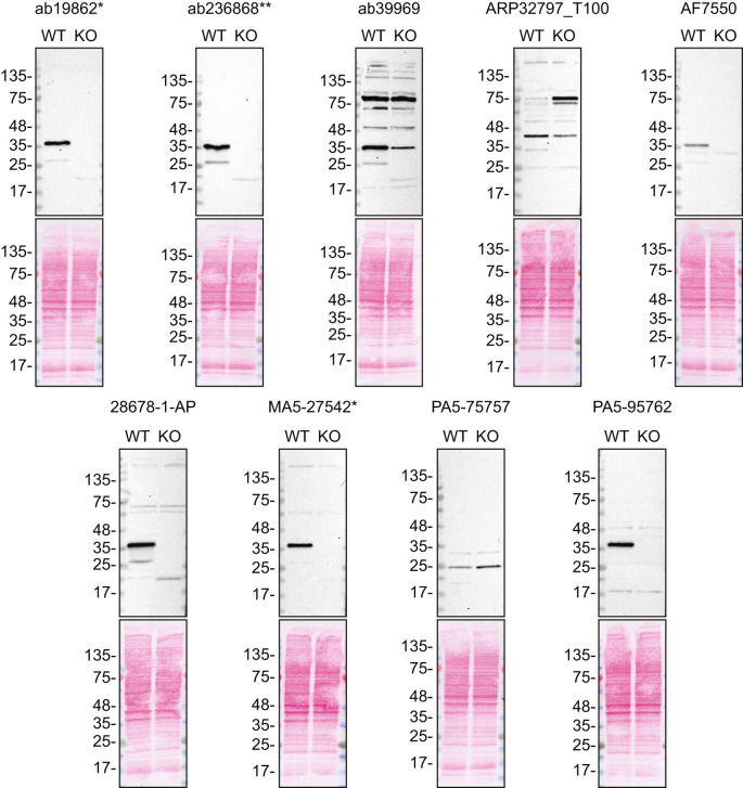 Figure 1
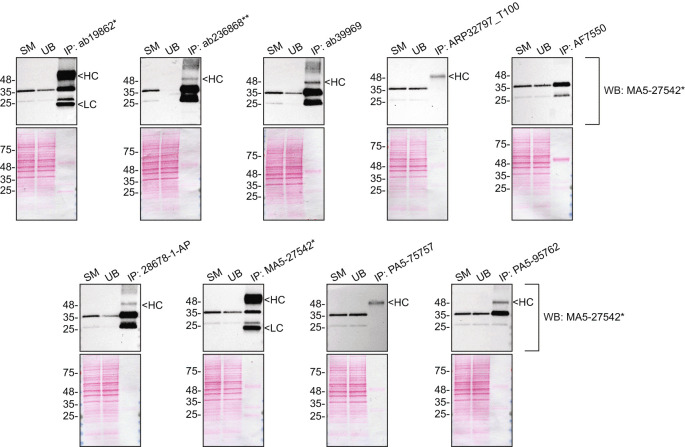 Figure 2
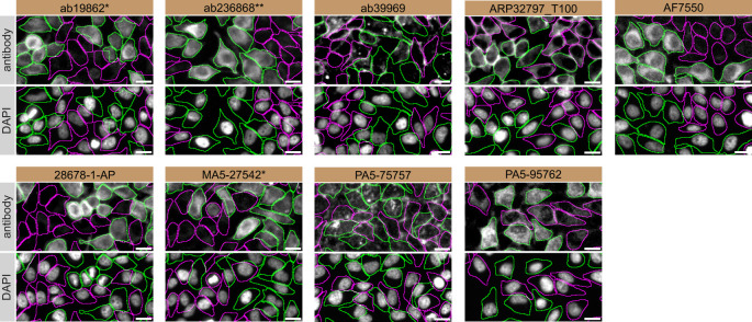 Figure 3
| Institution | Catalog number | RRID (Cellosaurus) | Cell line | Genotype |
|---|---|---|---|---|
| Abcam | ab255448 |
| HeLa | WT |
| Abcam | ab265220 |
| HeLa |
|
| Company | Catalog number | Lot number | RRID (Antibody Registry) | Clonality | Clone ID | Host | Concentration (μg/μl) | Vendors recommended applications |
|---|---|---|---|---|---|---|---|---|
| Abcam | ab19862
| 1057200-1 |
| monoclonal | CD.E10 | mouse | 1.00 | Wb, IP, IF |
| Abcam | ab236868
| 1007366-14 |
| recombinant mono | rabbit | 0.61 | Wb, IP, IF | |
| Abcam | ab39969 | 1036585-6 |
| polyclonal | rabbit | 0.90 | Wb | |
| Aviva Systems Biology | ARP32797_T100 | QC2226-43641 |
| polyclonal | rabbit | 1.00 | Wb | |
| Bio-Techne | AF7550 | CGOP0121061 |
| polyclonal | sheep | 0.20 | Wb | |
| Proteintech | 28678-1-AP | 00103543 |
| polyclonal | rabbit | 0.40 | Wb, IF | |
| Thermo Fisher Scientific | MA5-27542
| YH4004441A |
| monoclonal | CD.E10 | mouse | 1.00 | Wb, IP, IF |
| Thermo Fisher Scientific | PA5-75757 | YJ4089139 |
| polyclonal | rabbit | 1.00 | Wb, IF | |
| Thermo Fisher Scientific | PA5-95762 | YJ4090059A |
| polyclonal | rabbit | 1.35 | Wb, IF |
- —Michael J. Fox Foundation for Parkinson's Research
- —Mitacs
- —Ontario Genomics
- —Génome Québec
- —Genome Canada
Peer Reviews
No public reviews on file for this paper yet. If you reviewed it on a platform where reviews are public (OpenReview, ICLR, NeurIPS, ICML), you can paste yours below so the community can read it here.
Videos
No videos yet. Explain this paper in a talk, walkthrough, or lecture? Add one.
Taxonomy
TopicsPeroxisome Proliferator-Activated Receptors · Cancer, Lipids, and Metabolism · Cancer, Hypoxia, and Metabolism
Introduction
Stearoyl-CoA desaturase (SCD1) is a membrane-bound enzyme which catalyzes the rate-limiting step in the conversion of saturated fatty acids into mono-unsaturated fatty acids. ^ 1, 2 ^ The regulation of SCD1 is physiologically important as maintaining a proper ratio of saturated to monounsaturated fatty acids is essential for membrane fluidity. Disruption to this ratio can lead to pathological conditions, including cardiovascular disease, obesity, non-insulin dependent diabetes mellitus, hypertension, neurological diseases, immune disorders and cancer. ^ 2– 7 ^
SCD1 is of particular importance in Parkinson’s disease (PD) research as its inhibition has been found toto rescue α-Synuclein cytotoxicity and inclusion formation, both hallmarks of PD progression. The neurotoxic mechanisms underlying PD progression are not yet clearly defined. ^ 8– 10 ^ Mechanistic studies would be facilitated with the availability of high quality SCD1 antibodies.
This research is part of a broader collaborative initiative in which academics, funders and commercial antibody manufacturers are working together to address antibody reproducibility issues by characterizing commercial antibodies for human proteins using standardized protocols, and openly sharing the data. ^ 11– 13 ^ Here we evaluated the performance of nine commercial antibodies for SCD1 for use in western blot, immunoprecipitation, and immunofluorescence, enabling biochemical and cellular assessment of SCD1 properties and function. The platform for antibody characterization used to carry out this study was endorsed by a committee of industry academic representatives. It consists of identifying human cell lines with adequate target protein expression and the development/contribution of equivalent knockout (KO) cell lines, followed by antibody characterization procedures using most commercially available antibodies against the corresponding protein. The standardized consensus antibody characterization protocols are openly available on Protocol Exchange (DOI: 10.21203/rs.3.pex-2607/v1). ^ 14 ^
The authors do not engage in result analysis or offer explicit antibody recommendations. A limitation of this study is the use of universal protocols – any conclusions remain relevant within the confines of the experimental setup and cell line used in this study. Our primary aim is to deliver top-tier data to the scientific community, grounded in Open Science principles. This empowers experts to interpret the characterization data independently, enabling them to make informed choices regarding the most suitable antibodies for their specific experimental needs. Guidelines on how to interpret antibody characterization data found in this study are featured on the YCharOS gateway. ^ 15 ^
Results and discussion
Our standard protocol involves comparing readouts from WT (wild type) and KO cells. ^ 16, 17 ^ The first step was to identify a cell line(s) that expresses sufficient levels of a given protein to generate a measurable signal using antibodies. To this end, we examined the DepMap transcriptomics database to identify all cell lines that express the target at levels greater than 2.5 log 2 (transcripts per million “TPM” + 1), which we have found to be a suitable cut-off (Cancer Dependency Map Portal, RRID:SCR_017655). The HeLa expresses the SCD1 transcript at 6.7 log 2 (TPM+1) RNA levels, which is above the average range of cancer cells analyzed. A SCD KO HeLa cells were obtained from Abcam ( Table 1).
For western blot experiments, WT and SCD KO protein lysates were ran on SDS-PAGE, transferred onto nitrocellulose membranes, and then probed with nine antibodies in parallel ( Table 2, Figure 1).
SCD1 antibody screening by western blot.Lysates of HeLa WT and SCD KO were prepared, and 35 μg of protein were processed for western blot with the indicated SCD1 antibodies. The Ponceau stained transfers of each blot are presented to show equal loading of WT and KO lysates and protein transfer efficiency from the acrylamide gels to the nitrocellulose membrane. Tris-Glycine 4-20% gels were used. Antibody dilutions were chosen according to the recommendations of the antibody supplier. An exception was given for antibody AF7550 which was titrated because the signal was too weak when following the supplier’s recommendations. Antibody dilution used: ab19862 at 1/1000, ab236868** at 1/1000, ab39969 at 1/1000, ARP32797_T100 at 1/1000, AF7550 at 1/200, 28678-1-AP at 1/1000, MA5-27542* at 1/1000, PA5-75757 at 1/200 and PA5-95762 at 1/1000. Predicted band size: 41.5 kDa *Monoclonal antibody, *Recombinant antibody.
We then assessed the capability of all nine antibodies to capture SCD1 from HeLa protein extracts using immunoprecipitation techniques, followed by western blot analysis. For the immunoblot step, a specific SCD1 antibody identified previously (refer to Figure 1) was selected. Equal amounts of the starting material (SM), the unbound fraction (UB), as well as the whole immunoprecipitate (IP) eluates were separated by SDS-PAGE ( Figure 2).
SCD1 antibody screening by immunoprecipitation.HeLa lysates were prepared, and immunoprecipitation was performed using 1 mg of lysate and 2.0 μg of the indicated SCD1 antibodies pre-coupled to Dynabeads protein A or protein G. Samples were washed and processed for western blot with the indicated SCD1 antibody. For western blot, MA5-27542 was used at 1/1000. Tris-Glycine 4-20% gels were used. The Ponceau stained transfers of each blot are shown. Predicted band size: 41.5 kDa. SM=4% starting material; UB=4% unbound fraction; IP=immunoprecipitate, HC= antibody heavy chain, LC= antibody light chain. *Monoclonal antibody, *Recombinant antibody.
For immunofluorescence, nine antibodies were screened using a mosaic strategy. First, HeLa WT and SCD KO cells were labelled with different fluorescent dyes in order to distinguish the two cell lines, and the SCD1 antibodies were evaluated. Both WT and KO lines imaged in the same field of view to reduce staining, imaging and image analysis bias ( Figure 3). Quantification of immunofluorescence intensity in hundreds of WT and KO cells was performed for each antibody tested, and the images presented in Figure 3 are representative of this analysis. ^ 14 ^
SCD1 antibody screening by immunofluorescence.HeLa WT and SCD KO cells were labelled with a green or a far-red fluorescent dye, respectively. WT and KO cells were mixed and plated to a 1:1 ratio on coverslips. Cells were stained with the indicated SCD1 antibodies and with the corresponding Alexa-fluor 555 coupled secondary antibody including DAPI. Acquisition of the blue (nucleus-DAPI), green (WT), red (antibody staining) and far-red (KO) channels was performed. Representative images of the merged blue and red (grayscale) channels are shown. WT and KO cells are outlined with green and magenta dashed line, respectively. When an antibody was recommended for immunofluorescence by the supplier, we tested it at the recommended dilution. The rest of the antibodies were tested at 1 and 2 μg/mL and the final concentration was selected based on the detection range of the microscope used and a quantitative analysis not shown here. Antibody dilution used: ab19862 at 1/1000, ab236868** at 1/600, ab39969 at 1/150, ARP32797_T100 at 1/500, AF7550 at 1/200, 28678-1-AP at 1/400, MA5-27542* at 1/1000, PA5-75757 at 1/1000 and PA5-95762 at 1/1300. Bars = 10 μm. *Monoclonal antibody, *Recombinant antibody.
In conclusion, we have screened nine SCD1 commercial antibodies by western blot, immunoprecipitation, and immunofluorescence by comparing the signal produced by the antibodies in human HeLa WT and SCD KO cells. Several high-quality and renewable antibodies that successfully detect SCD1 were identified in all applications. Researchers who wish to study SCD1 in a different species are encouraged to select high-quality antibodies based on the results presented and investigate the predicted species reactivity of the manufacturer before extending their research.
Method
The standardized protocols used to carry out this KO cell line-based antibody characterization platform was established and approved by a collaborative group of academics, industry researchers and antibody manufacturers. The detailed materials and step-by-step protocols used to characterize antibodies in western blot, immunoprecipitation and immunofluorescence are openly available on Protocol Exchange (DOI: 10.21203/rs.3.pex-2607/v1). ^ 14 ^
Antibodies and cell line used
Cell lines used and primary antibodies tested in this study are listed in Tables 1 and 2, respectively. To ensure that the cell lines and antibodies are cited properly and can be easily identified, we have included their corresponding Research Resource Identifiers, or RRID. ^ 18, 19 ^
The reference list from the paper itself. Each links out to its DOI / PubMed record.
- 1Enoch HG CataláA Strittmatter P : Mechanism of rat liver microsomal stearyl-Co A desaturase. Studies of the substrate specificity, enzyme-substrate interactions, and the function of lipid. J. Biol. Chem. 1976;251(16):5095–5103. 10.1016/S 0021-9258(17)33223-4 8453 · doi ↗ · pubmed ↗
- 2Ntambi JM : Regulation of stearoyl-Co A desaturase by polyunsaturated fatty acids and cholesterol. J. Lipid Res. 1999;40(9):1549–1558. 10.1016/S 0022-2275(20)33401-5 10484602 · doi ↗ · pubmed ↗
- 3Kinsella JE Lokesh B Stone RA : Dietary n-3 polyunsaturated fatty acids and amelioration of cardiovascular disease: possible mechanisms. Am. J. Clin. Nutr. 1990;52(1):1–28. 10.1093/ajcn/52.1.1 2193500 · doi ↗ · pubmed ↗
- 4Jones BH Maher MA Banz WJ : Adipose tissue stearoyl-Co A desaturase m RNA is increased by obesity and decreased by polyunsaturated fatty acids. Am. J. Phys. 1996;271(1 Pt 1):E 44–E 49. 10.1152/ajpendo.1996.271.1.E 44 8760080 · doi ↗ · pubmed ↗
- 5Li J Ding SF Habib NA : Partial characterization of a c DNA for human stearoyl-Co A desaturase and changes in its m RNA expression in some normal and malignant tissues. Int. J. Cancer. 1994;57(3):348–352. 10.1002/ijc.2910570310 7909540 · doi ↗ · pubmed ↗
- 6Habib NA Wood CB Apostolov K : Stearic acid and carcinogenesis. Br. J. Cancer. 1987;56(4):455–458. 10.1038/bjc.1987.223 3689663 PMC 2001814 · doi ↗ · pubmed ↗
- 7Khoo DE Fermor B Miller J : Manipulation of body fat composition with sterculic acid can inhibit mammary carcinomas in vivo. Br. J. Cancer. 1991;63(1):97–101. 10.1038/bjc.1991.20 1989672 PMC 1971644 · doi ↗ · pubmed ↗
- 8Fanning S Haque A Imberdis T : Lipidomic Analysis of α-Synuclein Neurotoxicity Identifies Stearoyl Co A Desaturase as a Target for Parkinson Treatment. Mol. Cell. 2019;73(5):1001–14.e 8. 10.1016/j.molcel.2018.11.028 30527540 PMC 6408259 · doi ↗ · pubmed ↗