Wood in the Wound: Detecting an Intra-Articular Wooden Foreign Body on Ultrasound
Christopher Smilios, Amanda Dalpiaz, Alexander Nello, Allison Cohen, Mathew Nelson

Abstract
Genes, proteins, chemicals, diseases, species, mutations and cell lines named across the full text — each resolved to its canonical identifier and authoritative record.
Click any figure to enlarge with its caption.
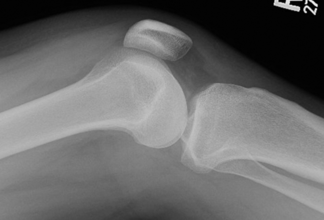 Figure 1
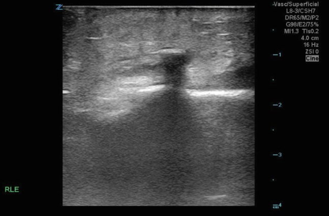 Figure 2
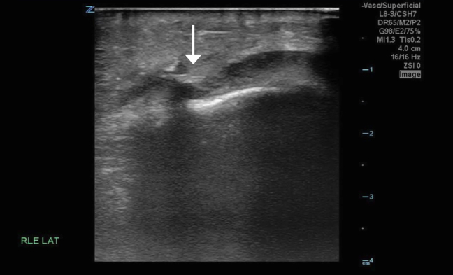 Figure 3
Peer Reviews
No public reviews on file for this paper yet. If you reviewed it on a platform where reviews are public (OpenReview, ICLR, NeurIPS, ICML), you can paste yours below so the community can read it here.
Videos
No videos yet. Explain this paper in a talk, walkthrough, or lecture? Add one.
Taxonomy
TopicsTraumatic Ocular and Foreign Body Injuries · Orthopedic Surgery and Rehabilitation · Foreign Body Medical Cases
Patient Presentation
1
A 34-year-old man with no significant medical history presented to the emergency department with a 3-day history of right knee pain after falling on wood chips. Although he initially removed visible debris from the puncture site, he continued to experience pain with movement and ambulation. On presentation, he reported increasing swelling and erythema around the area.
Diagnosis: Wooden Foreign Body Penetrating The Joint
2
Initial knee radiographs revealed a joint effusion without evidence of a radiopaque foreign body (Fig 1). A computed tomography scan similarly failed to detect any radiopaque material or soft tissue gas. Synovial fluid analysis was inconclusive. Given persistent clinical concern, a point-of-care ultrasound (POCUS) was performed. POCUS revealed a linear echogenic structure with posterior shadowing, consistent with a wooden foreign body (Fig 2). The foreign body was visualized penetrating the joint capsule (Fig 3), and the patient was taken to the operating room for irrigation and debridement.Figure 1. Lateral knee x-ray showing joint effusion with no visualized foreign body.Figure 2. Linear hyperechoic foreign body visualized in the longitudinal plane with posterior acoustic shadowing consistent with a foreign body.Figure 3. Radiopaque linear foreign body surrounded by hypoechoic synovial fluid, suggesting intra-articular intrusion. White arrow representing radiopaque linear foreign body surrounded by hypoechoic synovial fluid, suggesting intra-articular intrusion.
Wooden foreign bodies are notoriously difficult to detect on imaging. Only 7% to 15% are visible on plain radiographs, and computed tomography identifies just 63% to 70% overall.1, 2, 3 POCUS offers a radiation-free, accessible alternative with a reported sensitivity of 96.7% for detecting wooden foreign bodies.4^,^5 However, detection becomes more difficult over time, as wooden material becomes more isoechoic to surrounding tissue.3 In this case, POCUS was the only modality that visualized the foreign body and confirmed intra-articular extension, proving essential to timely diagnosis and management.
Funding and Support
By JACEP Open policy, all authors are required to disclose any and all commercial, financial, and other relationships in any way related to the subject of this article as per ICMJE conflict of interest guidelines (see www.icmje.org). The authors have stated that no such relationships exist.
Conflict of Interest
All authors have affirmed they have no conflicts of interest to declare.
The reference list from the paper itself. Each links out to its DOI / PubMed record.
- 1Graham D.D.Jr.Ultrasound in the emergency department: detection of wooden foreign bodies in the soft tissues J Emerg Med 221200275791180956010.1016/s 0736-4679(01)00440-1 · doi ↗ · pubmed ↗
- 2Pattamapaspong N.Srisuwan T.Sivasomboon C.Accuracy of radiography, computed tomography and magnetic resonance imaging in diagnosing foreign bodies in the foot Radiol Med 118220133033102274434910.1007/s 11547-012-0844-4 · doi ↗ · pubmed ↗
- 3Lewis D.Jivraj A.Atkinson P.Jarman R.My patient is injured: identifying foreign bodies with ultrasound Ultrasound 23320151741802743325410.1177/1742271 X 15579950 PMC 4760591 · doi ↗ · pubmed ↗
- 4Budhram G.R.Schmunk J.C.Bedside ultrasound AIDS identification and removal of cutaneous foreign bodies: a case series J Emerg Med 4722014 e 43e 482468545210.1016/j.jemermed.2014.01.033 · doi ↗ · pubmed ↗
- 5Davis J.Czerniski B.Au A.Adhikari S.Farrell I.Fields J.M.Diagnostic accuracy of ultrasonography in retained soft tissue foreign bodies: a systematic review and meta-analysis Acad Emerg Med 22720157777872611154510.1111/acem.12714 · doi ↗ · pubmed ↗